 Movie
Movie Controller

[English] 日本語
 Yorodumi
- PDB-8tc1: Cryo-EM Structure of Spike Glycoprotein from Civet Coronavirus 00... -
+ Open data

- Basic information
| Entry | Database: PDB / ID: 8tc1 | ||||||
|---|---|---|---|---|---|---|---|
| Title | Cryo-EM Structure of Spike Glycoprotein from Civet Coronavirus 007 in Closed Conformation | ||||||
 Components Components | Spike glycoprotein | ||||||
 Keywords Keywords | VIRAL PROTEIN / Spike / Glycoprotein / Coronavirus | ||||||
| Function / homology |  Function and homology information Function and homology informationpositive regulation of viral entry into host cell / host cell endoplasmic reticulum-Golgi intermediate compartment membrane / receptor-mediated virion attachment to host cell / host cell surface receptor binding / endocytosis involved in viral entry into host cell / fusion of virus membrane with host plasma membrane / fusion of virus membrane with host endosome membrane / viral envelope / host cell plasma membrane / virion membrane ...positive regulation of viral entry into host cell / host cell endoplasmic reticulum-Golgi intermediate compartment membrane / receptor-mediated virion attachment to host cell / host cell surface receptor binding / endocytosis involved in viral entry into host cell / fusion of virus membrane with host plasma membrane / fusion of virus membrane with host endosome membrane / viral envelope / host cell plasma membrane / virion membrane / membrane / identical protein binding Similarity search - Function | ||||||
| Biological species |  Civet SARS CoV 007/2004 (virus) Civet SARS CoV 007/2004 (virus) | ||||||
| Method | ELECTRON MICROSCOPY / single particle reconstruction / cryo EM / Resolution: 1.92 Å | ||||||
 Authors Authors | Bostina, M. / Hills, F.R. / Eruera, A.R. | ||||||
| Funding support |  New Zealand, 1items New Zealand, 1items
| ||||||
 Citation Citation | Journal: PLoS Pathog / Year: 2024 Title: Variation in structural motifs within SARS-related coronavirus spike proteins. Authors: Francesca R Hills / Alice-Roza Eruera / James Hodgkinson-Bean / Fátima Jorge / Richard Easingwood / Simon H J Brown / James C Bouwer / Yi-Ping Li / Laura N Burga / Mihnea Bostina /   Abstract: SARS-CoV-2 is the third known coronavirus (CoV) that has crossed the animal-human barrier in the last two decades. However, little structural information exists related to the close genetic species ...SARS-CoV-2 is the third known coronavirus (CoV) that has crossed the animal-human barrier in the last two decades. However, little structural information exists related to the close genetic species within the SARS-related coronaviruses. Here, we present three novel SARS-related CoV spike protein structures solved by single particle cryo-electron microscopy analysis derived from bat (bat SL-CoV WIV1) and civet (cCoV-SZ3, cCoV-007) hosts. We report complex glycan trees that decorate the glycoproteins and density for water molecules which facilitated modeling of the water molecule coordination networks within structurally important regions. We note structural conservation of the fatty acid binding pocket and presence of a linoleic acid molecule which are associated with stabilization of the receptor binding domains in the "down" conformation. Additionally, the N-terminal biliverdin binding pocket is occupied by a density in all the structures. Finally, we analyzed structural differences in a loop of the receptor binding motif between coronaviruses known to infect humans and the animal coronaviruses described in this study, which regulate binding to the human angiotensin converting enzyme 2 receptor. This study offers a structural framework to evaluate the close relatives of SARS-CoV-2, the ability to inform pandemic prevention, and aid in the development of pan-neutralizing treatments. | ||||||
| History |
|
- Structure visualization
| Structure viewer | Molecule: MolmilJmol/JSmol |
|---|
- Downloads & links
-Download
| PDBx/mmCIF format | 8tc1.cif.gz | 1.4 MB | Display | PDBx/mmCIF format |
|---|---|---|---|---|
| PDB format | pdb8tc1.ent.gz | 987.4 KB | Display | PDB format |
| PDBx/mmJSON format | 8tc1.json.gz | Tree view | PDBx/mmJSON format | |
| Others |  Other downloads Other downloads |
-Validation report
| Arichive directory | https://data.pdbj.org/pub/pdb/validation_reports/tc/8tc1ftp://data.pdbj.org/pub/pdb/validation_reports/tc/8tc1 | HTTPS FTP |
|---|
-Related structure data
| Related structure data |  41150MC 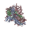 8tc0C  8tc5C M: map data used to model this data C: citing same article ( |
|---|---|
| Similar structure data |


-Links
 PDBj
PDBj
- Assembly
| Deposited unit | 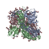
|
|---|---|
| 1 |
|
-Components
-Protein , 1 types, 3 molecules ABC
| #1: Protein | Mass: 122479.953 Da / Num. of mol.: 3 Source method: isolated from a genetically manipulated source Source: (gene. exp.) Civet SARS CoV 007/2004 (virus) / Production host:   Cricetulus griseus (Chinese hamster) / References: UniProt: Q3ZTF3 Cricetulus griseus (Chinese hamster) / References: UniProt: Q3ZTF3 |
|---|
-Sugars , 3 types, 45 molecules 
| #2: Polysaccharide | 2-acetamido-2-deoxy-beta-D-glucopyranose-(1-4)-2-acetamido-2-deoxy-beta-D-glucopyranose Source method: isolated from a genetically manipulated source #3: Polysaccharide | beta-D-mannopyranose-(1-4)-2-acetamido-2-deoxy-beta-D-glucopyranose-(1-4)-2-acetamido-2-deoxy-beta- ...beta-D-mannopyranose-(1-4)-2-acetamido-2-deoxy-beta-D-glucopyranose-(1-4)-2-acetamido-2-deoxy-beta-D-glucopyranose Source method: isolated from a genetically manipulated source #4: Sugar | ChemComp-NAG /  Type: D-saccharide, beta linking / Mass: 221.208 Da / Num. of mol.: 15 / Source method: obtained synthetically / Formula: C8H15NO6 Type: D-saccharide, beta linking / Mass: 221.208 Da / Num. of mol.: 15 / Source method: obtained synthetically / Formula: C8H15NO6 |
|---|
-Non-polymers , 2 types, 585 molecules 

| #5: Chemical |  Mass: 280.445 Da / Num. of mol.: 3 / Source method: obtained synthetically / Formula: C18H32O2 Mass: 280.445 Da / Num. of mol.: 3 / Source method: obtained synthetically / Formula: C18H32O2#6: Water | ChemComp-HOH / | Mass: 18.015 Da / Num. of mol.: 582 / Source method: isolated from a natural source / Formula: H2O |
|---|
-Details
| Has ligand of interest | N |
|---|---|
| Has protein modification | Y |
-Experimental details
-Experiment
| Experiment | Method: ELECTRON MICROSCOPY |
|---|---|
| EM experiment | Aggregation state: PARTICLE / 3D reconstruction method: single particle reconstruction |
- Sample preparation
| Component | Name: Civet SARS CoV 007/2004 / Type: VIRUS / Entity ID: #1 / Source: RECOMBINANT |
|---|---|
| Source (natural) | Organism: Civet SARS CoV 007/2004 (virus) |
| Source (recombinant) | Organism: Cricetulus griseus (Chinese hamster) |
| Details of virus | Empty: NO / Enveloped: YES / Isolate: STRAIN / Type: VIRION |
| Buffer solution | pH: 8 |
| Specimen | Embedding applied: NO / Shadowing applied: NO / Staining applied: NO / Vitrification applied: YES |
| Vitrification | Cryogen name: ETHANE |
- Electron microscopy imaging
| Experimental equipment |  Model: Titan Krios / Image courtesy: FEI Company |
|---|---|
| Microscopy | Model: FEI TITAN KRIOS |
| Electron gun | Electron source:  FIELD EMISSION GUN / Accelerating voltage: 300 kV / Illumination mode: SPOT SCAN FIELD EMISSION GUN / Accelerating voltage: 300 kV / Illumination mode: SPOT SCAN |
| Electron lens | Mode: OTHER / Nominal defocus max: 2000 nm / Nominal defocus min: 600 nm |
| Image recording | Electron dose: 59 e/Å2 / Film or detector model: GATAN K3 BIOQUANTUM (6k x 4k) |
- Processing
| EM software | Name: PHENIX / Version: 1.21_5207 / Category: model refinement | ||||||||||||||||||||||||
|---|---|---|---|---|---|---|---|---|---|---|---|---|---|---|---|---|---|---|---|---|---|---|---|---|---|
| CTF correction | Type: PHASE FLIPPING AND AMPLITUDE CORRECTION | ||||||||||||||||||||||||
| 3D reconstruction | Resolution: 1.92 Å / Resolution method: FSC 0.143 CUT-OFF / Num. of particles: 800000 / Symmetry type: POINT | ||||||||||||||||||||||||
| Refinement | Cross valid method: NONE Stereochemistry target values: GeoStd + Monomer Library + CDL v1.2 | ||||||||||||||||||||||||
| Displacement parameters | Biso mean: 66.08 Å2 | ||||||||||||||||||||||||
| Refine LS restraints |
|
