 Movie
Movie Controller

[English] 日本語
 Yorodumi
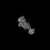
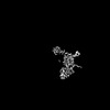
- PDB-8uo9: Structure of synaptic vesicle protein 2A in complex with a nanobody -
+ Open data

- Basic information
| Entry | Database: PDB / ID: 8uo9 | |||||||||
|---|---|---|---|---|---|---|---|---|---|---|
| Title | Structure of synaptic vesicle protein 2A in complex with a nanobody | |||||||||
 Components Components |
| |||||||||
 Keywords Keywords | TRANSPORT PROTEIN / Synaptic vesicle / SLC22 / Inhibitor / Nanobody / AEDs | |||||||||
| Function / homology |  Function and homology information Function and homology informationToxicity of botulinum toxin type F (botF) / Toxicity of botulinum toxin type D (botD) / Toxicity of botulinum toxin type E (botE) / Toxicity of botulinum toxin type A (botA) / presynaptic active zone / synaptic vesicle priming / transmembrane transporter activity / neuromuscular junction / GABA-ergic synapse / intracellular calcium ion homeostasis ...Toxicity of botulinum toxin type F (botF) / Toxicity of botulinum toxin type D (botD) / Toxicity of botulinum toxin type E (botE) / Toxicity of botulinum toxin type A (botA) / presynaptic active zone / synaptic vesicle priming / transmembrane transporter activity / neuromuscular junction / GABA-ergic synapse / intracellular calcium ion homeostasis / cell-cell junction / synaptic vesicle / synaptic vesicle membrane / neuron projection / protein kinase binding / glutamatergic synapse / endoplasmic reticulum / plasma membrane Similarity search - Function | |||||||||
| Biological species |  Homo sapiens (human) Homo sapiens (human) | |||||||||
| Method | ELECTRON MICROSCOPY / single particle reconstruction / cryo EM / Resolution: 3.3 Å | |||||||||
 Authors Authors | Mittal, A. / Martin, M.F. / Levin, E. / Adams, C. / Yang, M. / Ledecq, M. / Horanyi, P.S. / Coleman, J.A. | |||||||||
| Funding support |  United States, 1items United States, 1items
| |||||||||
 Citation Citation | Journal: Nat Struct Mol Biol / Year: 2024 Title: Structures of synaptic vesicle protein 2A and 2B bound to anticonvulsants. Authors: Anshumali Mittal / Matthew F Martin / Elena J Levin / Christopher Adams / Meng Yang / Laurent Provins / Adrian Hall / Martin Procter / Marie Ledecq / Alexander Hillisch / Christian Wolff / ...Authors: Anshumali Mittal / Matthew F Martin / Elena J Levin / Christopher Adams / Meng Yang / Laurent Provins / Adrian Hall / Martin Procter / Marie Ledecq / Alexander Hillisch / Christian Wolff / Michel Gillard / Peter S Horanyi / Jonathan A Coleman /    Abstract: Epilepsy is a common neurological disorder characterized by abnormal activity of neuronal networks, leading to seizures. The racetam class of anti-seizure medications bind specifically to a membrane ...Epilepsy is a common neurological disorder characterized by abnormal activity of neuronal networks, leading to seizures. The racetam class of anti-seizure medications bind specifically to a membrane protein found in the synaptic vesicles of neurons called synaptic vesicle protein 2 (SV2) A (SV2A). SV2A belongs to an orphan subfamily of the solute carrier 22 organic ion transporter family that also includes SV2B and SV2C. The molecular basis for how anti-seizure medications act on SV2s remains unknown. Here we report cryo-electron microscopy structures of SV2A and SV2B captured in a luminal-occluded conformation complexed with anticonvulsant ligands. The conformation bound by anticonvulsants resembles an inhibited transporter with closed luminal and intracellular gates. Anticonvulsants bind to a highly conserved central site in SV2s. These structures provide blueprints for future drug design and will facilitate future investigations into the biological function of SV2s. | |||||||||
| History |
|
- Structure visualization
| Structure viewer | Molecule: MolmilJmol/JSmol |
|---|
- Downloads & links
-Download
| PDBx/mmCIF format | 8uo9.cif.gz | 149.7 KB | Display | PDBx/mmCIF format |
|---|---|---|---|---|
| PDB format | pdb8uo9.ent.gz | 106.9 KB | Display | PDB format |
| PDBx/mmJSON format | 8uo9.json.gz | Tree view | PDBx/mmJSON format | |
| Others |  Other downloads Other downloads |
-Validation report
| Arichive directory | https://data.pdbj.org/pub/pdb/validation_reports/uo/8uo9ftp://data.pdbj.org/pub/pdb/validation_reports/uo/8uo9 | HTTPS FTP |
|---|
-Related structure data
| Related structure data | 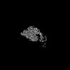 42431MC  8uo8C 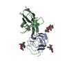 8uoaC M: map data used to model this data C: citing same article ( |
|---|---|
| Similar structure data |
-Links
 PDBj
PDBj


- Assembly
| Deposited unit | 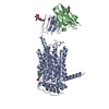
|
|---|---|
| 1 |
|
-Components
-Protein / Antibody , 2 types, 2 molecules AB
| #1: Protein | Mass: 75589.188 Da / Num. of mol.: 1 Source method: isolated from a genetically manipulated source Source: (gene. exp.) Homo sapiens (human) / Gene: SV2A / Cell line (production host): tsA201 / Production host: Homo sapiens (human) / References: UniProt: Q7L0J3 |
|---|---|
| #2: Antibody | Mass: 15469.103 Da / Num. of mol.: 1 Source method: isolated from a genetically manipulated source Source: (gene. exp.)  |
-Sugars , 2 types, 3 molecules
| #3: Polysaccharide | beta-D-mannopyranose-(1-4)-2-acetamido-2-deoxy-beta-D-glucopyranose-(1-4)-2-acetamido-2-deoxy-beta- ...beta-D-mannopyranose-(1-4)-2-acetamido-2-deoxy-beta-D-glucopyranose-(1-4)-2-acetamido-2-deoxy-beta-D-glucopyranose Source method: isolated from a genetically manipulated source |
|---|---|
| #4: Polysaccharide | Source method: isolated from a genetically manipulated source |
-Non-polymers , 3 types, 3 molecules 

| #5: Chemical | ChemComp-X49 / ( Mass: 444.395 Da / Num. of mol.: 1 / Source method: obtained synthetically / Formula: C16H18F6N4O2S / Feature type: SUBJECT OF INVESTIGATION |
|---|---|
| #6: Chemical | ChemComp-Y01 /  Mass: 486.726 Da / Num. of mol.: 1 / Source method: obtained synthetically / Formula: C31H50O4 Mass: 486.726 Da / Num. of mol.: 1 / Source method: obtained synthetically / Formula: C31H50O4 |
| #7: Chemical | ChemComp-PS1 /  Mass: 566.642 Da / Num. of mol.: 1 / Source method: obtained synthetically / Formula: C26H49NO10P Mass: 566.642 Da / Num. of mol.: 1 / Source method: obtained synthetically / Formula: C26H49NO10P |
-Details
| Has ligand of interest | Y |
|---|---|
| Has protein modification | Y |
-Experimental details
-Experiment
| Experiment | Method: ELECTRON MICROSCOPY |
|---|---|
| EM experiment | Aggregation state: PARTICLE / 3D reconstruction method: single particle reconstruction |
- Sample preparation
| Component | Name: Protein with a nanobody / Type: COMPLEX / Entity ID: #2 / Source: MULTIPLE SOURCES |
|---|---|
| Molecular weight | Value: 98.1 kDa/nm / Experimental value: NO |
| Source (natural) | Organism: Homo sapiens (human) |
| Source (recombinant) | Organism: Homo sapiens (human) |
| Buffer solution | pH: 8 |
| Specimen | Conc.: 4.38 mg/ml / Embedding applied: NO / Shadowing applied: NO / Staining applied: NO / Vitrification applied: YES |
| Specimen support | Grid material: GOLD / Grid mesh size: 200 divisions/in. / Grid type: Quantifoil R2/1 |
| Vitrification | Instrument: FEI VITROBOT MARK IV / Cryogen name: ETHANE-PROPANE / Humidity: 100 % / Chamber temperature: 298 K |
- Electron microscopy imaging
| Experimental equipment |  Model: Titan Krios / Image courtesy: FEI Company |
|---|---|
| Microscopy | Model: TFS KRIOS |
| Electron gun | Electron source:  FIELD EMISSION GUN / Accelerating voltage: 300 kV / Illumination mode: FLOOD BEAM FIELD EMISSION GUN / Accelerating voltage: 300 kV / Illumination mode: FLOOD BEAM |
| Electron lens | Mode: BRIGHT FIELD / Nominal magnification: 77160 X / Nominal defocus max: 2800 nm / Nominal defocus min: 750 nm |
| Image recording | Electron dose: 60 e/Å2 / Film or detector model: GATAN K3 BIOQUANTUM (6k x 4k) |
| EM imaging optics | Energyfilter name: GIF Bioquantum / Energyfilter slit width: 20 eV |
- Processing
| Image processing |
| |||||||||||||||||||||||||||||||||||
|---|---|---|---|---|---|---|---|---|---|---|---|---|---|---|---|---|---|---|---|---|---|---|---|---|---|---|---|---|---|---|---|---|---|---|---|---|
| CTF correction |
| |||||||||||||||||||||||||||||||||||
| 3D reconstruction |
| |||||||||||||||||||||||||||||||||||
| Atomic model building | Accession code: AF-Q7L0J3-F1 / Source name: AlphaFold / Type: in silico model | |||||||||||||||||||||||||||||||||||
| Refinement | Highest resolution: 3.3 Å |
