 Movie
Movie Controller
 Structure viewers
 About Yorodumi Papers


+Search query
-Structure paper
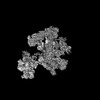
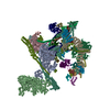
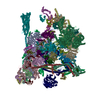
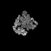
| Title | Mechanism for the initiation of spliceosome disassembly. |
|---|---|
| Journal, issue, pages | Nature, Vol. 632, Issue 8024, Page 443-450, Year 2024 |
| Publish date | Jun 26, 2024 |
 Authors Authors | Matthias K Vorländer / Patricia Rothe / Justus Kleifeld / Eric D Cormack / Lalitha Veleti / Daria Riabov-Bassat / Laura Fin / Alex W Phillips / Luisa Cochella / Clemens Plaschka /   |
| PubMed Abstract | Precursor-mRNA (pre-mRNA) splicing requires the assembly, remodelling and disassembly of the multi-megadalton ribonucleoprotein complex called the spliceosome. Recent studies have shed light on ...Precursor-mRNA (pre-mRNA) splicing requires the assembly, remodelling and disassembly of the multi-megadalton ribonucleoprotein complex called the spliceosome. Recent studies have shed light on spliceosome assembly and remodelling for catalysis, but the mechanism of disassembly remains unclear. Here we report cryo-electron microscopy structures of nematode and human terminal intron lariat spliceosomes along with biochemical and genetic data. Our results uncover how four disassembly factors and the conserved RNA helicase DHX15 initiate spliceosome disassembly. The disassembly factors probe large inner and outer spliceosome surfaces to detect the release of ligated mRNA. Two of these factors, TFIP11 and C19L1, and three general spliceosome subunits, SYF1, SYF2 and SDE2, then dock and activate DHX15 on the catalytic U6 snRNA to initiate disassembly. U6 therefore controls both the start and end of pre-mRNA splicing. Taken together, our results explain the molecular basis of the initiation of canonical spliceosome disassembly and provide a framework to understand general spliceosomal RNA helicase control and the discard of aberrant spliceosomes. |
 External links External links | Nature / PubMed:38925148 / PubMed Central |
| Methods | EM (single particle) |
| Resolution | 2.73 - 8.06 Å |
| Structure data | EMDB-19397: Composite map of the C. elegans Intron Lariat Spliceosome primed for disassembly (ILS') EMDB-19398, PDB-8ro1: EMDB-19399, PDB-8ro2:  EMDB-50447: Structure of the C. elegans Intron Lariat Spliceosome (Map 1)  EMDB-50449: Structure of the C. elegans Intron Lariat Spliceosome (Map 2)  EMDB-50450: Structure of the C. elegans Intron Lariat Spliceosome (Map 3)  EMDB-50451: Structure of the C. elegans Intron Lariat Spliceosome (Map 4)  EMDB-50452: Structure of the C. elegans Intron Lariat Spliceosome (Map 5)  EMDB-50453: Structure of the C. elegans Intron Lariat Spliceosome (Map 6)  EMDB-50454: Structure of the C. elegans Intron Lariat Spliceosome (Map 7)  EMDB-50455: Structure of the C. elegans Intron Lariat Spliceosome (Map 8)  EMDB-50456: Structure of the C. elegans Intron Lariat Spliceosome (Map 9)  EMDB-50457: Structure of the C. elegans Intron Lariat Spliceosome (Map 10)  EMDB-50458: Structure of the C. elegans Intron Lariat Spliceosome (Map 11)  EMDB-50459: Structure of the C. elegans Intron Lariat Spliceosome (Map 12)  EMDB-50460: Structure of the C. elegans Intron Lariat Spliceosome (Map 13)  EMDB-50461: Structure of the C. elegans Intron Lariat Spliceosome (Map 14)  EMDB-50462: Structure of the C. elegans Intron Lariat Spliceosome (Map 15)  EMDB-50463: Structure of the C. elegans Intron Lariat Spliceosome (Map 16)  EMDB-50464: Structure of the C. elegans Intron Lariat Spliceosome (Map 17)  EMDB-50465: Structure of the C. elegans Intron Lariat Spliceosome (Map 18)  EMDB-50466: Structure of the C. elegans Intron Lariat Spliceosome (Map 19)  EMDB-50467: Structure of the C. elegans Intron Lariat Spliceosome (Map 20)  EMDB-50468: Structure of the C. elegans Intron Lariat Spliceosome (Map 21) 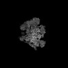 EMDB-50469: Structure of the C. elegans Intron Lariat Spliceosome (Map 22) 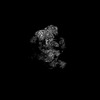 EMDB-50471: Structure of the C. elegans Intron Lariat Spliceosome (Map 23) 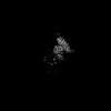 EMDB-50472: Structure of the C. elegans Intron Lariat Spliceosome (Map 24) 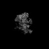 EMDB-50473: Structure of the C. elegans Intron Lariat Spliceosome (Map 25) 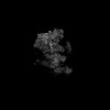 EMDB-50474: Structure of the C. elegans Intron Lariat Spliceosome (Map 27) 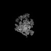 EMDB-50475: Structure of the C. elegans Intron Lariat Spliceosome (Map 26) 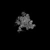 EMDB-50477: Integrative Structure of the human intron lariat Spliceosome (ILS'') (Map 1) 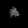 EMDB-50478: Integrative Structure of the human intron lariat Spliceosome (ILS'') (Map 2)  EMDB-50479: Integrative Structure of the human intron lariat Spliceosome (ILS'') (Map 3)  EMDB-50480: Integrative Structure of the human intron lariat Spliceosome (ILS'') (Map 4)  EMDB-50481: Integrative Structure of the human intron lariat Spliceosome (ILS'') (Map 5)  EMDB-50482: Integrative Structure of the human intron lariat Spliceosome (ILS'')(Map 6)  EMDB-50483: Integrative Structure of the human intron lariat Spliceosome (ILS'') (Map 7)  EMDB-50484: Integrative Structure of the human intron lariat Spliceosome (ILS'') (Map 8)  EMDB-50485: Integrative Structure of the human intron lariat Spliceosome (ILS'') (Map 9)  EMDB-50486: Integrative Structure of the human intron lariat Spliceosome (ILS'') (Map 10) 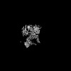 EMDB-50487: Integrative Structure of the human intron lariat Spliceosome (ILS'') (Map 11) 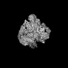 EMDB-50488: Integrative Structure of the human intron lariat Spliceosome (ILS'') (Map 12) 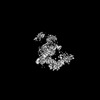 EMDB-50489: Integrative Structure of the human intron lariat Spliceosome (ILS'') (Map 13) 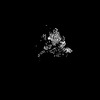 EMDB-50490: Integrative Structure of the human intron lariat Spliceosome (ILS'')(Map14)  PDB-9fmd: |
| Chemicals |  ChemComp-MG:  ChemComp-IHP:  ChemComp-GTP:  ChemComp-ZN:  ChemComp-K:  ChemComp-ATP: |
| Source |
|
 Keywords Keywords | SPLICING / mRNA / Intorn Lariat spliceosome / ILS / pre-mRNA / pre-mRNA splicing / intron lariat spliceosome / gene expression / spliceosome / P-complex |


 homo sapiens (human)
 human adenovirus 2