6GZ6
 
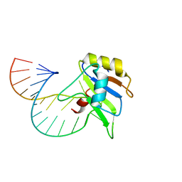
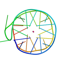 | | Structure of a left-handed G-quadruplex | | 分子名称: | DNA (27-MER), POTASSIUM ION | | 著者 | Bakalar, B, Heddi, B, Schmitt, E, Mechulam, Y, Phan, A.T. | | 登録日 | 2018-07-03 | | 公開日 | 2019-04-24 | | 最終更新日 | 2024-01-17 | | 実験手法 | X-RAY DIFFRACTION (2.006 Å) | | 主引用文献 | A Minimal Sequence for Left-Handed G-Quadruplex Formation.
Angew.Chem.Int.Ed.Engl., 58, 2019
|
|
6D37
 | |
6CVO
 | | Human Aprataxin (Aptx) bound to nicked RNA-DNA, AMP and Zn product complex | | 分子名称: | ADENOSINE MONOPHOSPHATE, Aprataxin, BETA-MERCAPTOETHANOL, ... | | 著者 | Schellenberg, M.J, Tumbale, P.P, Williams, R.S. | | 登録日 | 2018-03-28 | | 公開日 | 2018-08-29 | | 最終更新日 | 2023-10-04 | | 実験手法 | X-RAY DIFFRACTION (2.4 Å) | | 主引用文献 | Mechanism of APTX nicked DNA sensing and pleiotropic inactivation in neurodegenerative disease.
EMBO J., 37, 2018
|
|
6GB1
 | |
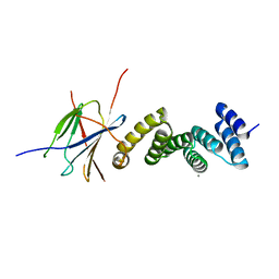
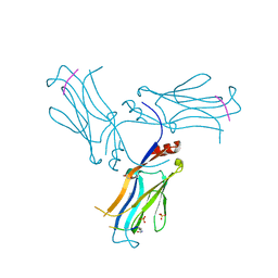
6GBM
 | | Solution structure of FUS-RRM bound to stem-loop RNA | | 分子名称: | RNA (5'-R(*GP*GP*CP*AP*GP*AP*UP*UP*AP*CP*AP*AP*UP*UP*CP*UP*AP*UP*UP*UP*GP*CP*C)-3'), RNA-binding protein FUS | | 著者 | Loughlin, F.E, Allain, F.H.-T. | | 登録日 | 2018-04-15 | | 公開日 | 2019-02-20 | | 最終更新日 | 2024-05-15 | | 実験手法 | SOLUTION NMR | | 主引用文献 | The Solution Structure of FUS Bound to RNA Reveals a Bipartite Mode of RNA Recognition with Both Sequence and Shape Specificity.
Mol. Cell, 73, 2019
|
|
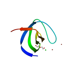
6CGX
 | | Backbone cyclised conotoxin Vc1.1 mutant - D11A, E14A | | 分子名称: | Alpha-conotoxin Vc1A | | 著者 | Clark, R.J. | | 登録日 | 2018-02-21 | | 公開日 | 2018-05-23 | | 最終更新日 | 2023-06-14 | | 実験手法 | SOLUTION NMR | | 主引用文献 | Structure-Activity Studies Reveal the Molecular Basis for GABAB-Receptor Mediated Inhibition of High Voltage-Activated Calcium Channels by alpha-Conotoxin Vc1.1.
ACS Chem. Biol., 13, 2018
|
|
6COQ
 | | CSP1-K6A | | 分子名称: | Competence-stimulating peptide type 1 | | 著者 | Yang, Y. | | 登録日 | 2018-03-12 | | 公開日 | 2018-08-29 | | 最終更新日 | 2024-05-15 | | 実験手法 | SOLUTION NMR | | 主引用文献 | Structural Characterization of Competence-Stimulating Peptide Analogues Reveals Key Features for ComD1 and ComD2 Receptor Binding in Streptococcus pneumoniae.
Biochemistry, 57, 2018
|
|
6G4J
 | |
6COP
 | | CSP1-R3A | | 分子名称: | Competence-stimulating peptide type 1 | | 著者 | Yang, Y. | | 登録日 | 2018-03-12 | | 公開日 | 2018-08-29 | | 最終更新日 | 2024-05-15 | | 実験手法 | SOLUTION NMR | | 主引用文献 | Structural Characterization of Competence-Stimulating Peptide Analogues Reveals Key Features for ComD1 and ComD2 Receptor Binding in Streptococcus pneumoniae.
Biochemistry, 57, 2018
|
|
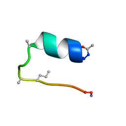
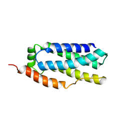
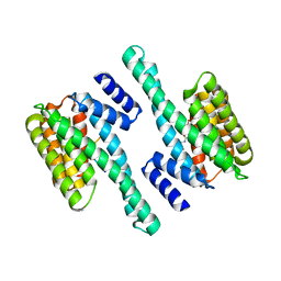
6BZL
 | | Solution structure of VEK75 | | 分子名称: | M protein | | 著者 | Qiu, C, Yuan, Y, Castellino, F.J. | | 登録日 | 2017-12-24 | | 公開日 | 2019-01-16 | | 最終更新日 | 2024-05-01 | | 実験手法 | SOLUTION NMR | | 主引用文献 | Contributions of different modules of the plasminogen-binding Streptococcus pyogenes M-protein that mediate its functional dimerization.
J.Struct.Biol., 204, 2018
|
|
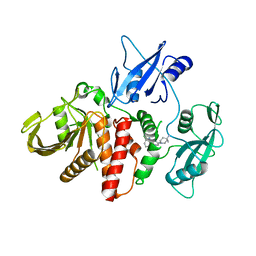
6BPU
 | | Crystal structure of ferrous form of the F2-Tyr157 human cysteine dioxygenase with both uncrosslinked and crosslinked cofactor | | 分子名称: | Cysteine dioxygenase type 1, FE (II) ION, GLYCEROL, ... | | 著者 | Liu, A, Li, J, Shin, I. | | 登録日 | 2017-11-26 | | 公開日 | 2018-07-04 | | 最終更新日 | 2023-11-15 | | 実験手法 | X-RAY DIFFRACTION (1.8 Å) | | 主引用文献 | Cleavage of a carbon-fluorine bond by an engineered cysteine dioxygenase.
Nat. Chem. Biol., 14, 2018
|
|
6GWS
 | | Crystal structure of human PCNA in complex with three p15 peptides | | 分子名称: | PCNA-associated factor, Proliferating cell nuclear antigen | | 著者 | De March, M, Merino, N, Gonzalez-Magana, A, Romano-Moreno, M, Onesti, S, Blanco, F.J, De Biasio, A. | | 登録日 | 2018-06-25 | | 公開日 | 2018-08-22 | | 最終更新日 | 2024-01-17 | | 実験手法 | X-RAY DIFFRACTION (2.9 Å) | | 主引用文献 | p15PAF binding to PCNA modulates the DNA sliding surface.
Nucleic Acids Res., 46, 2018
|
|
6D74
 | |
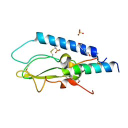
6H5T
 | | Intersectin SH3A short isoform | | 分子名称: | 2,5,8,11,14,17,20,23-OCTAOXAPENTACOSAN-25-OL, ACETATE ION, CHLORIDE ION, ... | | 著者 | Driller, J.H, Gerth, F, Freund, C, Wahl, M.C, Loll, B. | | 登録日 | 2018-07-25 | | 公開日 | 2019-02-27 | | 最終更新日 | 2024-05-15 | | 実験手法 | X-RAY DIFFRACTION (1.689 Å) | | 主引用文献 | Exon Inclusion Modulates Conformational Plasticity and Autoinhibition of the Intersectin 1 SH3A Domain.
Structure, 27, 2019
|
|
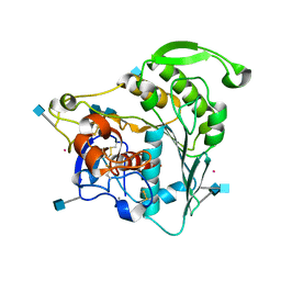
6GXG
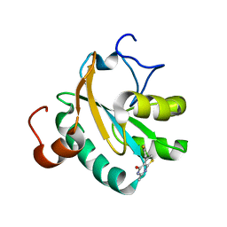 | | Tryparedoxin from Trypanosoma brucei in complex with CFT | | 分子名称: | 5-(4-fluorophenyl)-2-methyl-3~{H}-thieno[2,3-d]pyrimidin-4-one, GLYCEROL, Tryparedoxin | | 著者 | Bader, N, Wagner, A, Hellmich, U, Schindelin, H. | | 登録日 | 2018-06-27 | | 公開日 | 2019-01-16 | | 最終更新日 | 2024-01-17 | | 実験手法 | X-RAY DIFFRACTION (1.6 Å) | | 主引用文献 | Inhibitor-Induced Dimerization of an Essential Oxidoreductase from African Trypanosomes.
Angew. Chem. Int. Ed. Engl., 58, 2019
|
|
6DFA
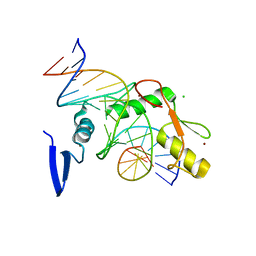 | | Kaiso (ZBTB33) E535A zinc finger DNA binding domain in complex with the specific Kaiso binding sequence (KBS) | | 分子名称: | CHLORIDE ION, DNA (5'-D(*CP*GP*TP*TP*AP*TP*TP*GP*GP*CP*AP*GP*GP*AP*AP*GP*CP*A)-3'), DNA (5'-D(*TP*GP*CP*TP*TP*CP*CP*TP*GP*CP*CP*AP*AP*TP*AP*AP*CP*G)-3'), ... | | 著者 | Nikolova, E.N, Stanfield, R.L, Dyson, H.J, Wright, P.E. | | 登録日 | 2018-05-14 | | 公開日 | 2019-05-22 | | 最終更新日 | 2023-10-11 | | 実験手法 | X-RAY DIFFRACTION (1.908 Å) | | 主引用文献 | A conformational switch in the zinc finger protein Kaiso mediates differential readout of specific and methylated DNA sequences.
Biochemistry, 2020
|
|
6GXZ
 | | Crystal structure of the human RPAP3(TPR2)-PIH1D1(CS) complex | | 分子名称: | CALCIUM ION, PIH1 domain-containing protein 1, RNA polymerase II-associated protein 3 | | 著者 | Henri, J, Quinternet, M, Manival, X, Chagot, M.-E, Charpentier, B, Meyer, P. | | 登録日 | 2018-06-27 | | 公開日 | 2018-08-08 | | 最終更新日 | 2024-01-17 | | 実験手法 | X-RAY DIFFRACTION (2.965 Å) | | 主引用文献 | Deep Structural Analysis of RPAP3 and PIH1D1, Two Components of the HSP90 Co-chaperone R2TP Complex.
Structure, 26, 2018
|
|
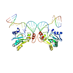
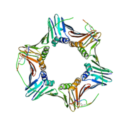
6GIS
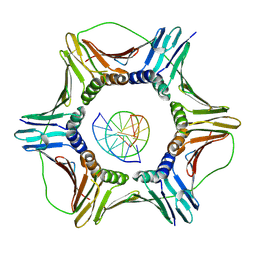 | | Structural basis of human clamp sliding on DNA | | 分子名称: | DNA (5'-D(P*AP*TP*AP*CP*GP*AP*TP*GP*GP*G)-3'), DNA (5'-D(P*CP*CP*CP*AP*TP*CP*GP*TP*AP*T)-3'), Proliferating cell nuclear antigen | | 著者 | De March, M, Merino, N, Barrera-Vilarmau, S, Crehuet, R, Onesti, S, Blanco, F.J, De Biasio, A. | | 登録日 | 2018-05-15 | | 公開日 | 2018-05-30 | | 最終更新日 | 2024-01-17 | | 実験手法 | X-RAY DIFFRACTION (2.82 Å) | | 主引用文献 | Structural basis of human PCNA sliding on DNA.
Nat Commun, 8, 2017
|
|
6CMS
 | | Closed structure of active SHP2 mutant E76K bound to SHP099 inhibitor | | 分子名称: | 6-(4-azanyl-4-methyl-piperidin-1-yl)-3-[2,3-bis(chloranyl)phenyl]pyrazin-2-amine, Tyrosine-protein phosphatase non-receptor type 11 | | 著者 | Padua, R.A.P, Sun, Y, Marko, I, Pitsawong, W, Kern, D. | | 登録日 | 2018-03-06 | | 公開日 | 2018-11-14 | | 最終更新日 | 2023-10-04 | | 実験手法 | X-RAY DIFFRACTION (2.68 Å) | | 主引用文献 | Mechanism of activating mutations and allosteric drug inhibition of the phosphatase SHP2.
Nat Commun, 9, 2018
|
|
6COT
 | | CSP2-d10 | | 分子名称: | Competence-stimulating peptide type 2 | | 著者 | Yang, Y. | | 登録日 | 2018-03-12 | | 公開日 | 2018-08-29 | | 最終更新日 | 2018-09-19 | | 実験手法 | SOLUTION NMR | | 主引用文献 | Structural Characterization of Competence-Stimulating Peptide Analogues Reveals Key Features for ComD1 and ComD2 Receptor Binding in Streptococcus pneumoniae.
Biochemistry, 57, 2018
|
|
6CBI
 | | PCNA in complex with inhibitor | | 分子名称: | GLY-ARG-LYS-ARG-ARG-GLN-DAB-SER-MET-THR-GLU-PHE-TYR-HIS, Proliferating cell nuclear antigen, SULFATE ION | | 著者 | Bruning, J.B, Wegener, K.L. | | 登録日 | 2018-02-03 | | 公開日 | 2018-07-04 | | 最終更新日 | 2020-01-29 | | 実験手法 | X-RAY DIFFRACTION (2.75 Å) | | 主引用文献 | Rational Design of a 310-Helical PIP-Box Mimetic Targeting PCNA, the Human Sliding Clamp.
Chemistry, 24, 2018
|
|
6CCI
 | | The Crystal Structure of XOAT1 | | 分子名称: | 2-acetamido-2-deoxy-beta-D-glucopyranose, 2-acetamido-2-deoxy-beta-D-glucopyranose-(1-4)-2-acetamido-2-deoxy-beta-D-glucopyranose, CALCIUM ION, ... | | 著者 | Alahuhta, P.M, Lunin, V.V. | | 登録日 | 2018-02-07 | | 公開日 | 2019-02-20 | | 最終更新日 | 2020-09-02 | | 実験手法 | X-RAY DIFFRACTION (1.85 Å) | | 主引用文献 | Molecular Mechanism of Polysaccharide Acetylation by the Arabidopsis XylanO-acetyltransferase XOAT1.
Plant Cell, 32, 2020
|
|
6GKG
 | |
6AXJ
 | |
6AVI
 | |


