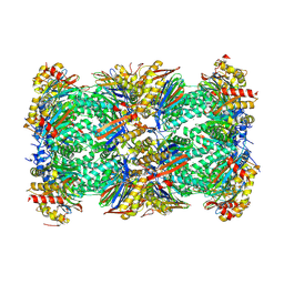
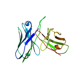
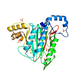
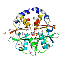
3K8Q
 
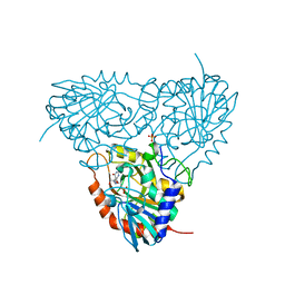 | | Crystal structure of human purine nucleoside phosphorylase in complex with SerMe-Immucillin H | | 分子名称: | 7-({[2-hydroxy-1-(hydroxymethyl)ethyl]amino}methyl)-3,5-dihydro-4H-pyrrolo[3,2-d]pyrimidin-4-one, PHOSPHATE ION, Purine nucleoside phosphorylase | | 著者 | Ho, M, Almo, S.C, Scharmm, V.L. | | 登録日 | 2009-10-14 | | 公開日 | 2010-03-02 | | 最終更新日 | 2024-02-21 | | 実験手法 | X-RAY DIFFRACTION (2.5 Å) | | 主引用文献 | Crystal structure of human nucleoside phosphorylase in complex with SerMe-ImmH
Proc.Natl.Acad.Sci.USA, 2010
|
|
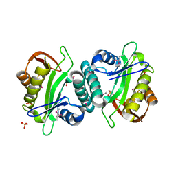
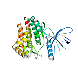
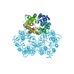
6ZZQ
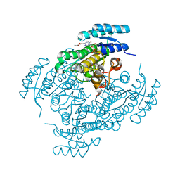 | | Crystal structure of (R)-3-hydroxybutyrate dehydrogenase from Acinetobacter baumannii complexed with NAD+ and acetoacetate | | 分子名称: | 3-hydroxybutyrate dehydrogenase, ACETOACETIC ACID, NICOTINAMIDE-ADENINE-DINUCLEOTIDE | | 著者 | Machado, T.F.G, da Silva, R.G, Gloster, T.M, McMahon, S.A, Oehler, V. | | 登録日 | 2020-08-05 | | 公開日 | 2020-10-07 | | 最終更新日 | 2024-01-31 | | 実験手法 | X-RAY DIFFRACTION (1.93 Å) | | 主引用文献 | Dissecting the Mechanism of ( R )-3-Hydroxybutyrate Dehydrogenase by Kinetic Isotope Effects, Protein Crystallography, and Computational Chemistry.
Acs Catalysis, 10, 2020
|
|
6ZT1
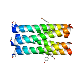 | |
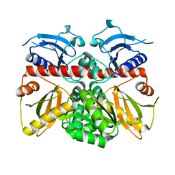
3JV7
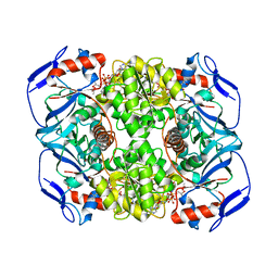 | | Structure of ADH-A from Rhodococcus ruber | | 分子名称: | (4S)-2-METHYL-2,4-PENTANEDIOL, ACETIC ACID, ADH-A, ... | | 著者 | Karabec, M, Lyskowski, A, Gruber, K. | | 登録日 | 2009-09-16 | | 公開日 | 2010-08-25 | | 最終更新日 | 2023-11-01 | | 実験手法 | X-RAY DIFFRACTION (2 Å) | | 主引用文献 | Structural insights into substrate specificity and solvent tolerance in alcohol dehydrogenase ADH-'A' from Rhodococcus ruber DSM 44541.
Chem.Commun.(Camb.), 2010
|
|
8BZL
 | | Human 20S Proteasome in complex with peptide activator peptide BLM42 | | 分子名称: | 1-ETHYL-PYRROLIDINE-2,5-DIONE, ARG-SER-TYR-TYR-SER, CHLORIDE ION, ... | | 著者 | Henneberg, F, Chari, A, Jankowska, E, Witkowska, J. | | 登録日 | 2022-12-15 | | 公開日 | 2023-12-27 | | 最終更新日 | 2025-02-26 | | 実験手法 | X-RAY DIFFRACTION (2.14 Å) | | 主引用文献 | Blm10-Based Compounds Add to the Knowledge of How Allosteric Modulators Influence Human 20S Proteasome.
Acs Chem.Biol., 2025
|
|
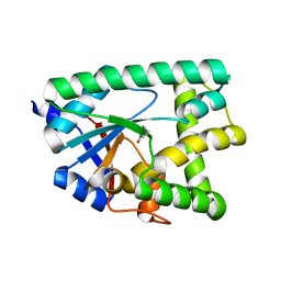
3K0D
 | | Crystal Structure of CNG mimicking NaK mutant, NaK-ETPP, K+ complex | | 分子名称: | (4S)-2-METHYL-2,4-PENTANEDIOL, POTASSIUM ION, Potassium channel protein NaK | | 著者 | Jiang, Y, Derebe, M.G. | | 登録日 | 2009-09-24 | | 公開日 | 2011-01-12 | | 最終更新日 | 2024-02-21 | | 実験手法 | X-RAY DIFFRACTION (1.95 Å) | | 主引用文献 | Structural studies of ion permeation and Ca2+ blockage of a bacterial channel mimicking the cyclic nucleotide-gated channel pore.
Proc.Natl.Acad.Sci.USA, 108, 2011
|
|
8C6R
 | | PBP AccA from A. tumefaciens Bo542 in apoform 4 | | 分子名称: | 1,2-ETHANEDIOL, Agrocinopine utilization periplasmic binding protein AccA, DI(HYDROXYETHYL)ETHER, ... | | 著者 | Morera, S, Vigouroux, A. | | 登録日 | 2023-01-12 | | 公開日 | 2024-01-24 | | 最終更新日 | 2024-02-07 | | 実験手法 | X-RAY DIFFRACTION (1.884 Å) | | 主引用文献 | A highly conserved ligand-binding site for AccA transporters of antibiotic and quorum-sensing regulator in Agrobacterium leads to a different specificity.
Biochem.J., 481, 2024
|
|
3JTR
 | | Mutations in Cephalosporin Acylase Affecting Stability and Autoproteolysis | | 分子名称: | GLYCEROL, Glutaryl 7-aminocephalosporanic acid acylase | | 著者 | Cho, K.J, Kim, J.K, Lee, J.H, Shin, H.J, Park, S.S, Kim, K.H. | | 登録日 | 2009-09-14 | | 公開日 | 2010-01-26 | | 最終更新日 | 2023-11-01 | | 実験手法 | X-RAY DIFFRACTION (2.5 Å) | | 主引用文献 | Structural features of cephalosporin acylase reveal the basis of autocatalytic activation.
Biochem.Biophys.Res.Commun., 390, 2009
|
|
3JUY
 | |
3JUW
 | |
3JV3
 | |
3JWJ
 | |
7A15
 | |
3JY9
 | | Janus Kinase 2 Inhibitors | | 分子名称: | (3S)-3-(4-hydroxyphenyl)-1,5-dihydro-1,5,12-triazabenzo[4,5]cycloocta[1,2,3-cd]inden-4(3H)-one, SODIUM ION, Tyrosine-protein kinase JAK2 | | 著者 | Zuccola, H.J, Ledeboer, M.W, Pierce, A.C. | | 登録日 | 2009-09-21 | | 公開日 | 2009-12-01 | | 最終更新日 | 2024-11-20 | | 実験手法 | X-RAY DIFFRACTION (2.1 Å) | | 主引用文献 | Janus kinase 2 inhibitors. Synthesis and characterization of a novel polycyclic azaindole.
J.Med.Chem., 52, 2009
|
|
7A79
 | | Crystal structure of RXR gamma LBD in complexes with palmitic acid and GRIP-1 peptide | | 分子名称: | Nuclear receptor coactivator 2, PALMITIC ACID, Retinoic acid receptor RXR-gamma | | 著者 | Chaikuad, A, Merk, D, Knapp, S, Structural Genomics Consortium (SGC) | | 登録日 | 2020-08-27 | | 公開日 | 2020-10-21 | | 最終更新日 | 2024-01-31 | | 実験手法 | X-RAY DIFFRACTION (2.05 Å) | | 主引用文献 | Comprehensive Set of Tertiary Complex Structures and Palmitic Acid Binding Provide Molecular Insights into Ligand Design for RXR Isoforms.
Int J Mol Sci, 21, 2020
|
|
3JXB
 | |
3JXJ
 | |
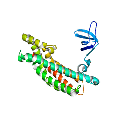
3K29
 | | Structure of a putative YscO homolog CT670 from Chlamydia trachomatis | | 分子名称: | Putative uncharacterized protein | | 著者 | Lam, R, Singer, A, Skarina, T, Onopriyenko, O, Bochkarev, A, Brunzelle, J.S, Edwards, A.M, Anderson, W.F, Chirgadze, N.Y, Savchenko, A, Center for Structural Genomics of Infectious Diseases (CSGID) | | 登録日 | 2009-09-29 | | 公開日 | 2009-10-13 | | 最終更新日 | 2024-11-20 | | 実験手法 | X-RAY DIFFRACTION (2 Å) | | 主引用文献 | Structure and protein-protein interaction studies on Chlamydia trachomatis protein CT670 (YscO Homolog).
J.Bacteriol., 192, 2010
|
|
7ABP
 | |
6ZPE
 | |
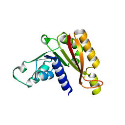
3K2L
 | | Crystal Structure of dual-specificity tyrosine phosphorylation regulated kinase 2 (DYRK2) | | 分子名称: | CHLORIDE ION, Dual specificity tyrosine-phosphorylation-regulated kinase 2, SODIUM ION, ... | | 著者 | Filippakopoulos, P, Myrianthopoulos, V, Soundararajan, M, Krojer, T, Hapka, E, Fedorov, O, Berridge, G, Wang, J, Shrestha, L, Pike, A.C.W, Ugochukwu, E, von Delft, F, Arrowsmith, C.H, Edwards, A, Weigelt, J, Bountra, C, Mikros, E, Knapp, S, Structural Genomics Consortium (SGC) | | 登録日 | 2009-09-30 | | 公開日 | 2009-10-13 | | 最終更新日 | 2024-11-20 | | 実験手法 | X-RAY DIFFRACTION (2.36 Å) | | 主引用文献 | Structures of Down Syndrome Kinases, DYRKs, Reveal Mechanisms of Kinase Activation and Substrate Recognition.
Structure, 21, 2013
|
|
3K2V
 | | Structure of the CBS pair of a putative D-arabinose 5-phosphate isomerase from Klebsiella pneumoniae subsp. pneumoniae. | | 分子名称: | CYTIDINE 5'-MONOPHOSPHATE 3-DEOXY-BETA-D-GULO-OCT-2-ULO-PYRANOSONIC ACID, GLYCEROL, SULFATE ION, ... | | 著者 | Cuff, M.E, Volkart, L, Bearden, J, Joachimiak, A, Midwest Center for Structural Genomics (MCSG) | | 登録日 | 2009-09-30 | | 公開日 | 2009-12-22 | | 最終更新日 | 2024-11-20 | | 実験手法 | X-RAY DIFFRACTION (1.95 Å) | | 主引用文献 | Structure of the CBS pair of a putative D-arabinose 5-phosphate isomerase from Klebsiella pneumoniae subsp. pneumoniae.
TO BE PUBLISHED
|
|
3K3F
 | |
3K3P
 | |
6ZTU
 | |


