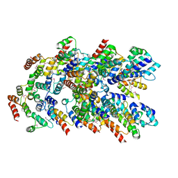
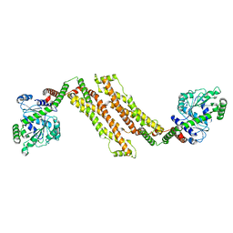
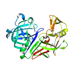
5LYQ
 
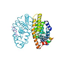 | | Crystal structure of the Retinoic Acid Receptor alpha in complex with a synthetic spiroketal agonist and a fragment of the TIF2 co-activator. | | 分子名称: | (2~{R})-6,6,9,9-tetramethylspiro[3,4,7,8-tetrahydrobenzo[g]chromene-2,2'-3,4-dihydrochromene]-6'-carboxylic acid, HIS-LYS-ILE-LEU-HIS-ARG-LEU-LEU-GLN-ASP, Retinoic acid receptor RXR-alpha | | 著者 | Andrei, S.A, Ottmann, C. | | 登録日 | 2016-09-28 | | 公開日 | 2017-04-26 | | 最終更新日 | 2024-01-17 | | 実験手法 | X-RAY DIFFRACTION (2.17 Å) | | 主引用文献 | Designed Spiroketal Protein Modulation.
Angew. Chem. Int. Ed. Engl., 56, 2017
|
|
4M7I
 | | Crystal Structure of GSK6157 Bound to PERK (R587-R1092, delete A660-T867) at 2.34A Resolution | | 分子名称: | 1-[5-(4-amino-7-methyl-7H-pyrrolo[2,3-d]pyrimidin-5-yl)-4-fluoro-1H-indol-1-yl]-2-(6-methylpyridin-2-yl)ethanone, Eukaryotic translation initiation factor 2-alpha kinase 3 | | 著者 | Gampe, R.T, Axten, J.M. | | 登録日 | 2013-08-12 | | 公開日 | 2014-09-03 | | 最終更新日 | 2023-09-20 | | 実験手法 | X-RAY DIFFRACTION (2.34 Å) | | 主引用文献 | Discovery of 5-{4-fluoro-1-[(6-methyl-2-pyridinyl)acetyl]-2,3-dihydro-1H-indol-5-yl}-7-methyl-7H-pyrrolo[2,3-d]pyrimidin-4-amine (GSK2656157), a Potent and Selective PERK Inhibitor Selected for Preclinical Development
To be Published
|
|
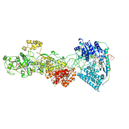
3EQM
 | |
7LVO
 | | Cryptococcus neoformans GAR synthetase | | 分子名称: | 1,2-ETHANEDIOL, phosphoribosyl-glycinamide (GAR) synthetase | | 著者 | Chua, S.M.H, Luo, Z, Lim, B.Y.J, Kobe, B, Fraser, J.A. | | 登録日 | 2021-02-26 | | 公開日 | 2021-08-25 | | 最終更新日 | 2023-10-18 | | 実験手法 | X-RAY DIFFRACTION (2 Å) | | 主引用文献 | Structural features of Cryptococcus neoformans bifunctional GAR/AIR synthetase may present novel antifungal drug targets.
J.Biol.Chem., 297, 2021
|
|
6NSU
 | | Crystallographic Capture of Quinolinate Synthase (NadA) from Pyrococcus horikoshii in its Substrates and Product-Bound States | | 分子名称: | DIDEHYDROASPARTATE, IRON/SULFUR CLUSTER, Quinolinate synthase A | | 著者 | Esakova, O.A, Grove, T.L, Silakov, A, Yennawar, N.H, Booker, S.J. | | 登録日 | 2019-01-25 | | 公開日 | 2019-08-21 | | 最終更新日 | 2024-03-13 | | 実験手法 | X-RAY DIFFRACTION (2.15 Å) | | 主引用文献 | An Unexpected Species Determined by X-ray Crystallography that May Represent an Intermediate in the Reaction Catalyzed by Quinolinate Synthase.
J.Am.Chem.Soc., 141, 2019
|
|
8YD7
 | |
7LU6
 | | Crystal structure of the mouse Kirrel3 D1 homodimer | | 分子名称: | Kin of IRRE-like protein 3, SODIUM ION | | 著者 | Roman, C.A, Pak, J.S, Wang, J, Ozkan, E. | | 登録日 | 2021-02-21 | | 公開日 | 2021-11-24 | | 最終更新日 | 2023-10-18 | | 実験手法 | X-RAY DIFFRACTION (1.95 Å) | | 主引用文献 | Molecular and structural basis of olfactory sensory neuron axon coalescence by Kirrel receptors.
Cell Rep, 37, 2021
|
|
7LTW
 | | Crystal structure of the mouse Kirrel2 D1 homodimer | | 分子名称: | Kin of IRRE-like protein 2, SODIUM ION | | 著者 | Roman, C.A, Pak, J.S, Wang, J, Ozkan, E. | | 登録日 | 2021-02-20 | | 公開日 | 2021-11-24 | | 最終更新日 | 2023-10-18 | | 実験手法 | X-RAY DIFFRACTION (1.8 Å) | | 主引用文献 | Molecular and structural basis of olfactory sensory neuron axon coalescence by Kirrel receptors.
Cell Rep, 37, 2021
|
|
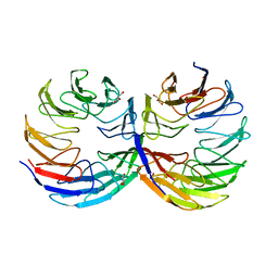
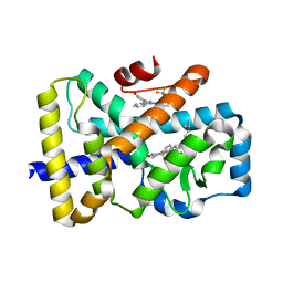
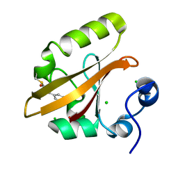
9AME
 | | TYPE III ANTIFREEZE PROTEIN ISOFORM HPLC 12 S42G | | 分子名称: | PROTEIN (ANTIFREEZE PROTEIN TYPE III) | | 著者 | Graether, S.P, Deluca, C.I, Baardsnes, J, Hill, G.A, Davies, P.L, Jia, Z. | | 登録日 | 1999-01-24 | | 公開日 | 1999-04-29 | | 最終更新日 | 2023-09-20 | | 実験手法 | X-RAY DIFFRACTION (1.8 Å) | | 主引用文献 | Quantitative and qualitative analysis of type III antifreeze protein structure and function.
J.Biol.Chem., 274, 1999
|
|
8T1H
 | |
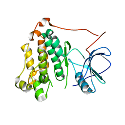
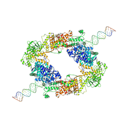
9ANT
 | | ANTENNAPEDIA HOMEODOMAIN-DNA COMPLEX | | 分子名称: | ANTENNAPEDIA HOMEODOMAIN, DNA (5'-D(*AP*GP*AP*AP*AP*GP*CP*CP*AP*TP*TP*AP*GP*AP*G)-3'), DNA (5'-D(*TP*CP*TP*CP*TP*AP*AP*TP*GP*GP*CP*TP*TP*TP*C)-3'), ... | | 著者 | Fraenkel, E, Pabo, C.O. | | 登録日 | 1998-07-02 | | 公開日 | 1998-10-14 | | 最終更新日 | 2024-02-14 | | 実験手法 | X-RAY DIFFRACTION (2.4 Å) | | 主引用文献 | Comparison of X-ray and NMR structures for the Antennapedia homeodomain-DNA complex.
Nat.Struct.Biol., 5, 1998
|
|
7LML
 | | Receptor for Advanced Glycation End Products VC1 domain in complex with 3-(3-(((3-(4-Carboxyphenoxy)benzyl)oxy)methyl)phenyl)-1H-indole-2-carboxylic acid | | 分子名称: | 6-iodanyl-1~{H}-indole-2-carboxylic acid, ACETATE ION, Advanced glycosylation end product-specific receptor, ... | | 著者 | Salay, L.E, Kozlyuk, N, Gilston, B.A, Gogliotti, R.D, Christov, P.P, Kim, K, Ovee, M, Waterson, A.G, Chazin, W.J. | | 登録日 | 2021-02-05 | | 公開日 | 2021-12-15 | | 最終更新日 | 2023-10-18 | | 実験手法 | X-RAY DIFFRACTION (2.15 Å) | | 主引用文献 | A fragment-based approach to discovery of Receptor for Advanced Glycation End products inhibitors.
Proteins, 89, 2021
|
|
3EUG
 | | CRYSTAL STRUCTURE OF ESCHERICHIA COLI URACIL DNA GLYCOSYLASE AND ITS COMPLEXES WITH URACIL AND GLYCEROL: STRUCTURE AND GLYCOSYLASE MECHANISM REVISITED | | 分子名称: | GLYCEROL, PROTEIN (GLYCOSYLASE) | | 著者 | Xiao, G, Tordova, M, Jagadeesh, J, Drohat, A.C, Stivers, J.T, Gilliland, G.L. | | 登録日 | 1998-10-13 | | 公開日 | 1999-10-13 | | 最終更新日 | 2023-09-06 | | 実験手法 | X-RAY DIFFRACTION (1.43 Å) | | 主引用文献 | Crystal structure of Escherichia coli uracil DNA glycosylase and its complexes with uracil and glycerol: structure and glycosylase mechanism revisited.
Proteins, 35, 1999
|
|
5MB7
 | |
6TJF
 | | Crystal structure of the computationally designed Cake6 protein | | 分子名称: | Cake6, GLYCEROL | | 著者 | Mylemans, B, Laier, I, Voet, A.R.D, Noguchi, H. | | 登録日 | 2019-11-26 | | 公開日 | 2020-05-06 | | 最終更新日 | 2024-05-01 | | 実験手法 | X-RAY DIFFRACTION (2.4 Å) | | 主引用文献 | Structural plasticity of a designer protein sheds light on beta-propeller protein evolution.
Febs J., 288, 2021
|
|
7W0B
 | | Dicer2-LoqsPD complex at apo status | | 分子名称: | Dicer-2, isoform A, Loquacious, ... | | 著者 | Su, S, Wang, J, Wang, H.W, Ma, J. | | 登録日 | 2021-11-18 | | 公開日 | 2022-04-27 | | 最終更新日 | 2024-06-26 | | 実験手法 | ELECTRON MICROSCOPY (3.33 Å) | | 主引用文献 | Structural insights into dsRNA processing by Drosophila Dicer-2-Loqs-PD.
Nature, 607, 2022
|
|
8XZF
 | | Cryo-EM structure of the WN561-bound human APLNR-Gi complex | | 分子名称: | Apelin receptor, Guanine nucleotide-binding protein G(I)/G(S)/G(O) subunit gamma-2, Guanine nucleotide-binding protein G(I)/G(S)/G(T) subunit beta-1, ... | | 著者 | Wang, W, Ji, S, Zhang, Y. | | 登録日 | 2024-01-21 | | 公開日 | 2024-03-20 | | 最終更新日 | 2024-03-27 | | 実験手法 | ELECTRON MICROSCOPY (3 Å) | | 主引用文献 | Structure-based design of non-hypertrophic apelin receptor modulator.
Cell, 187, 2024
|
|
7W0E
 | | dmDicer2-LoqsPD-dsRNA Active-dicing status | | 分子名称: | ADENOSINE-5'-DIPHOSPHATE, Dicer-2, isoform A, ... | | 著者 | Su, S, Wang, J, Wang, H.W, Ma, J. | | 登録日 | 2021-11-18 | | 公開日 | 2022-04-27 | | 最終更新日 | 2024-06-26 | | 実験手法 | ELECTRON MICROSCOPY (4.03 Å) | | 主引用文献 | Structural insights into dsRNA processing by Drosophila Dicer-2-Loqs-PD.
Nature, 607, 2022
|
|
6T4U
 | | ROR(gamma)t ligand binding domain in complex with 20-alpha-hydroxycholesterol and allosteric ligand MRL871 | | 分子名称: | (3alpha,8alpha)-cholest-5-ene-3,20-diol, 4-{1-[2-chloro-6-(trifluoromethyl)benzoyl]-1H-indazol-3-yl}benzoic acid, GLYCEROL, ... | | 著者 | de Vries, R.M.J.M, Meijer, F.A, Brunsveld, L. | | 登録日 | 2019-10-15 | | 公開日 | 2020-11-18 | | 最終更新日 | 2024-01-24 | | 実験手法 | X-RAY DIFFRACTION (2 Å) | | 主引用文献 | Cooperativity between the orthosteric and allosteric ligand binding sites of ROR gamma t.
Proc.Natl.Acad.Sci.USA, 118, 2021
|
|
7TVD
 | |
8XZJ
 | | Cryo-EM structure of the WN353-bound human APLNR-Gi complex | | 分子名称: | Apelin receptor, Guanine nucleotide-binding protein G(I)/G(S)/G(O) subunit gamma-2, Guanine nucleotide-binding protein G(I)/G(S)/G(T) subunit beta-1, ... | | 著者 | Wang, W, Ji, S, Zhang, Y. | | 登録日 | 2024-01-21 | | 公開日 | 2024-03-20 | | 最終更新日 | 2024-03-27 | | 実験手法 | ELECTRON MICROSCOPY (3 Å) | | 主引用文献 | Structure-based design of non-hypertrophic apelin receptor modulator.
Cell, 187, 2024
|
|
7W0A
 | | dmDicer2-LoqsPD-dsRNA Dimer status | | 分子名称: | Dicer-2, isoform A, Loquacious, ... | | 著者 | Su, S, Wang, J, Wang, H.W, Ma, J. | | 登録日 | 2021-11-18 | | 公開日 | 2022-04-27 | | 最終更新日 | 2024-06-26 | | 実験手法 | ELECTRON MICROSCOPY (3.12 Å) | | 主引用文献 | Structural insights into dsRNA processing by Drosophila Dicer-2-Loqs-PD.
Nature, 607, 2022
|
|
9AZ7
 | | Chloride Sites in Photoactive Yellow Protein | | 分子名称: | 4'-HYDROXYCINNAMIC ACID, CHLORIDE ION, Photoactive yellow protein | | 著者 | Dyda, F, Schotte, F, Anfinrud, P, Cho, H.S. | | 登録日 | 2024-03-10 | | 公開日 | 2024-03-20 | | 最終更新日 | 2024-04-24 | | 実験手法 | X-RAY DIFFRACTION (2 Å) | | 主引用文献 | Watching a signaling protein function: What has been learned over four decades of time-resolved studies of photoactive yellow protein.
Struct Dyn., 11, 2024
|
|
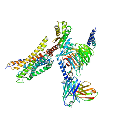
7W39
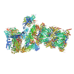 | | Structure of USP14-bound human 26S proteasome in state EA2.1_UBL | | 分子名称: | 26S protease regulatory subunit 4, 26S protease regulatory subunit 6A, 26S protease regulatory subunit 6B, ... | | 著者 | Zhang, S, Zou, S, Yin, D, Wu, Z, Mao, Y. | | 登録日 | 2021-11-25 | | 公開日 | 2022-05-04 | | 最終更新日 | 2022-06-01 | | 実験手法 | ELECTRON MICROSCOPY (3.2 Å) | | 主引用文献 | USP14-regulated allostery of the human proteasome by time-resolved cryo-EM.
Nature, 605, 2022
|
|
8YBE
 | |


