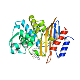
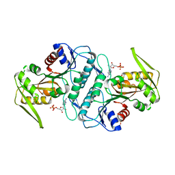
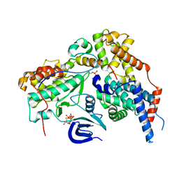
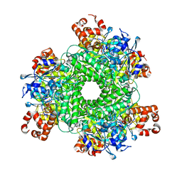
2RGJ
 
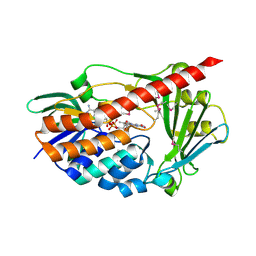 | | Crystal structure of flavin-containing monooxygenase PhzS | | 分子名称: | FLAVIN-ADENINE DINUCLEOTIDE, Flavin-containing monooxygenase | | 著者 | Ladner, J.E, Parsons, J.F, Greenhagen, B.T, Robinson, H. | | 登録日 | 2007-10-03 | | 公開日 | 2008-05-20 | | 最終更新日 | 2017-10-25 | | 実験手法 | X-RAY DIFFRACTION (2.4 Å) | | 主引用文献 | Crystal Structure of the Pyocyanin Biosynthetic Protein PhzS.
Biochemistry, 47, 2008
|
|
5HNS
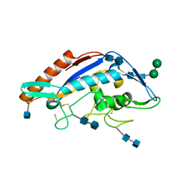 | | Structure of glycosylated NPC1 luminal domain C | | 分子名称: | 2-acetamido-2-deoxy-beta-D-glucopyranose, 2-acetamido-2-deoxy-beta-D-glucopyranose-(1-4)-2-acetamido-2-deoxy-beta-D-glucopyranose, Niemann-Pick C1 protein, ... | | 著者 | Zhao, Y, Ren, J, Harlos, K, Stuart, D.I. | | 登録日 | 2016-01-18 | | 公開日 | 2016-02-10 | | 最終更新日 | 2020-07-29 | | 実験手法 | X-RAY DIFFRACTION (2.45 Å) | | 主引用文献 | Structure of glycosylated NPC1 luminal domain C reveals insights into NPC2 and Ebola virus interactions.
Febs Lett., 590, 2016
|
|
7RUO
 | | Crystal structure of human UTP15 | | 分子名称: | U3 small nucleolar RNA-associated protein 15 homolog, UNKNOWN ATOM OR ION | | 著者 | Dehghani-Tafti, S, Dong, A, Zeng, H, Hutchinson, A, Seitova, A, Loppnau, P, Arrowsmith, C.H, Edwards, A.M, Halabelian, L, Structural Genomics Consortium (SGC) | | 登録日 | 2021-08-17 | | 公開日 | 2021-11-10 | | 最終更新日 | 2024-05-22 | | 実験手法 | X-RAY DIFFRACTION (1.8 Å) | | 主引用文献 | Crystal structure of human UTP15
To Be Published
|
|
6XIR
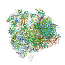 | | Cryo-EM Structure of K63 Ubiquitinated Yeast Translocating Ribosome under Oxidative Stress | | 分子名称: | 18S ribosomal RNA, 35S ribosomal RNA, 40S ribosomal protein S0-A, ... | | 著者 | Zhou, Y, Bartesaghi, A, Silva, G.M. | | 登録日 | 2020-06-21 | | 公開日 | 2020-08-26 | | 最終更新日 | 2020-09-23 | | 実験手法 | ELECTRON MICROSCOPY (3.2 Å) | | 主引用文献 | Structural impact of K63 ubiquitin on yeast translocating ribosomes under oxidative stress.
Proc.Natl.Acad.Sci.USA, 117, 2020
|
|
7S0V
 | |
7S8O
 | | CryoEM structure of Gi-coupled MRGPRX2 with small molecule agonist (R)-Zinc-3573 | | 分子名称: | (3R)-N,N-dimethyl-1-[(8S)-5-phenylpyrazolo[1,5-a]pyrimidin-7-yl]pyrrolidin-3-amine, Guanine nucleotide-binding protein G(I)/G(S)/G(O) subunit gamma-2, Guanine nucleotide-binding protein G(I)/G(S)/G(T) subunit beta-1, ... | | 著者 | Cao, C, Fay, J.F, Gumpper, R.H, Roth, B.L. | | 登録日 | 2021-09-18 | | 公開日 | 2021-11-17 | | 最終更新日 | 2021-12-15 | | 実験手法 | ELECTRON MICROSCOPY (2.58 Å) | | 主引用文献 | Structure, function and pharmacology of human itch GPCRs.
Nature, 600, 2021
|
|
2RDO
 | | 50S subunit with EF-G(GDPNP) and RRF bound | | 分子名称: | 23S RIBOSOMAL RNA, 50S ribosomal protein L1, 50S ribosomal protein L11, ... | | 著者 | Gao, N, Zavialov, A.V, Ehrenberg, M, Frank, J. | | 登録日 | 2007-09-24 | | 公開日 | 2008-03-04 | | 最終更新日 | 2024-02-21 | | 実験手法 | ELECTRON MICROSCOPY (9.1 Å) | | 主引用文献 | Specific interaction between EF-G and RRF and its implication for GTP-dependent ribosome splitting into subunits.
J.Mol.Biol., 374, 2007
|
|
2RHU
 | |
7RWS
 | |
3LRT
 | | Crystal structure of the phosphoribosyl pyrophosphate (PRPP) synthetase from Thermoplasma volcanium in complex with ADP. | | 分子名称: | ADENOSINE-5'-DIPHOSPHATE, Ribose-phosphate pyrophosphokinase, SULFATE ION | | 著者 | Cherney, M.M, Cherney, L.T, Garen, C.R, James, M.N.G. | | 登録日 | 2010-02-11 | | 公開日 | 2011-02-16 | | 最終更新日 | 2023-09-06 | | 実験手法 | X-RAY DIFFRACTION (1.534 Å) | | 主引用文献 | The structures of Thermoplasma volcanium phosphoribosyl pyrophosphate synthetase bound to ribose-5-phosphate and ATP analogs.
J.Mol.Biol., 413, 2011
|
|
2RI7
 | |
5HOT
 | | Structural Basis for Inhibitor-Induced Aggregation of HIV-1 Integrase | | 分子名称: | (2S)-tert-butoxy[4-(8-fluoro-5-methyl-3,4-dihydro-2H-chromen-6-yl)-2-methyl-1-oxo-1,2-dihydroisoquinolin-3-yl]ethanoic acid, Integrase | | 著者 | Gupta, K, Turkki, V, Sherrill-Mix, S, Hwang, Y, Eilers, G, Taylor, L, McDanal, C, Wang, P, Temelkoff, D, Nolte, R, Velthuisen, E, Jeffrey, J, Van Duyne, G.D, Bushman, F.D. | | 登録日 | 2016-01-19 | | 公開日 | 2016-12-14 | | 最終更新日 | 2023-09-27 | | 実験手法 | X-RAY DIFFRACTION (4.4 Å) | | 主引用文献 | Structural Basis for Inhibitor-Induced Aggregation of HIV Integrase.
PLoS Biol., 14, 2016
|
|
2G57
 | | Structure of the Phosphorylation Motif of the oncogenic Protein beta-Catenin Recognized By a Selective Monoclonal Antibody | | 分子名称: | Beta-catenin | | 著者 | Megy, S, Bertho, G, Gharbi-Benarous, J, Baleux, F, Benarous, R, Girault, J.P. | | 登録日 | 2006-02-22 | | 公開日 | 2006-03-28 | | 最終更新日 | 2022-03-09 | | 実験手法 | SOLUTION NMR | | 主引用文献 | STD and TRNOESY NMR studies for the epitope mapping of the phosphorylation motif of the oncogenic protein beta-catenin recognized by a selective monoclonal antibody
Febs Lett., 580, 2006
|
|
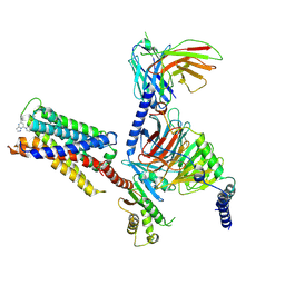
6XBZ
 | | Structure of the human CDK-activating kinase | | 分子名称: | CDK-activating kinase assembly factor MAT1, Cyclin-H, Cyclin-dependent kinase 7, ... | | 著者 | Greber, B.J, Perez-Bertoldi, J.M, Lim, K, Iavarone, A.T, Toso, D.B, Nogales, E. | | 登録日 | 2020-06-07 | | 公開日 | 2020-09-09 | | 最終更新日 | 2020-09-30 | | 実験手法 | ELECTRON MICROSCOPY (2.8 Å) | | 主引用文献 | The cryoelectron microscopy structure of the human CDK-activating kinase.
Proc.Natl.Acad.Sci.USA, 117, 2020
|
|
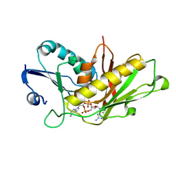
2RG6
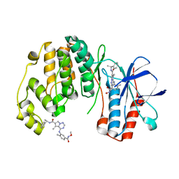 | | Phenylalanine pyrrolotriazine p38 alpha map kinase inhibitor compound 11J | | 分子名称: | 4-{[5-(methoxycarbamoyl)-2-methylphenyl]amino}-5-methyl-N-[(1S)-1-phenylethyl]pyrrolo[2,1-f][1,2,4]triazine-6-carboxamide, Mitogen-activated protein kinase 14 | | 著者 | Sack, J.S. | | 登録日 | 2007-10-02 | | 公開日 | 2008-01-15 | | 最終更新日 | 2024-02-21 | | 実験手法 | X-RAY DIFFRACTION (1.72 Å) | | 主引用文献 | Design, Synthesis, and Anti-inflammatory Properties of Orally Active 4-(Phenylamino)-pyrrolo[2,1-f][1,2,4]triazine p38alpha Mitogen-Activated Protein Kinase Inhibitors
J.Med.Chem., 51, 2008
|
|
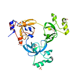
5HQ0
 | | Ternary complex of human proteins CDK1, Cyclin B and CKS2, bound to an inhibitor | | 分子名称: | Cyclin-dependent kinase 1, Cyclin-dependent kinases regulatory subunit 2, G2/mitotic-specific cyclin-B1, ... | | 著者 | Noble, M.E, Martin, M.P, Korolchuk, S, Brown, N.R, Moukhametzianov, R, Stanley, W.A. | | 登録日 | 2016-01-21 | | 公開日 | 2016-03-02 | | 最終更新日 | 2024-05-08 | | 実験手法 | X-RAY DIFFRACTION (2.3 Å) | | 主引用文献 | CDK1 structures reveal conserved and unique features of the essential cell cycle CDK.
Nat Commun, 6, 2015
|
|
2G5M
 | | Spinophilin PDZ domain | | 分子名称: | Neurabin-2 | | 著者 | Kelker, M.S, Peti, W. | | 登録日 | 2006-02-23 | | 公開日 | 2007-01-09 | | 最終更新日 | 2024-05-29 | | 実験手法 | SOLUTION NMR | | 主引用文献 | Structural basis for spinophilin-neurabin receptor interaction.
Biochemistry, 46, 2007
|
|
5HQL
 | | Structure function studies of R. palustris RubisCO (A47V-M331A mutant; CABP-bound; no expression tag) | | 分子名称: | 2-CARBOXYARABINITOL-1,5-DIPHOSPHATE, MAGNESIUM ION, Ribulose bisphosphate carboxylase | | 著者 | Arbing, M.A, Shin, A, Cascio, D, Satagopan, S, North, J.A, Tabita, F.R. | | 登録日 | 2016-01-21 | | 公開日 | 2017-01-25 | | 最終更新日 | 2023-11-15 | | 実験手法 | X-RAY DIFFRACTION (2.53 Å) | | 主引用文献 | Structure function studies of R. palustris RubisCO
To Be Published
|
|
7D4U
 | | ATP complex with double mutant cyclic trinucleotide synthase CdnD | | 分子名称: | ADENOSINE-5'-TRIPHOSPHATE, Cyclic AMP-AMP-GMP synthase | | 著者 | Yang, C.-S, Hou, M.-H, Tsai, C.-L, Wang, Y.-C, Ko, T.-P, Chen, Y. | | 登録日 | 2020-09-24 | | 公開日 | 2021-03-17 | | 最終更新日 | 2023-11-29 | | 実験手法 | X-RAY DIFFRACTION (2.7 Å) | | 主引用文献 | Crystal structure and functional implication of a bacterial cyclic AMP-AMP-GMP synthetase.
Nucleic Acids Res., 49, 2021
|
|
2G6P
 | | Crystal structure of truncated (delta 1-89) human methionine aminopeptidase Type 1 in complex with Pyridyl pyrimidine derivative | | 分子名称: | 4-(2-HYDROXYETHYL)-1-PIPERAZINE ETHANESULFONIC ACID, 5-CHLORO-6-METHYL-N-(2-PHENYLETHYL)-2-PYRIDIN-2-YLPYRIMIDIN-4-AMINE, COBALT (II) ION, ... | | 著者 | Addlagatta, A, Hu, X, Liu, J.O, Matthews, B.W. | | 登録日 | 2006-02-24 | | 公開日 | 2006-06-20 | | 最終更新日 | 2024-02-14 | | 実験手法 | X-RAY DIFFRACTION (1.9 Å) | | 主引用文献 | Identification of Pyridinylpyrimidines as Inhibitors of Human Methionine Aminopeptidases.
Angew.Chem.Int.Ed.Engl., 45, 2006
|
|
5HW5
 | |
2RLQ
 | | NMR structure of CCP modules 2-3 of complement factor H | | 分子名称: | Complement factor H | | 著者 | Hocking, H.G, Herbert, A.P, Pangburn, M.K, Kavanagh, D, Barlow, P.N, Uhrin, D. | | 登録日 | 2007-07-29 | | 公開日 | 2008-02-19 | | 最終更新日 | 2022-03-16 | | 実験手法 | SOLUTION NMR | | 主引用文献 | Structure of the N-terminal region of complement factor H and conformational implications of disease-linked sequence variations.
J.Biol.Chem., 283, 2008
|
|
2RMV
 | |
5HWX
 | | Structural mechanisms of extracellular ion exchange and induced binding-site occlusion in the sodium-calcium exchanger NCX_Mj soaked with 2.5 mM Na+ and zero Ca2+ | | 分子名称: | CHLORIDE ION, DI(HYDROXYETHYL)ETHER, PENTADECANE, ... | | 著者 | Liao, J, Jiang, Y.X, Faraldo-Gomez, J.D. | | 登録日 | 2016-01-29 | | 公開日 | 2016-05-11 | | 最終更新日 | 2023-11-08 | | 実験手法 | X-RAY DIFFRACTION (2.4 Å) | | 主引用文献 | Mechanism of extracellular ion exchange and binding-site occlusion in a sodium/calcium exchanger
Nat.Struct.Mol.Biol., 23, 2016
|
|
2RMZ
 | |


