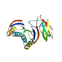
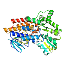
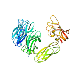
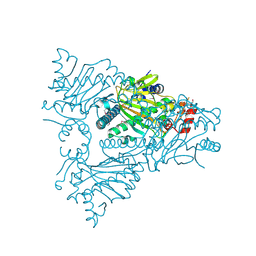
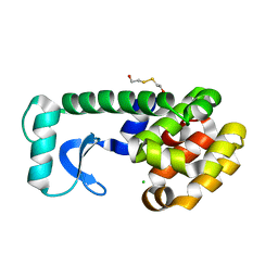
5H3H
 
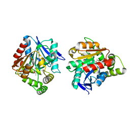 | | Esterase (EaEST) from Exiguobacterium antarcticum | | 分子名称: | Abhydrolase domain-containing protein, ETHANEPEROXOIC ACID | | 著者 | Lee, J.H, Lee, C.W. | | 登録日 | 2016-10-24 | | 公開日 | 2017-01-11 | | 最終更新日 | 2024-05-29 | | 実験手法 | X-RAY DIFFRACTION (1.9 Å) | | 主引用文献 | Crystal Structure and Functional Characterization of an Esterase (EaEST) from Exiguobacterium antarcticum.
Plos One, 12, 2017
|
|
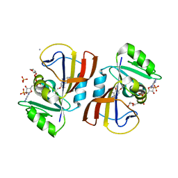
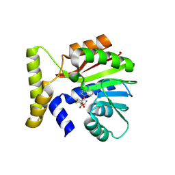
1W1Q
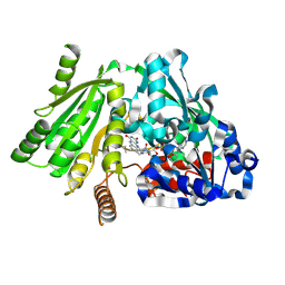 | | Plant Cytokinin Dehydrogenase in Complex with Isopentenyladenine | | 分子名称: | 2-acetamido-2-deoxy-beta-D-glucopyranose, 2-acetamido-2-deoxy-beta-D-glucopyranose-(1-4)-2-acetamido-2-deoxy-beta-D-glucopyranose, CYTOKININ DEHYDROGENASE 1, ... | | 著者 | Malito, E, Mattevi, A. | | 登録日 | 2004-06-23 | | 公開日 | 2004-08-26 | | 最終更新日 | 2020-07-29 | | 実験手法 | X-RAY DIFFRACTION (1.8 Å) | | 主引用文献 | Structures of Michaelis and Product Complexes of Plant Cytokinin Dehydrogenase: Implications for Flavoenzyme Catalysis
J.Mol.Biol., 341, 2004
|
|
7RK6
 | | Aplysia Slo1 with Barium | | 分子名称: | (1R)-2-{[(S)-{[(2S)-2,3-dihydroxypropyl]oxy}(hydroxy)phosphoryl]oxy}-1-[(hexadecanoyloxy)methyl]ethyl (9Z)-octadec-9-enoate, BARIUM ION, BK channel, ... | | 著者 | Zhu, J, Srivastava, S, Cachau, R, Holmgren, M. | | 登録日 | 2021-07-22 | | 公開日 | 2022-06-22 | | 最終更新日 | 2023-04-05 | | 実験手法 | ELECTRON MICROSCOPY (2.91 Å) | | 主引用文献 | CryoEM structure of Aplysia Slo1 with 0 mM Ba2+ at 2.91 A
To Be Published
|
|
5H3Q
 | | Crystal Structure of TrkA kinase with ligand | | 分子名称: | 1-[(3S,4R)-4-[3,4-bis(fluoranyl)phenyl]-1-(2-methoxyethyl)pyrrolidin-3-yl]-3-(5-ethoxy-4-methyl-2-phenyl-pyrazol-3-yl)urea, 2,3-DIHYDROXY-1,4-DITHIOBUTANE, High affinity nerve growth factor receptor, ... | | 著者 | Noritaka, F. | | 登録日 | 2016-10-26 | | 公開日 | 2017-02-22 | | 最終更新日 | 2023-11-08 | | 実験手法 | X-RAY DIFFRACTION (2.1 Å) | | 主引用文献 | The juxtamembrane region of TrkA kinase is critical for inhibitor selectivity
Bioorg. Med. Chem. Lett., 27, 2017
|
|
7QZD
 | | Complex of rice blast (Magnaporthe oryzae) effector protein AVR-PikF with an engineered HMA domain of Pikp-1 (Pikp-SNK-EKE) from rice (Oryza sativa) | | 分子名称: | Avr-Pik, Resistance protein Pikp-1 | | 著者 | Maidment, J.H.R, Franceschetti, M, Longya, A, Banfield, M.J. | | 登録日 | 2022-01-31 | | 公開日 | 2022-06-22 | | 最終更新日 | 2024-02-07 | | 実験手法 | X-RAY DIFFRACTION (2.2 Å) | | 主引用文献 | Effector target-guided engineering of an integrated domain expands the disease resistance profile of a rice NLR immune receptor.
Elife, 12, 2023
|
|
7RJT
 | | Aplysia Slo1 with Barium | | 分子名称: | (1R)-2-{[(S)-{[(2S)-2,3-dihydroxypropyl]oxy}(hydroxy)phosphoryl]oxy}-1-[(hexadecanoyloxy)methyl]ethyl (9Z)-octadec-9-enoate, BARIUM ION, BK channel, ... | | 著者 | Zhu, J, Srivastava, S, Cachau, R, Holmgren, M. | | 登録日 | 2021-07-21 | | 公開日 | 2022-06-22 | | 最終更新日 | 2024-06-05 | | 実験手法 | ELECTRON MICROSCOPY (2.93 Å) | | 主引用文献 | CryoEM structure of Aplysia Slo1 with 10 mM Ba2+ at 2.93 A
To Be Published
|
|
1W53
 | | Kinase recruitment domain of the stress phosphatase RsbU | | 分子名称: | GLYCEROL, PHOSPHOSERINE PHOSPHATASE RSBU, XENON | | 著者 | Delumeau, O, Dutta, S, Brigulla, M, Kuhnke, G, Hardwick, S.W, Voelker, U, Yudkin, M.D, Lewis, R.J. | | 登録日 | 2004-08-05 | | 公開日 | 2004-08-05 | | 最終更新日 | 2024-05-08 | | 実験手法 | X-RAY DIFFRACTION (1.6 Å) | | 主引用文献 | Functional and Structural Characterization of Rsbu, a Stress Signaling Protein Phosphatase 2C
J.Biol.Chem., 279, 2004
|
|
7R5O
 | |
1W4X
 | | Phenylacetone Monooxygenase, a Baeyer-Villiger Monooxygenase | | 分子名称: | FLAVIN-ADENINE DINUCLEOTIDE, PHENYLACETONE MONOOXYGENASE, SULFATE ION | | 著者 | Malito, E, Alfieri, A, Mattevi, A. | | 登録日 | 2004-08-03 | | 公開日 | 2004-09-02 | | 最終更新日 | 2024-05-08 | | 実験手法 | X-RAY DIFFRACTION (1.7 Å) | | 主引用文献 | Crystal Structure of a Baeyer-Villiger Monooxygenase
Proc.Natl.Acad.Sci.USA, 101, 2004
|
|
5GT6
 | | Apo structure of Aldehyde Dehydrogenase from Bacillus cereus | | 分子名称: | Betaine-aldehyde dehydrogenase, SODIUM ION | | 著者 | Ngo, H.P.T, Hong, S.H, Ho, T.H, Oh, D.K, Kang, L.W. | | 登録日 | 2016-08-18 | | 公開日 | 2017-09-06 | | 最終更新日 | 2023-11-08 | | 実験手法 | X-RAY DIFFRACTION (2.6 Å) | | 主引用文献 | crystal structures of aldehyde dehydrogenase from Bacillus cereus having atypical bidirectional oxidizing and reducing activities for all-trans-retinal
To Be Published
|
|
1VCL
 | | Crystal Structure of Hemolytic Lectin CEL-III | | 分子名称: | 2-[BIS-(2-HYDROXY-ETHYL)-AMINO]-2-HYDROXYMETHYL-PROPANE-1,3-DIOL, CALCIUM ION, CHLORIDE ION, ... | | 著者 | Uchida, T, Yamasaki, T, Eto, S, Sugawara, H, Kurisu, G, Nakagawa, A, Kusunoki, M, Hatakeyama, T. | | 登録日 | 2004-03-09 | | 公開日 | 2004-09-07 | | 最終更新日 | 2023-12-27 | | 実験手法 | X-RAY DIFFRACTION (1.7 Å) | | 主引用文献 | Crystal Structure of the Hemolytic Lectin CEL-III Isolated from the Marine Invertebrate Cucumaria echinata: IMPLICATIONS OF DOMAIN STRUCTURE FOR ITS MEMBRANE PORE-FORMATION MECHANISM
J.Biol.Chem., 279, 2004
|
|
1W82
 | | p38 Kinase crystal structure in complex with small molecule inhibitor | | 分子名称: | MITOGEN-ACTIVATED PROTEIN KINASE 14, N-[(3Z)-5-TERT-BUTYL-2-PHENYL-1,2-DIHYDRO-3H-PYRAZOL-3-YLIDENE]-N'-(4-CHLOROPHENYL)UREA | | 著者 | Tickle, J, Jhoti, H, Cleasby, A, Devine, L. | | 登録日 | 2004-09-16 | | 公開日 | 2005-02-08 | | 最終更新日 | 2024-05-08 | | 実験手法 | X-RAY DIFFRACTION (2.2 Å) | | 主引用文献 | Identification of Novel P38Alpha Map Kinase Inhibitors Using Fragment-Based Lead Generation
J.Med.Chem., 48, 2005
|
|
1W8I
 | | The Structure of gene product af1683 from Archaeoglobus fulgidus. | | 分子名称: | PUTATIVE VAPC RIBONUCLEASE AF_1683 | | 著者 | Midwest Center for Structural Genomics (MCSG), Cuff, M.E, Zhang, R, Ginell, S.L, Xu, X, Savchenko, A, Edwards, A, Joachimiak, A. | | 登録日 | 2004-09-22 | | 公開日 | 2004-11-16 | | 最終更新日 | 2017-06-28 | | 実験手法 | X-RAY DIFFRACTION (2.1 Å) | | 主引用文献 | The Structure of Gene Product Af1683 from Archaeoglobus Fulgidus
To be Published
|
|
1W8O
 | | Contribution of the Active Site Aspartic Acid to Catalysis in the Bacterial Neuraminidase from Micromonospora viridifaciens | | 分子名称: | BACTERIAL SIALIDASE, CITRIC ACID, GLYCEROL, ... | | 著者 | Newstead, S, Watson, J.N, Dookhun, V, Bennet, A.J, Taylor, G. | | 登録日 | 2004-09-24 | | 公開日 | 2004-09-29 | | 最終更新日 | 2023-12-13 | | 実験手法 | X-RAY DIFFRACTION (1.7 Å) | | 主引用文献 | Contribution of the Active Site Aspartic Acid to Catalysis in the Bacterial Neuraminidase from Micromonospora Viridifaciens
FEBS Lett., 577, 2004
|
|
7R6G
 | | Crystal structure of DfrA5 dihydrofolate reductase in complex with TRIMETHOPRIM and NADPH | | 分子名称: | Dihydrofolate reductase type 5, NADPH DIHYDRO-NICOTINAMIDE-ADENINE-DINUCLEOTIDE PHOSPHATE, SULFATE ION, ... | | 著者 | Estrada, A, Wright, D, Krucinska, J, Erlandsen, H. | | 登録日 | 2021-06-22 | | 公開日 | 2022-06-29 | | 最終更新日 | 2023-10-18 | | 実験手法 | X-RAY DIFFRACTION (2.61 Å) | | 主引用文献 | Structure-guided functional studies of plasmid-encoded dihydrofolate reductases reveal a common mechanism of trimethoprim resistance in Gram-negative pathogens.
Commun Biol, 5, 2022
|
|
9FCE
 | | BelI in complex with SAM from Streptomyces sp. UCK14 | | 分子名称: | (R,R)-2,3-BUTANEDIOL, 1,2-ETHANEDIOL, CALCIUM ION, ... | | 著者 | Kuttenlochner, W, Beller, P, Kaysser, L, Groll, M. | | 登録日 | 2024-05-15 | | 公開日 | 2024-08-21 | | 実験手法 | X-RAY DIFFRACTION (1.95 Å) | | 主引用文献 | Deciphering the SAM- and Metal-Dependent Mechanism of O-Methyltransferases in Cystargolide and Belactosin Biosynthesis: A Structure-Activity Relationship Study.
J.Biol.Chem., 2024
|
|
9FCX
 | | CysG(N-16)-H121N mutant in complex with SAH from Kitasatospora cystarginea | | 分子名称: | 1,2-ETHANEDIOL, S-ADENOSYL-L-HOMOCYSTEINE, SAM-dependent methyltransferase | | 著者 | Kuttenlochner, W, Beller, P, Kaysser, L, Groll, M. | | 登録日 | 2024-05-16 | | 公開日 | 2024-08-21 | | 実験手法 | X-RAY DIFFRACTION (1.8 Å) | | 主引用文献 | Deciphering the SAM- and Metal-Dependent Mechanism of O-Methyltransferases in Cystargolide and Belactosin Biosynthesis: A Structure-Activity Relationship Study.
J.Biol.Chem., 2024
|
|
1W9Y
 | | The structure of ACC oxidase | | 分子名称: | 1-AMINOCYCLOPROPANE-1-CARBOXYLATE OXIDASE 1, SULFATE ION | | 著者 | Zhang, Z, Ren, J.-S, Clifton, I.J, Schofield, C.J. | | 登録日 | 2004-10-20 | | 公開日 | 2005-10-26 | | 最終更新日 | 2011-07-13 | | 実験手法 | X-RAY DIFFRACTION (2.1 Å) | | 主引用文献 | Crystal Structure and Mechanistic Implications of 1-Aminocyclopropane-1-Carboxylic Acid Oxidase (the Ethyling Forming Enzyme)
Chem.Biol., 11, 2004
|
|
9FCS
 | | CysG(N-16)-H122N mutant in complex with SAH from Kitasatospora cystarginea | | 分子名称: | 1,2-ETHANEDIOL, S-ADENOSYL-L-HOMOCYSTEINE, SAM-dependent methyltransferase | | 著者 | Kuttenlochner, W, Beller, P, Kaysser, L, Groll, M. | | 登録日 | 2024-05-15 | | 公開日 | 2024-08-21 | | 実験手法 | X-RAY DIFFRACTION (1.6 Å) | | 主引用文献 | Deciphering the SAM- and Metal-Dependent Mechanism of O-Methyltransferases in Cystargolide and Belactosin Biosynthesis: A Structure-Activity Relationship Study.
J.Biol.Chem., 2024
|
|
8R86
 | |
8VLL
 | | Crystal structure of the yeast cytosine deaminase (yCD) M100W mutant | | 分子名称: | 1,2-ETHANEDIOL, Cytosine deaminase, PHOSPHATE ION, ... | | 著者 | Picard, M.-E, Grenier, J, Despres, P.C, Dube, A.K, Landry, C.R, Shi, R. | | 登録日 | 2024-01-11 | | 公開日 | 2024-08-21 | | 実験手法 | X-RAY DIFFRACTION (1.67 Å) | | 主引用文献 | Compensatory mutations potentiate constructive neutral evolution by gene duplication
Science, 2024
|
|
1QUG
 | | E108V MUTANT OF T4 LYSOZYME | | 分子名称: | 2-HYDROXYETHYL DISULFIDE, CHLORIDE ION, PROTEIN (LYSOZYME) | | 著者 | Wray, J, Baase, W.A, Lindstrom, J.D, Poteete, A.R, Matthews, B.W. | | 登録日 | 1999-07-01 | | 公開日 | 1999-07-08 | | 最終更新日 | 2024-02-14 | | 実験手法 | X-RAY DIFFRACTION (1.9 Å) | | 主引用文献 | Structural analysis of a non-contiguous second-site revertant in T4 lysozyme shows that increasing the rigidity of a protein can enhance its stability.
J.Mol.Biol., 292, 1999
|
|
6D67
 | | Crystal structure of the human dual specificity phosphatase 1 catalytic domain (C258S) as a maltose binding protein fusion (maltose bound form) in complex with the designed AR protein mbp3_16 | | 分子名称: | 1,2-ETHANEDIOL, DI(HYDROXYETHYL)ETHER, Designed AR protein mbp3_16, ... | | 著者 | Gumpena, R, Lountos, G.T, Waugh, D.S. | | 登録日 | 2018-04-20 | | 公開日 | 2018-09-19 | | 最終更新日 | 2023-10-04 | | 実験手法 | X-RAY DIFFRACTION (2.55 Å) | | 主引用文献 | MBP-binding DARPins facilitate the crystallization of an MBP fusion protein.
Acta Crystallogr F Struct Biol Commun, 74, 2018
|
|
5GHX
 | | Crystal structure of beta-lactamase PenP mutant-E166H | | 分子名称: | 1,2-ETHANEDIOL, ACETIC ACID, Beta-lactamase | | 著者 | Pan, X, Zhao, Y. | | 登録日 | 2016-06-21 | | 公開日 | 2017-01-25 | | 最終更新日 | 2024-03-20 | | 実験手法 | X-RAY DIFFRACTION (1.24 Å) | | 主引用文献 | Crystallographic Snapshots of Class A beta-Lactamase Catalysis Reveal Structural Changes That Facilitate beta-Lactam Hydrolysis
J. Biol. Chem., 292, 2017
|
|
3L5W
 | | Crystal structure of the complex between IL-13 and C836 FAB | | 分子名称: | C836 HEAVY CHAIN, C836 LIGHT CHAIN, GLYCEROL, ... | | 著者 | Teplyakov, A, Obmolova, G, Malia, T, Gilliland, G.L. | | 登録日 | 2009-12-22 | | 公開日 | 2010-04-14 | | 最終更新日 | 2023-09-06 | | 実験手法 | X-RAY DIFFRACTION (2 Å) | | 主引用文献 | Human framework adaptation of a mouse anti-human IL-13 antibody.
J.Mol.Biol., 398, 2010
|
|


