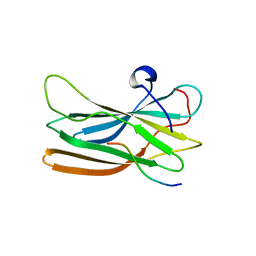
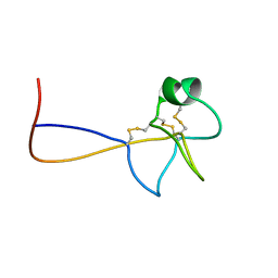
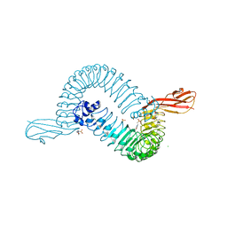
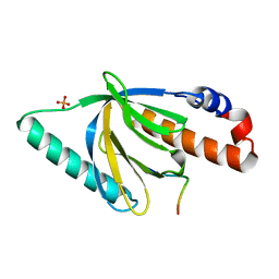
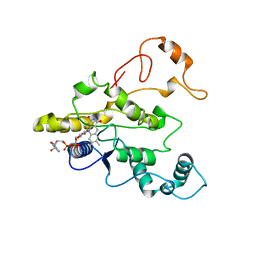
1OXZ
 
 | | Crystal Structure of the Human GGA1 GAT domain | | 分子名称: | ADP-ribosylation factor binding protein GGA1 | | 著者 | Zhu, G, Zhai, P, He, X, Terzyan, S, Zhang, R, Joachimiak, A, Tang, J, Zhang, X.C. | | 登録日 | 2003-04-03 | | 公開日 | 2003-04-15 | | 最終更新日 | 2024-02-14 | | 実験手法 | X-RAY DIFFRACTION (2.8 Å) | | 主引用文献 | Crystal Structure of Human GGA1 GAT Domain
Biochemistry, 42, 2003
|
|
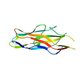
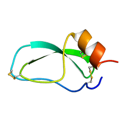
1OM9
 | | Structure of the GGA1-appendage in complex with the p56 binding peptide | | 分子名称: | 15-mer peptide fragment of p56, ADP-ribosylation factor binding protein GGA1 | | 著者 | Collins, B.M, Praefcke, G.J.K, Robinson, M.S, Owen, D.J. | | 登録日 | 2003-02-25 | | 公開日 | 2003-07-29 | | 最終更新日 | 2023-08-16 | | 実験手法 | X-RAY DIFFRACTION (2.5 Å) | | 主引用文献 | Structural basis for binding of accessory proteins by the appendage domain of GGAs
Nat.Struct.Biol., 10, 2003
|
|
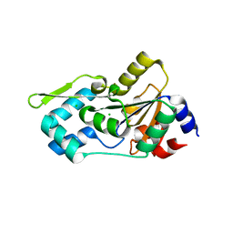
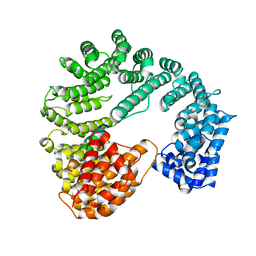
1P5R
 | | Formyl-CoA Transferase in complex with Coenzyme A | | 分子名称: | COENZYME A, Formyl-coenzyme A transferase | | 著者 | Ricagno, S, Jonsson, S, Richards, N, Lindqvist, Y. | | 登録日 | 2003-04-28 | | 公開日 | 2003-07-29 | | 最終更新日 | 2023-08-16 | | 実験手法 | X-RAY DIFFRACTION (2.5 Å) | | 主引用文献 | Formyl-CoA Transferase encloses the CoA binding site at the interface of an interlocked dimer
Embo J., 22, 2003
|
|
1OUM
 | | M64V PNP +Talo | | 分子名称: | 9-(6-DEOXY-ALPHA-L-TALOFURANOSYL)-6-METHYLPURINE, PHOSPHATE ION, Purine nucleoside phosphorylase | | 著者 | Ealick, S.E, Bennett, E.M, Anand, R, Secrist, J.A, Parker, W.B, Hassan, A.E, Allan, P.W, McPherson, D.T, Sorscher, E.J. | | 登録日 | 2003-03-24 | | 公開日 | 2004-02-17 | | 最終更新日 | 2024-02-14 | | 実験手法 | X-RAY DIFFRACTION (2.4 Å) | | 主引用文献 | Designer gene therapy using an Escherichia coli purine nucleoside phosphorylase/prodrug system.
Chem.Biol., 10, 2003
|
|
1PRX
 | | HORF6 A NOVEL HUMAN PEROXIDASE ENZYME | | 分子名称: | HORF6 | | 著者 | Choi, H.-J, Kang, S.W, Yang, C.-H, Rhee, S.G, Ryu, S.-E. | | 登録日 | 1998-04-03 | | 公開日 | 1998-06-17 | | 最終更新日 | 2021-11-03 | | 実験手法 | X-RAY DIFFRACTION (2 Å) | | 主引用文献 | Crystal structure of a novel human peroxidase enzyme at 2.0 A resolution.
Nat.Struct.Biol., 5, 1998
|
|
1PS2
 | | HIGH RESOLUTION NMR SOLUTION STRUCTURE OF HUMAN PS2, 19 STRUCTURES | | 分子名称: | PS2 | | 著者 | Williams, M.A, Polshakov, V.I, Gargaro, A.R, Feeney, J. | | 登録日 | 1997-01-07 | | 公開日 | 1997-07-07 | | 最終更新日 | 2021-11-03 | | 実験手法 | SOLUTION NMR | | 主引用文献 | High-resolution solution structure of human pNR-2/pS2: a single trefoil motif protein.
J.Mol.Biol., 267, 1997
|
|
1POV
 | |
1O9W
 | | F17-aG lectin domain from Escherichia coli in complex with N-acetyl-glucosamine | | 分子名称: | 2-acetamido-2-deoxy-beta-D-glucopyranose, F17A-G FIMBRIAL ADHESIN | | 著者 | Buts, L, De Genst, E, Loris, R, Oscarson, S, Lahmann, M, Messens, J, Brosens, E, Wyns, L, Bouckaert, J, De Greve, H. | | 登録日 | 2002-12-20 | | 公開日 | 2003-05-29 | | 最終更新日 | 2023-12-13 | | 実験手法 | X-RAY DIFFRACTION (1.65 Å) | | 主引用文献 | The Fimbrial Adhesin F17-G of Enterotoxigenic Escherichia Coli Has an Immunoglobulin-Like Lectin Domain that Binds N-Acetylglucosamine
Mol.Microbiol., 49, 2003
|
|
1NNL
 | | Crystal structure of Human Phosphoserine Phosphatase | | 分子名称: | CALCIUM ION, CHLORIDE ION, L-3-phosphoserine phosphatase | | 著者 | Peeraer, Y, Rabijns, A, Verboven, C, Collet, J.F, Van Schaftingen, E, De Ranter, C. | | 登録日 | 2003-01-14 | | 公開日 | 2003-06-03 | | 最終更新日 | 2024-03-13 | | 実験手法 | X-RAY DIFFRACTION (1.53 Å) | | 主引用文献 | High-resolution structure of human phosphoserine phosphatase in open conformation.
Acta Crystallogr.,Sect.D, 59, 2003
|
|
1O6T
 | | Internalin (INLA, Listeria monocytogenes) - functional domain, uncomplexed | | 分子名称: | 2-(N-MORPHOLINO)-ETHANESULFONIC ACID, CALCIUM ION, CHLORIDE ION, ... | | 著者 | Schubert, W.-D, Urbanke, C, Ziehm, T, Beier, V, Machner, M.P, Domann, E, Wehland, J, Chakraborty, T, Heinz, D.W. | | 登録日 | 2002-10-15 | | 公開日 | 2002-12-23 | | 最終更新日 | 2024-05-08 | | 実験手法 | X-RAY DIFFRACTION (1.6 Å) | | 主引用文献 | Structure of Internalin, a Major Invasion Protein of Listeria Monocytogenes, in Complex with its Human Receptor E-Cadherin
Cell(Cambridge,Mass.), 111, 2002
|
|
1NY3
 | | Crystal structure of ADP bound to MAP KAP kinase 2 | | 分子名称: | ADENOSINE-5'-DIPHOSPHATE, MAP kinase-activated protein kinase 2 | | 著者 | Underwood, K.W, Parris, K.D, Federico, E, Mosyak, L, Shane, T, Taylor, M, Svenson, K, Liu, Y, Hsiao, C.L, Wolfrom, S, Maguire, M, Malakian, K, Telliez, J.B, Lin, L.L, Kriz, R.W, Seehra, J, Somers, W.S, Stahl, M.L. | | 登録日 | 2003-02-11 | | 公開日 | 2003-10-14 | | 最終更新日 | 2023-08-16 | | 実験手法 | X-RAY DIFFRACTION (3 Å) | | 主引用文献 | Catalytically active MAP KAP kinase 2 structures in complex with staurosporine and ADP reveal differences with the autoinhibited enzyme
Structure, 11, 2003
|
|
1OA5
 | |
1NPW
 | | Crystal structure of HIV protease complexed with LGZ479 | | 分子名称: | CARBAMIC ACID 1-{5-BENZYL-5-[2-HYDROXY-4-PHENYL-3-(TETRAHYDRO-FURAN- 3-YLOXYCARBONYLAMINO)-BUTYL]-4-OXO-4,5-DIHYDRO-1H-PYRROL-3-YL}- INDAN-2-YL ESTER, POL polyprotein | | 著者 | Smith III, A.B. | | 登録日 | 2003-01-20 | | 公開日 | 2003-02-04 | | 最終更新日 | 2024-02-14 | | 実験手法 | X-RAY DIFFRACTION (2 Å) | | 主引用文献 | Design, synthesis, and biological evaluation of monopyrrolinone-based HIV-1 protease inhibitors.
J.Med.Chem., 46, 2003
|
|
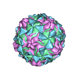
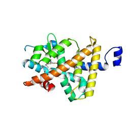
3SEJ
 | | Structural characterization of a GII.4 2004 norovirus variant (TCH05) bound to Secretor Lewis HBGA (LeB) | | 分子名称: | Capsid, alpha-L-fucopyranose-(1-2)-beta-D-galactopyranose-(1-3)-[alpha-L-fucopyranose-(1-4)]2-acetamido-2-deoxy-beta-D-glucopyranose-(1-3)-beta-D-galactopyranose-(1-4)-beta-D-glucopyranose | | 著者 | Shanker, S, Choi, J.-M, Sankaran, B, Atmar, R.L, Estes, M.K, Prasad, B.V.V. | | 登録日 | 2011-06-10 | | 公開日 | 2011-07-13 | | 最終更新日 | 2024-02-28 | | 実験手法 | X-RAY DIFFRACTION (3.041 Å) | | 主引用文献 | Structural Analysis of Histo-Blood Group Antigen Binding Specificity in a Norovirus GII.4 Epidemic Variant: Implications for Epochal Evolution.
J.Virol., 85, 2011
|
|
1NTV
 | | Crystal Structure of the Disabled-1 (Dab1) PTB domain-ApoER2 peptide complex | | 分子名称: | Apolipoprotein E Receptor-2 peptide, Disabled homolog 1, PHOSPHATE ION | | 著者 | Stolt, P.C, Jeon, H, Song, H.K, Herz, J, Eck, M.J, Blacklow, S.C. | | 登録日 | 2003-01-30 | | 公開日 | 2003-04-15 | | 最終更新日 | 2024-02-14 | | 実験手法 | X-RAY DIFFRACTION (1.5 Å) | | 主引用文献 | Origins of Peptide Selectivity and Phosphoinositide Binding Revealed by Structures of Disabled-1 PTB Domain Complexes
Structure, 11, 2003
|
|
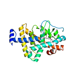
1NUO
 | | Two RTH Mutants with Impaired Hormone Binding | | 分子名称: | Thyroid hormone receptor beta-1, [4-(4-HYDROXY-3-IODO-PHENOXY)-3,5-DIIODO-PHENYL]-ACETIC ACID | | 著者 | Huber, B.R, Sandler, B, West, B.L, Cunha-Lima, S.T, Nguyen, H.T, Apriletti, J.W, Baxter, J.D, Fletterick, R.J. | | 登録日 | 2003-01-31 | | 公開日 | 2003-04-15 | | 最終更新日 | 2024-04-03 | | 実験手法 | X-RAY DIFFRACTION (3.1 Å) | | 主引用文献 | Two resistance to thyroid hormone mutants with impaired hormone binding
Mol.Endocrinol., 17, 2003
|
|
1NQ1
 | | TR Receptor Mutations Conferring Hormone Resistance and Reduced Corepressor Release Exhibit Decreased Stability in the Nterminal LBD | | 分子名称: | ARSENIC, Thyroid hormone receptor beta-1, [4-(4-HYDROXY-3-IODO-PHENOXY)-3,5-DIIODO-PHENYL]-ACETIC ACID | | 著者 | Huber, B.R, Desclozeaux, M, West, B.L, Cunha-Lima, S.T, Nguyen, H.T, Baxter, J.D, Ingraham, H.A, Fletterick, R.J. | | 登録日 | 2003-01-20 | | 公開日 | 2003-04-15 | | 最終更新日 | 2024-04-03 | | 実験手法 | X-RAY DIFFRACTION (2.9 Å) | | 主引用文献 | Thyroid hormone receptor-beta mutations conferring hormone resistance and reduced corepressor release exhibit decreased stability in the N-terminal ligand-binding domain
Mol.Endocrinol., 17, 2003
|
|
1PSJ
 | |
1POE
 | |
1Q9M
 | | Three dimensional structures of PDE4D in complex with roliprams and implication on inhibitor selectivity | | 分子名称: | ROLIPRAM, ZINC ION, cAMP-specific phosphodiesterase PDE4D2 | | 著者 | Huai, Q, Wang, H, Sun, Y, Kim, H.Y, Liu, Y, Ke, H. | | 登録日 | 2003-08-25 | | 公開日 | 2003-09-02 | | 最終更新日 | 2024-04-03 | | 実験手法 | X-RAY DIFFRACTION (2.3 Å) | | 主引用文献 | Three dimensional structures of PDE4D in complex with roliprams and implication on inhibitor selectivity
Structure, 11, 2003
|
|
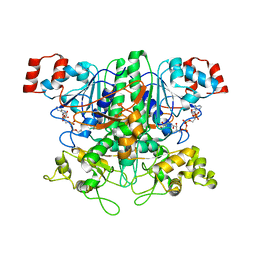
1QBG
 | | CRYSTAL STRUCTURE OF HUMAN DT-DIAPHORASE (NAD(P)H OXIDOREDUCTASE) | | 分子名称: | FLAVIN-ADENINE DINUCLEOTIDE, NAD(P)H DEHYDROGENASE [QUINONE] 1 | | 著者 | Skelly, J.V, Sanderson, M.R, Suter, D.A, Baumann, U, Gregory, D.S, Bennett, M, Hobbs, S.M, Neidle, S. | | 登録日 | 1999-04-20 | | 公開日 | 2000-04-24 | | 最終更新日 | 2024-02-14 | | 実験手法 | X-RAY DIFFRACTION (2.3 Å) | | 主引用文献 | Crystal structure of human DT-diaphorase: a model for interaction with the cytotoxic prodrug 5-(aziridin-1-yl)-2,4-dinitrobenzamide (CB1954).
J.Med.Chem., 42, 1999
|
|
1QGR
 | | STRUCTURE OF IMPORTIN BETA BOUND TO THE IBB DOMAIN OF IMPORTIN ALPHA (II CRYSTAL FORM, GROWN AT LOW PH) | | 分子名称: | PROTEIN (IMPORTIN ALPHA-2 SUBUNIT), PROTEIN (IMPORTIN BETA SUBUNIT) | | 著者 | Cingolani, G, Petosa, C, Weis, K, Muller, C.W. | | 登録日 | 1999-05-04 | | 公開日 | 1999-05-24 | | 最終更新日 | 2023-08-16 | | 実験手法 | X-RAY DIFFRACTION (2.3 Å) | | 主引用文献 | Structure of importin-beta bound to the IBB domain of importin-alpha.
Nature, 399, 1999
|
|
1Q8G
 | | NMR structure of human Cofilin | | 分子名称: | Cofilin, non-muscle isoform | | 著者 | Pope, B.J, Zierler-Gould, K.M, Kuhne, R, Weeds, A.G, Ball, L.J. | | 登録日 | 2003-08-21 | | 公開日 | 2004-07-06 | | 最終更新日 | 2024-05-22 | | 実験手法 | SOLUTION NMR | | 主引用文献 | The solution structure of human cofilin: rationalizing actin binding and pH sensitivity
J.Biol.Chem., 279, 2004
|
|
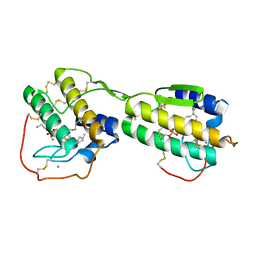
1QC6
 | | EVH1 domain from ENA/VASP-like protein in complex with ACTA peptide | | 分子名称: | EVH1 DOMAIN FROM ENA/VASP-LIKE PROTEIN, PHE-GLU-PHE-PRO-PRO-PRO-PRO-THR-ASP-GLU-GLU | | 著者 | Fedorov, A.A, Fedorov, E.V, Gertler, F.B, Almo, S.C. | | 登録日 | 1999-05-17 | | 公開日 | 1999-05-25 | | 最終更新日 | 2018-02-07 | | 実験手法 | X-RAY DIFFRACTION (2.6 Å) | | 主引用文献 | Structure of EVH1, a novel proline-rich ligand-binding module involved in cytoskeletal dynamics and neural function
Nat.Struct.Biol., 6, 1999
|
|
1QIB
 | | CRYSTAL STRUCTURE OF GELATINASE A CATALYTIC DOMAIN | | 分子名称: | 72 kDa type IV collagenase, CALCIUM ION, ZINC ION | | 著者 | Dhanaraj, V, Williams, M.G, Ye, Q.-Z, Molina, F, Johnson, L.L, Ortwine, D.F, Pavlovsky, A, Rubin, J.R, Skeean, R.W, White, A.D, Humblet, C, Hupe, D.J, Blundell, T.L. | | 登録日 | 1999-06-11 | | 公開日 | 1999-11-19 | | 最終更新日 | 2023-12-27 | | 実験手法 | X-RAY DIFFRACTION (2.8 Å) | | 主引用文献 | X-ray structure of gelatinase A catalytic domain complexed with a hydroxamate inhibitor
Croatica Chemica Acta, 72, 1999
|
|


