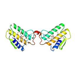
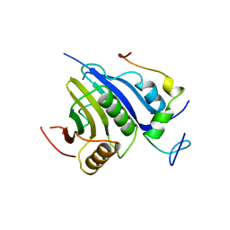
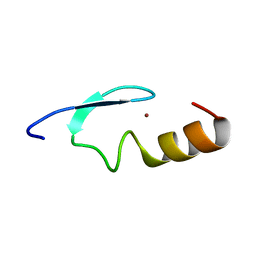
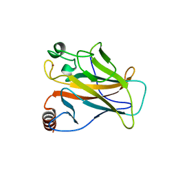
2JXZ
 
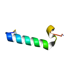 | | Solution Conformation of A Non-Amyloidogenic Analogue of Human Calcitonin in Sodium Dodecyl Sulfate Micelles | | 分子名称: | Calcitonin | | 著者 | Andreotti, G, Vitale, R, Avidan-Shpalter, C, Amodeo, P, Gazit, E, Motta, A. | | 登録日 | 2007-12-03 | | 公開日 | 2008-11-25 | | 最終更新日 | 2011-07-13 | | 実験手法 | SOLUTION NMR | | 主引用文献 | Converting the highly amyloidogenic human calcitonin into a powerful fibril inhibitor by 3D structure homology with a non-amyloidogenic analogue
J.Biol.Chem., 286, 2011
|
|
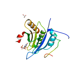
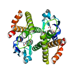
4LY9
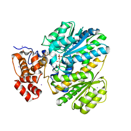 | | Human GKRP complexed to AMG-1694 [(2R)-1,1,1-trifluoro-2-{4-[(2S)-2-{[(3S)-3-methylmorpholin-4-yl]methyl}-4-(thiophen-2-ylsulfonyl)piperazin-1-yl]phenyl}propan-2-ol] and sorbitol-6-phosphate | | 分子名称: | (2R)-1,1,1-trifluoro-2-{4-[(2S)-2-{[(3S)-3-methylmorpholin-4-yl]methyl}-4-(thiophen-2-ylsulfonyl)piperazin-1-yl]phenyl}propan-2-ol, D-SORBITOL-6-PHOSPHATE, GLYCEROL, ... | | 著者 | Jordan, S.R. | | 登録日 | 2013-07-30 | | 公開日 | 2013-11-20 | | 最終更新日 | 2024-02-28 | | 実験手法 | X-RAY DIFFRACTION (2.35 Å) | | 主引用文献 | Antidiabetic effects of glucokinase regulatory protein small-molecule disruptors.
Nature, 504, 2013
|
|
2GR2
 | |
2JVI
 | |
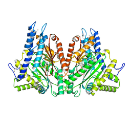
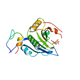
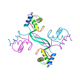
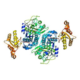
1POB
 | | CRYSTAL STRUCTURE OF COBRA-VENOM PHOSPHOLIPASE A2 IN A COMPLEX WITH A TRANSITION-STATE ANALOGUE | | 分子名称: | 1-O-OCTYL-2-HEPTYLPHOSPHONYL-SN-GLYCERO-3-PHOSPHOETHANOLAMINE, CALCIUM ION, PHOSPHOLIPASE A2 | | 著者 | White, S.P, Scott, D.L, Otwinowski, Z, Sigler, P.B. | | 登録日 | 1992-09-07 | | 公開日 | 1993-10-31 | | 最終更新日 | 2019-08-14 | | 実験手法 | X-RAY DIFFRACTION (2 Å) | | 主引用文献 | Crystal structure of cobra-venom phospholipase A2 in a complex with a transition-state analogue.
Science, 250, 1990
|
|
2MKA
 | |
2MK9
 | |
2FGY
 | |
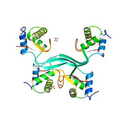
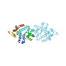
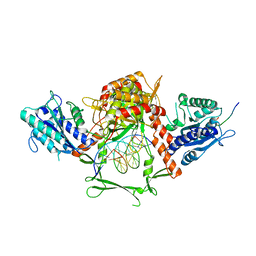
2QP2
 | | Structure of a MACPF/perforin-like protein | | 分子名称: | CALCIUM ION, Unknown protein | | 著者 | Rosado, C.J, Buckle, A.M, Law, R.H.P, Butcher, R.E, Kan, W.T, Bird, C.H, Ung, K, Browne, K.A, Baran, K, Bashtannyk-Puhalovich, T.A, Faux, N.G, Wong, W, Porter, C.J, Pike, R.N, Ellisdon, A.M, Pearce, M.C, Bottomley, S.P, Emsley, J, Smith, A.I, Rossjohn, J, Hartland, E.L, Voskoboinik, I, Trapani, J.A, Bird, P.I, Dunstone, M.A, Whisstock, J.C. | | 登録日 | 2007-07-22 | | 公開日 | 2007-09-04 | | 最終更新日 | 2024-03-13 | | 実験手法 | X-RAY DIFFRACTION (2 Å) | | 主引用文献 | A common fold mediates vertebrate defense and bacterial attack
Science, 317, 2007
|
|
5T47
 | | Crystal structure of the D. melanogaster eIF4E-eIF4G complex | | 分子名称: | Eukaryotic translation initiation factor 4E, Eukaryotic translation initiation factor 4G, isoform A, ... | | 著者 | Gruener, S, Peter, D, Weber, R, Wohlbold, L, Chung, M.-Y, Weichenrieder, O, Valkov, E, Igreja, C, Izaurralde, E. | | 登録日 | 2016-08-29 | | 公開日 | 2016-10-26 | | 最終更新日 | 2024-01-17 | | 実験手法 | X-RAY DIFFRACTION (2.2 Å) | | 主引用文献 | The Structures of eIF4E-eIF4G Complexes Reveal an Extended Interface to Regulate Translation Initiation.
Mol.Cell, 64, 2016
|
|
5T46
 | | Crystal structure of the human eIF4E-eIF4G complex | | 分子名称: | 7-METHYL-GUANOSINE-5'-TRIPHOSPHATE, Eukaryotic translation initiation factor 4 gamma 1, Eukaryotic translation initiation factor 4E, ... | | 著者 | Gruener, S, Peter, D, Weber, R, Wohlbold, L, Chung, M.-Y, Weichenrieder, O, Valkov, E, Igreja, C, Izaurralde, E. | | 登録日 | 2016-08-29 | | 公開日 | 2016-10-26 | | 最終更新日 | 2024-01-17 | | 実験手法 | X-RAY DIFFRACTION (1.53 Å) | | 主引用文献 | The Structures of eIF4E-eIF4G Complexes Reveal an Extended Interface to Regulate Translation Initiation.
Mol.Cell, 64, 2016
|
|
5T48
 | | Crystal structure of the D. melanogaster eIF4E-eIF4G complex without lateral contact | | 分子名称: | 7-METHYL-GUANOSINE-5'-TRIPHOSPHATE, Eukaryotic translation initiation factor 4E, Eukaryotic translation initiation factor 4G, ... | | 著者 | Gruener, S, Peter, D, Weber, R, Wohlbold, L, Chung, M.-Y, Weichenrieder, O, Valkov, E, Igreja, C, Izaurralde, E. | | 登録日 | 2016-08-29 | | 公開日 | 2016-10-26 | | 最終更新日 | 2024-01-17 | | 実験手法 | X-RAY DIFFRACTION (2.19 Å) | | 主引用文献 | The Structures of eIF4E-eIF4G Complexes Reveal an Extended Interface to Regulate Translation Initiation.
Mol.Cell, 64, 2016
|
|
5SUY
 | |
5SUZ
 | |
5THL
 | |
5THH
 | |
2W0T
 | |
2VUK
 | |
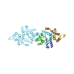
2X0W
 | | STRUCTURE OF THE P53 CORE DOMAIN MUTANT Y220C BOUND TO 5,6-dimethoxy- 2-methylbenzothiazole | | 分子名称: | 5,6-DIMETHOXY-2-METHYL-1,3-BENZOTHIAZOLE, CELLULAR TUMOR ANTIGEN P53, ZINC ION | | 著者 | Kaar, J.L, Basse, N, Joerger, A.C, Fersht, A.R. | | 登録日 | 2009-12-17 | | 公開日 | 2010-01-26 | | 最終更新日 | 2023-12-20 | | 実験手法 | X-RAY DIFFRACTION (2.1 Å) | | 主引用文献 | Toward the Rational Design of P53-Stabilizing Drugs: Probing the Surface of the Oncogenic Y220C Mutant.
Chem.Biol., 17, 2010
|
|
8ERF
 | |
3ERF
 | | Crystal structure of Gtt2 from Saccharomyces cerevisiae | | 分子名称: | Glutathione S-transferase 2 | | 著者 | Ma, X.X, Jiang, Y.L, He, Y.X, Chen, Y.X, Zhou, C.Z. | | 登録日 | 2008-10-02 | | 公開日 | 2009-10-13 | | 最終更新日 | 2023-12-27 | | 実験手法 | X-RAY DIFFRACTION (2.23 Å) | | 主引用文献 | Structures of yeast glutathione-S-transferase Gtt2 reveal a new catalytic type of GST family.
Embo Rep., 10, 2009
|
|
5ERF
 | |
6ERF
 | | Complex of APLF factor and Ku heterodimer bound to DNA | | 分子名称: | Aprataxin and PNK-like factor, DNA (34-MER), DNA (5'-D(*GP*TP*TP*TP*TP*TP*AP*GP*TP*TP*TP*AP*TP*TP*GP*GP*GP*CP*GP*CP*G)-3'), ... | | 著者 | Nemoz, C, Legrand, P, Ropars, V, Charbonnier, J.B. | | 登録日 | 2017-10-18 | | 公開日 | 2018-10-17 | | 最終更新日 | 2024-01-17 | | 実験手法 | X-RAY DIFFRACTION (3.01 Å) | | 主引用文献 | XLF and APLF bind Ku80 at two remote sites to ensure DNA repair by non-homologous end joining.
Nat.Struct.Mol.Biol., 25, 2018
|
|
3ERG
 | | Crystal structure of Gtt2 from Saccharomyces cerevisiae in complex with glutathione sulfnate | | 分子名称: | GLUTATHIONE SULFONIC ACID, Glutathione S-transferase 2 | | 著者 | Ma, X.X, Jiang, Y.L, He, Y.X, Chen, Y.X, Zhou, C.Z. | | 登録日 | 2008-10-02 | | 公開日 | 2009-10-13 | | 最終更新日 | 2023-12-27 | | 実験手法 | X-RAY DIFFRACTION (2.2 Å) | | 主引用文献 | Structures of yeast glutathione-S-transferase Gtt2 reveal a new catalytic type of GST family.
Embo Rep., 10, 2009
|
|
4ERF
 | | crystal structure of MDM2 (17-111) in complex with compound 29 (AM-8553) | | 分子名称: | E3 ubiquitin-protein ligase Mdm2, {(3R,5R,6S)-5-(3-chlorophenyl)-6-(4-chlorophenyl)-1-[(2S,3S)-2-hydroxypentan-3-yl]-3-methyl-2-oxopiperidin-3-yl}acetic acid | | 著者 | Huang, X. | | 登録日 | 2012-04-20 | | 公開日 | 2012-05-23 | | 最終更新日 | 2024-02-28 | | 実験手法 | X-RAY DIFFRACTION (2 Å) | | 主引用文献 | Structure-based design of novel inhibitors of the MDM2-p53 interaction.
J.Med.Chem., 55, 2012
|
|


