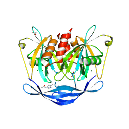
3DK5
 
 | |
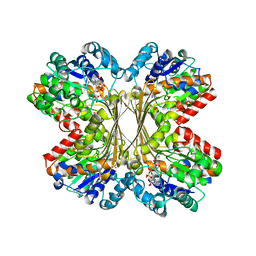
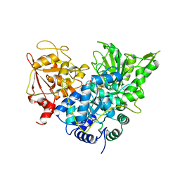
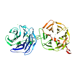
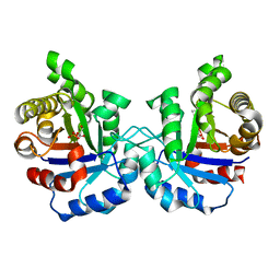
3NDW
 | | HIV-1 Protease Saquinavir:Ritonavir 1:15 complex structure | | 分子名称: | ACETATE ION, DIMETHYL SULFOXIDE, Protease, ... | | 著者 | Geremia, S, Olajuyigbe, F.M, Demitri, N. | | 登録日 | 2010-06-08 | | 公開日 | 2011-07-20 | | 最終更新日 | 2024-02-21 | | 実験手法 | X-RAY DIFFRACTION (1.14 Å) | | 主引用文献 | Investigation of 2-Fold Disorder of Inhibitors and Relative Potency by Crystallizations of HIV-1 Protease in Ritonavir and Saquinavir Mixtures
Cryst.Growth Des., 11, 2011
|
|
3LPP
 | | Crystal complex of N-terminal sucrase-isomaltase with kotalanol | | 分子名称: | (1S,2R,3R,4S)-1-{(1S)-2-[(2R,3S,4S)-3,4-dihydroxy-2-(hydroxymethyl)tetrahydrothiophenium-1-yl]-1-hydroxyethyl}-2,3,4,5-tetrahydroxypentyl sulfate, 2-AMINO-2-HYDROXYMETHYL-PROPANE-1,3-DIOL, 2-acetamido-2-deoxy-beta-D-glucopyranose, ... | | 著者 | Sim, L, Rose, D.R. | | 登録日 | 2010-02-05 | | 公開日 | 2010-03-31 | | 最終更新日 | 2024-10-30 | | 実験手法 | X-RAY DIFFRACTION (2.15 Å) | | 主引用文献 | Structural basis for substrate selectivity in human maltase-glucoamylase and sucrase-isomaltase N-terminal domains.
J.Biol.Chem., 285, 2010
|
|
2HT8
 | | N8 neuraminidase in complex with oseltamivir | | 分子名称: | (3R,4R,5S)-4-(acetylamino)-5-amino-3-(pentan-3-yloxy)cyclohex-1-ene-1-carboxylic acid, Neuraminidase | | 著者 | Russell, R.J, Haire, L.F, Stevens, D.J, Collins, P.J, Lin, Y.P, Blackburn, G.M, Hay, A.J, Gamblin, S.J, Skehel, J.J. | | 登録日 | 2006-07-25 | | 公開日 | 2006-09-05 | | 最終更新日 | 2024-11-20 | | 実験手法 | X-RAY DIFFRACTION (2.4 Å) | | 主引用文献 | The structure of H5N1 avian influenza neuraminidase suggests new opportunities for drug design.
Nature, 443, 2006
|
|
1U1I
 | | Myo-inositol phosphate synthase mIPS from A. fulgidus | | 分子名称: | NICOTINAMIDE-ADENINE-DINUCLEOTIDE, PHOSPHATE ION, POTASSIUM ION, ... | | 著者 | Stieglitz, K.A, Yang, H, Roberts, M.F, Stec, B. | | 登録日 | 2004-07-15 | | 公開日 | 2004-08-10 | | 最終更新日 | 2023-08-23 | | 実験手法 | X-RAY DIFFRACTION (1.9 Å) | | 主引用文献 | Reaching for Mechanistic Consensus Across Life Kingdoms: Structure and Insights into Catalysis of the myo-Inositol-1-phosphate Synthase (mIPS) from Archaeoglobus fulgidus
Biochemistry, 44, 2005
|
|
3I2H
 | | Cocaine Esterase with mutation L169K, bound to DTT adduct | | 分子名称: | (4S,5S)-4,5-BIS(MERCAPTOMETHYL)-1,3-DIOXOLAN-2-OL, CHLORIDE ION, Cocaine esterase, ... | | 著者 | Tesmer, J.J.G, Nance, M.R. | | 登録日 | 2009-06-29 | | 公開日 | 2010-06-16 | | 最終更新日 | 2024-11-27 | | 実験手法 | X-RAY DIFFRACTION (1.65 Å) | | 主引用文献 | Structural analysis of thermostabilizing mutations of cocaine esterase.
Protein Eng.Des.Sel., 23, 2010
|
|
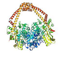
3N8I
 | |
3NF1
 | | Crystal structure of the TPR domain of kinesin light chain 1 | | 分子名称: | Kinesin light chain 1 | | 著者 | Tong, Y, Tempel, W, Shen, L, Shen, Y, Nedyalkova, L, Arrowsmith, C.H, Edwards, A.M, Bountra, C, Weigelt, J, Bochkarev, A, Park, H, Structural Genomics Consortium (SGC) | | 登録日 | 2010-06-09 | | 公開日 | 2011-05-25 | | 最終更新日 | 2023-09-06 | | 実験手法 | X-RAY DIFFRACTION (2.8 Å) | | 主引用文献 | Crystal structure of the TPR domain of kinesin light chain 1
to be published
|
|
6F7K
 | | Crystal structure of Dettilon tailspike protein (gp208) | | 分子名称: | 1,2-ETHANEDIOL, Tailspike, alpha-D-galactopyranose-(1-6)-beta-D-mannopyranose-(1-4)-alpha-L-rhamnopyranose-(1-3)-alpha-D-galactopyranose, ... | | 著者 | Roske, Y, Heinemann, U. | | 登録日 | 2017-12-11 | | 公開日 | 2018-12-19 | | 最終更新日 | 2024-01-17 | | 実験手法 | X-RAY DIFFRACTION (2.1 Å) | | 主引用文献 | Time-resolved DNA release from an O-antigen-specificSalmonellabacteriophage with a contractile tail.
J.Biol.Chem., 294, 2019
|
|
1B0M
 | | ACONITASE R644Q:FLUOROCITRATE COMPLEX | | 分子名称: | CITRATE ANION, IRON/SULFUR CLUSTER, PROTEIN (ACONITASE) | | 著者 | Lloyd, S.J, Lauble, H, Prasad, G.S, Stout, C.D. | | 登録日 | 1998-11-11 | | 公開日 | 1998-11-18 | | 最終更新日 | 2023-08-09 | | 実験手法 | X-RAY DIFFRACTION (2.5 Å) | | 主引用文献 | The mechanism of aconitase: 1.8 A resolution crystal structure of the S642a:citrate complex.
Protein Sci., 8, 1999
|
|
5QK3
 | | PanDDA analysis group deposition of models with modelled events (e.g. bound ligands) -- Crystal Structure of NUDT5 in complex with Z239136710 | | 分子名称: | 1,2-ETHANEDIOL, ADP-sugar pyrophosphatase, CHLORIDE ION, ... | | 著者 | Dubianok, Y, Collins, P, Krojer, T, Wright, N, Strain-Damerell, C, Burgess-Brown, N, Bountra, C, Arrowsmith, C.H, Edwards, A, Huber, K, von Delft, F. | | 登録日 | 2018-10-31 | | 公開日 | 2018-12-19 | | 最終更新日 | 2024-10-30 | | 実験手法 | X-RAY DIFFRACTION (1.71 Å) | | 主引用文献 | PanDDA analysis group deposition of models with modelled events (e.g. bound ligands)
To Be Published
|
|
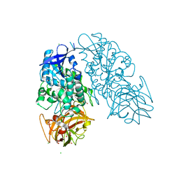
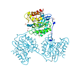
2XCS
 | | The 2.1A crystal structure of S. aureus Gyrase complex with GSK299423 and DNA | | 分子名称: | 5'-5UA*D(GP*CP*CP*GP*TP*AP*GP*GP*GP*CP*CP *CP*TP*AP*CP*GP*GP*CP*T)-3', 6-METHOXY-4-(2-{4-[([1,3]OXATHIOLO[5,4-C]PYRIDIN-6-YLMETHYL)AMINO]PIPERIDIN-1-YL}ETHYL)QUINOLINE-3-CARBONITRILE, DNA GYRASE SUBUNIT B, ... | | 著者 | Bax, B.D, Chan, P.F, Eggleston, D.S, Fosberry, A, Gentry, D.R, Gorrec, F, Giordano, I, Hann, M.M, Hennessy, A, Hibbs, M, Huang, J, Jones, E, Jones, J, Brown, K.K, Lewis, C.J, May, E.W, Singh, O, Spitzfaden, C, Shen, C, Shillings, A, Theobald, A.F, Wohlkonig, A, Pearson, N.D, Gwynn, M.N. | | 登録日 | 2010-04-25 | | 公開日 | 2010-08-04 | | 最終更新日 | 2023-12-20 | | 実験手法 | X-RAY DIFFRACTION (2.1 Å) | | 主引用文献 | Type Iia Topoisomerase Inhibition by a New Class of Antibacterial Agents.
Nature, 466, 2010
|
|
4OPY
 | | Room temperature crystal structure of stabilized TEM-1 beta-lactamase variant v.13 carrying R164S mutation | | 分子名称: | CALCIUM ION, Extended spectrum beta-lactamase TEM-63, SULFATE ION, ... | | 著者 | Dellus-Gur, E, Elias, M, Fraser, J.S, Tawfik, D.S. | | 登録日 | 2014-02-07 | | 公開日 | 2015-05-20 | | 最終更新日 | 2024-11-20 | | 実験手法 | X-RAY DIFFRACTION (1.75 Å) | | 主引用文献 | Negative Epistasis and Evolvability in TEM-1 beta-Lactamase--The Thin Line between an Enzyme's Conformational Freedom and Disorder.
J. Mol. Biol., 427, 2015
|
|
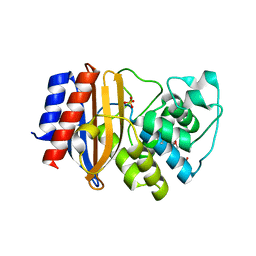
1XIF
 | |
4H1E
 | |
4H48
 | | 1.45 angstrom CyPet Structure at pH7.0 | | 分子名称: | 2-AMINO-2-HYDROXYMETHYL-PROPANE-1,3-DIOL, Green fluorescent protein | | 著者 | Hu, X.-J, Liu, R. | | 登録日 | 2012-09-17 | | 公開日 | 2013-09-18 | | 最終更新日 | 2024-10-30 | | 実験手法 | X-RAY DIFFRACTION (1.45 Å) | | 主引用文献 | Structure insight of the fluorescent state of CyPet
To be Published
|
|
3NNA
 | | Crystal Structure of CUGBP1 RRM1/2-RNA Complex | | 分子名称: | CUGBP Elav-like family member 1, RNA (5'-R(*GP*UP*UP*GP*UP*UP*UP*UP*GP*UP*UP*U)-3') | | 著者 | Teplova, M, Song, J, Gaw, H, Teplov, A, Patel, D.J. | | 登録日 | 2010-06-23 | | 公開日 | 2010-10-27 | | 最終更新日 | 2024-10-30 | | 実験手法 | X-RAY DIFFRACTION (1.899 Å) | | 主引用文献 | Structural Insights into RNA Recognition by the Alternate-Splicing Regulator CUG-Binding Protein 1.
Structure, 18, 2010
|
|
1BA4
 | | THE SOLUTION STRUCTURE OF AMYLOID BETA-PEPTIDE (1-40) IN A WATER-MICELLE ENVIRONMENT. IS THE MEMBRANE-SPANNING DOMAIN WHERE WE THINK IT IS? NMR, 10 STRUCTURES | | 分子名称: | AMYLOID BETA-PEPTIDE | | 著者 | Coles, M, Bicknell, W, Watson, A.A, Fairlie, D.P, Craik, D.J. | | 登録日 | 1998-04-07 | | 公開日 | 1998-06-17 | | 最終更新日 | 2024-05-22 | | 実験手法 | SOLUTION NMR | | 主引用文献 | Solution structure of amyloid beta-peptide(1-40) in a water-micelle environment. Is the membrane-spanning domain where we think it is?
Biochemistry, 37, 1998
|
|
4A0P
 | | Crystal structure of LRP6P3E3P4E4 | | 分子名称: | 1,2-ETHANEDIOL, 2-acetamido-2-deoxy-beta-D-glucopyranose, 2-acetamido-2-deoxy-beta-D-glucopyranose-(1-4)-2-acetamido-2-deoxy-beta-D-glucopyranose, ... | | 著者 | Chen, S, Malinauskas, T, Aricescu, A.R, Siebold, C, Jones, E.Y. | | 登録日 | 2011-09-11 | | 公開日 | 2011-10-26 | | 最終更新日 | 2024-11-06 | | 実験手法 | X-RAY DIFFRACTION (1.9 Å) | | 主引用文献 | Structural and Functional Studies of Lrp6 Ectodomain Reveal a Platform for Wnt Signaling.
Dev.Cell, 21, 2011
|
|
4OWN
 | | Anthranilate phosphoribosyl transferase from Mycobacterium tuberculosis in complex with 5-fluoroanthranilate, PRPP and Magnesium | | 分子名称: | 1-O-pyrophosphono-5-O-phosphono-alpha-D-ribofuranose, 2-amino-5-fluorobenzoic acid, Anthranilate phosphoribosyltransferase, ... | | 著者 | Castell, A, Cookson, T.V.M, Short, F.L, Lott, J.S. | | 登録日 | 2014-02-02 | | 公開日 | 2014-04-23 | | 最終更新日 | 2023-12-27 | | 実験手法 | X-RAY DIFFRACTION (2.11 Å) | | 主引用文献 | Alternative substrates reveal catalytic cycle and key binding events in the reaction catalysed by anthranilate phosphoribosyltransferase from Mycobacterium tuberculosis.
Biochem.J., 461, 2014
|
|
7E2E
 | | Crystal structure of the Estrogen-Related Receptor alpha (ERRalpha) ligand-binding domain (LBD) in complex with an agonist DS45500853 and a PGC-1alpha peptide | | 分子名称: | 1-[4-(3-tert-butyl-4-oxidanyl-phenoxy)phenyl]ethanone, IODIDE ION, Peroxisome proliferator-activated receptor gamma coactivator 1-alpha, ... | | 著者 | Ito, S, Shinozuka, T, Kimura, T, Izumi, M, Wakabayashi, K. | | 登録日 | 2021-02-05 | | 公開日 | 2021-06-30 | | 最終更新日 | 2023-11-29 | | 実験手法 | X-RAY DIFFRACTION (2.7 Å) | | 主引用文献 | Discovery of a Novel Class of ERR alpha Agonists.
Acs Med.Chem.Lett., 12, 2021
|
|
3N9W
 | | Crystal structure of 2-C-methyl-D-erythritol 4-phosphate cytidylyltransferase (IspD) in complex with 1,2-Propanediol | | 分子名称: | 2-C-methyl-D-erythritol 4-phosphate cytidylyltransferase, R-1,2-PROPANEDIOL, S-1,2-PROPANEDIOL | | 著者 | Behnen, J, Heine, A, Klebe, G. | | 登録日 | 2010-05-31 | | 公開日 | 2011-05-25 | | 最終更新日 | 2023-09-06 | | 実験手法 | X-RAY DIFFRACTION (1.9 Å) | | 主引用文献 | Experimental and computational active site mapping as a starting point to fragment-based lead discovery.
Chemmedchem, 7, 2012
|
|
1O5X
 | | Plasmodium falciparum TIM complexed to 2-phosphoglycerate | | 分子名称: | 2-PHOSPHOGLYCERIC ACID, 3-HYDROXYPYRUVIC ACID, PHOSPHITE ION, ... | | 著者 | Parthasarathy, S, Eaazhisai, K, Balaram, H, Balaram, P, Murthy, M.R. | | 登録日 | 2003-10-06 | | 公開日 | 2004-01-13 | | 最終更新日 | 2024-12-25 | | 実験手法 | X-RAY DIFFRACTION (1.1 Å) | | 主引用文献 | Structure of Plasmodium falciparum Triose-phosphate Isomerase-2-Phosphoglycerate Complex at 1.1-A Resolution
J.Biol.Chem., 278, 2003
|
|
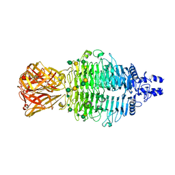
5A8P
 | | Crystal structure beta-glucanase SdGluc5_26A from Saccharophagus degradans in complex with tetrasaccharide B | | 分子名称: | CHLORIDE ION, GLYCEROL, MAGNESIUM ION, ... | | 著者 | Sulzenbacher, G, Lafond, M, Freyd, T, Henrissat, B, Coutinho, R.M, Berrin, J.G, Garron, M.L. | | 登録日 | 2015-07-16 | | 公開日 | 2016-01-20 | | 最終更新日 | 2024-01-10 | | 実験手法 | X-RAY DIFFRACTION (2.2 Å) | | 主引用文献 | The Quaternary Structure of a Glycoside Hydrolase Dictates Specificity Towards Beta-Glucans
J.Biol.Chem., 291, 2016
|
|
6HHQ
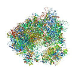 | | Crystal structure of compound C45 bound to the yeast 80S ribosome | | 分子名称: | (3~{R})-3-[(1~{S})-2-[(1~{S},4~{a}~{R},6~{S},7~{S},8~{a}~{R})-6,7-bis(chloranyl)-5,5,8~{a}-trimethyl-2-methylidene-3,4,4~{a},6,7,8-hexahydro-1~{H}-naphthalen-1-yl]-1-oxidanyl-ethyl]pyrrolidine-2,5-dione, 18S ribosomal RNA, 25S ribosomal RNA, ... | | 著者 | Pellegrino, S, Vanderwal, C.D, Yusupov, M. | | 登録日 | 2018-08-28 | | 公開日 | 2019-02-20 | | 最終更新日 | 2024-05-15 | | 実験手法 | X-RAY DIFFRACTION (3.10000038 Å) | | 主引用文献 | Understanding the role of intermolecular interactions between lissoclimides and the eukaryotic ribosome.
Nucleic Acids Res., 47, 2019
|
|


