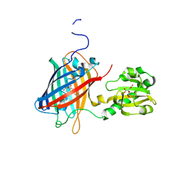
6WVD
 
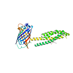 | | Human JAGN1 | | 分子名称: | (2R)-2,3-dihydroxypropyl (9Z)-octadec-9-enoate, Green fluorescent protein, Protein jagunal homolog 1 chimera | | 著者 | Yang, Y, Liu, S, Li, W. | | 登録日 | 2020-05-05 | | 公開日 | 2021-01-13 | | 最終更新日 | 2023-11-15 | | 実験手法 | X-RAY DIFFRACTION (2.25 Å) | | 主引用文献 | Termini restraining of small membrane proteins enables structure determination at near-atomic resolution.
Sci Adv, 6, 2020
|
|
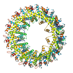
6WV5
 | | Human VKOR C43S mutant with vitamin K1 epoxide | | 分子名称: | (2R,3R)-2-hydroxy-3-methyl-2-[(2E,7S)-3,7,11,15-tetramethylhexadec-2-en-1-yl]-2,3-dihydronaphthalene-1,4-dione, Vitamin K epoxide reductase Cys43Ser mutant, termini restrained by green fluorescent protein | | 著者 | Liu, S, Sukumar, N, Li, W. | | 登録日 | 2020-05-05 | | 公開日 | 2020-11-11 | | 最終更新日 | 2023-11-15 | | 実験手法 | X-RAY DIFFRACTION (2.8 Å) | | 主引用文献 | Structural basis of antagonizing the vitamin K catalytic cycle for anticoagulation.
Science, 371, 2021
|
|
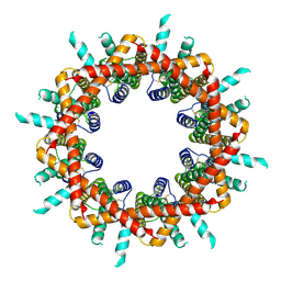
6WVE
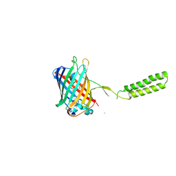 | | Chicken SPCS1 | | 分子名称: | Green fluorescent protein,Chicken Signal Peptidase Complex Subunit 1,Green fluorescent protein | | 著者 | Liu, S, Li, W. | | 登録日 | 2020-05-05 | | 公開日 | 2021-01-13 | | 最終更新日 | 2023-11-15 | | 実験手法 | X-RAY DIFFRACTION (2.429 Å) | | 主引用文献 | Termini restraining of small membrane proteins enables structure determination at near-atomic resolution.
Sci Adv, 6, 2020
|
|
6WSG
 | |
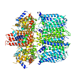
6WRF
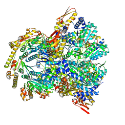
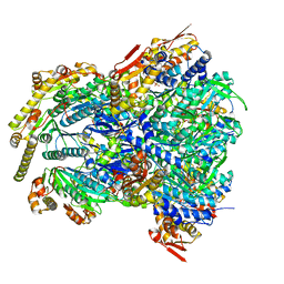 | | ClpX-ClpP complex bound to GFP-ssrA, recognition complex | | 分子名称: | ADENOSINE-5'-DIPHOSPHATE, ATP-dependent Clp protease ATP-binding subunit ClpX, ATP-dependent Clp protease proteolytic subunit, ... | | 著者 | Fei, X, Sauer, R.T. | | 登録日 | 2020-04-29 | | 公開日 | 2020-11-04 | | 最終更新日 | 2024-03-06 | | 実験手法 | ELECTRON MICROSCOPY (3.14 Å) | | 主引用文献 | Structural basis of ClpXP recognition and unfolding of ssrA-tagged substrates.
Elife, 9, 2020
|
|
6WQK
 | | hnRNPA2 Low complexity domain (LCD) determined by cryoEM | | 分子名称: | MCherry fluorescent protein,Heterogeneous nuclear ribonucleoproteins A2/B1 chimera | | 著者 | Lu, J, Cao, Q, Hughes, M.P, Sawaya, M.R, Boyer, D.R, Cascio, D, Eisenberg, D.S. | | 登録日 | 2020-04-29 | | 公開日 | 2020-08-26 | | 最終更新日 | 2024-03-06 | | 実験手法 | ELECTRON MICROSCOPY (3.1 Å) | | 主引用文献 | CryoEM structure of the low-complexity domain of hnRNPA2 and its conversion to pathogenic amyloid.
Nat Commun, 11, 2020
|
|
7BYM
 | | Cryo-EM structure of human KCNQ4 with retigabine | | 分子名称: | Calmodulin-3, Green fluorescent protein,Potassium voltage-gated channel subfamily KQT member 4, POTASSIUM ION, ... | | 著者 | Shen, H, Li, T, Yue, Z. | | 登録日 | 2020-04-23 | | 公開日 | 2020-12-02 | | 最終更新日 | 2024-03-27 | | 実験手法 | ELECTRON MICROSCOPY (3.1 Å) | | 主引用文献 | Structural Basis for the Modulation of Human KCNQ4 by Small-Molecule Drugs.
Mol.Cell, 81, 2021
|
|
7BYL
 | | Cryo-EM structure of human KCNQ4 | | 分子名称: | Calmodulin-3, Green fluorescent protein,Potassium voltage-gated channel subfamily KQT member 4, POTASSIUM ION, ... | | 著者 | Shen, H, Li, T, Yue, Z. | | 登録日 | 2020-04-23 | | 公開日 | 2020-12-02 | | 最終更新日 | 2024-03-27 | | 実験手法 | ELECTRON MICROSCOPY (2.5 Å) | | 主引用文献 | Structural Basis for the Modulation of Human KCNQ4 by Small-Molecule Drugs.
Mol.Cell, 81, 2021
|
|
7BYN
 | | Cryo-EM structure of human KCNQ4 with linopirdine | | 分子名称: | 1-phenyl-3,3-bis(pyridin-4-ylmethyl)indol-2-one, Calmodulin-3, Green fluorescent protein,Potassium voltage-gated channel subfamily KQT member 4, ... | | 著者 | Shen, H, Li, T, Yue, Z. | | 登録日 | 2020-04-23 | | 公開日 | 2020-12-02 | | 最終更新日 | 2024-03-27 | | 実験手法 | ELECTRON MICROSCOPY (3.3 Å) | | 主引用文献 | Structural Basis for the Modulation of Human KCNQ4 by Small-Molecule Drugs.
Mol.Cell, 81, 2021
|
|
6YOV
 | | OCT4-SOX2-bound nucleosome - SHL+6 | | 分子名称: | DNA (142-MER), Green fluorescent protein,POU domain, class 5, ... | | 著者 | Michael, A.K, Kempf, G, Cavadini, S, Bunker, R.D, Thoma, N.H. | | 登録日 | 2020-04-15 | | 公開日 | 2020-05-06 | | 最終更新日 | 2020-07-08 | | 実験手法 | ELECTRON MICROSCOPY (3.42 Å) | | 主引用文献 | Mechanisms of OCT4-SOX2 motif readout on nucleosomes.
Science, 368, 2020
|
|
7BWN
 | | Crystal Structure of a Designed Protein Heterocatenane | | 分子名称: | Cellular tumor antigen p53, Chimera of Green fluorescent protein and p53dim | | 著者 | Liu, Y.J, Duan, Z.L, Fang, J, Zhang, F, Xiao, J.Y, Zhang, W.B. | | 登録日 | 2020-04-15 | | 公開日 | 2020-06-17 | | 最終更新日 | 2023-11-29 | | 実験手法 | X-RAY DIFFRACTION (2.396 Å) | | 主引用文献 | Cellular Synthesis and X-ray Crystal Structure of a Designed Protein Heterocatenane.
Angew.Chem.Int.Ed.Engl., 59, 2020
|
|
6YLM
 | | mCherry | | 分子名称: | CHLORIDE ION, mCherry | | 著者 | Myskova, J, Rybakova, M, Brynda, J, Lazar, J. | | 登録日 | 2020-04-07 | | 公開日 | 2020-12-16 | | 最終更新日 | 2024-01-24 | | 実験手法 | X-RAY DIFFRACTION (1.6 Å) | | 主引用文献 | Directionality of light absorption and emission in representative fluorescent proteins.
Proc.Natl.Acad.Sci.USA, 117, 2020
|
|
6YLS
 | | mEos4b - Directionality of Optical Properties of Fluorescent Proteins | | 分子名称: | CHLORIDE ION, Green to red photoconvertible GFP-like protein EosFP, SODIUM ION | | 著者 | Myskova, J, Rybakova, O, Brynda, J, Lazar, J. | | 登録日 | 2020-04-07 | | 公開日 | 2020-12-16 | | 最終更新日 | 2024-01-24 | | 実験手法 | X-RAY DIFFRACTION (1.55 Å) | | 主引用文献 | Directionality of light absorption and emission in representative fluorescent proteins.
Proc.Natl.Acad.Sci.USA, 117, 2020
|
|
6M63
 | | Crystal structure of a cAMP sensor G-Flamp1. | | 分子名称: | ADENOSINE-3',5'-CYCLIC-MONOPHOSPHATE, Chimera of Cyclic nucleotide-gated potassium channel mll3241 and Yellow fluorescent protein | | 著者 | Zhou, Z, Chen, S, Wang, L, Chu, J. | | 登録日 | 2020-03-12 | | 公開日 | 2021-09-22 | | 最終更新日 | 2023-11-29 | | 実験手法 | X-RAY DIFFRACTION (2.25 Å) | | 主引用文献 | A high-performance genetically encoded fluorescent indicator for in vivo cAMP imaging.
Nat Commun, 13, 2022
|
|
6Y1G
 | | Photoconverted HcRed in its optoacoustic state | | 分子名称: | 1,2-ETHANEDIOL, GFP-like non-fluorescent chromoprotein | | 著者 | Janowski, R, Fuenzalida-Werner, J.P, Mishra, K, Stiel, A.C, Niessing, D. | | 登録日 | 2020-02-12 | | 公開日 | 2020-07-29 | | 最終更新日 | 2024-01-24 | | 実験手法 | X-RAY DIFFRACTION (2.3 Å) | | 主引用文献 | Challenging a Preconception: Optoacoustic Spectrum Differs from the Optical Absorption Spectrum of Proteins and Dyes for Molecular Imaging.
Anal.Chem., 92, 2020
|
|
6XZF
 | |
6XWY
 | | Highly pH-resistant long stokes-shift, red fluorescent protein mCRISPRed | | 分子名称: | 1,2-ETHANEDIOL, MALONIC ACID, Red fluorescent protein eqFP611 | | 著者 | Erdogan, M, Fabritius, A, Basquin, J, Griesbeck, O. | | 登録日 | 2020-01-24 | | 公開日 | 2020-02-05 | | 最終更新日 | 2024-01-24 | | 実験手法 | X-RAY DIFFRACTION (1.75 Å) | | 主引用文献 | Targeted In Situ Protein Diversification and Intra-organelle Validation in Mammalian Cells.
Cell Chem Biol, 27, 2020
|
|
6LR7
 | |
6VIO
 | |
6LOF
 | | Crystal structure of ZsYellow soaked by Cu2+ | | 分子名称: | GFP-like fluorescent chromoprotein FP538 | | 著者 | Nam, K.H. | | 登録日 | 2020-01-05 | | 公開日 | 2020-01-22 | | 最終更新日 | 2023-11-29 | | 実験手法 | X-RAY DIFFRACTION (2.6 Å) | | 主引用文献 | Spectroscopic and Structural Analysis of Cu 2+ -Induced Fluorescence Quenching of ZsYellow.
Biosensors (Basel), 10, 2020
|
|
6LNP
 | | Crystal structure of citrate Biosensor | | 分子名称: | CITRIC ACID, Fusion protein of Green fluorescent protein and Sensor histidine kinase CitA | | 著者 | Wen, Y, Campbell, R. | | 登録日 | 2019-12-31 | | 公開日 | 2020-05-06 | | 最終更新日 | 2023-11-22 | | 実験手法 | X-RAY DIFFRACTION (2.993 Å) | | 主引用文献 | High-Performance Intensiometric Direct- and Inverse-Response Genetically Encoded Biosensors for Citrate.
Acs Cent.Sci., 6, 2020
|
|
6VAL
 | |
6VAM
 | |
6V00
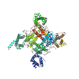
 | | structure of human KCNQ1-KCNE3-CaM complex | | 分子名称: | CALCIUM ION, Calmodulin-1, MCherry fluorescent protein,Potassium voltage-gated channel subfamily E member 3, ... | | 著者 | Mackinnon, R, Sun, J. | | 登録日 | 2019-11-16 | | 公開日 | 2019-12-04 | | 最終更新日 | 2024-03-06 | | 実験手法 | ELECTRON MICROSCOPY (3.1 Å) | | 主引用文献 | Structural Basis of Human KCNQ1 Modulation and Gating.
Cell, 180, 2020
|
|
6UZ3
 | | Cardiac sodium channel | | 分子名称: | (3beta,14beta,17beta,25R)-3-[4-methoxy-3-(methoxymethyl)butoxy]spirost-5-en, 2-acetamido-2-deoxy-beta-D-glucopyranose, 2-acetamido-2-deoxy-beta-D-glucopyranose-(1-4)-2-acetamido-2-deoxy-beta-D-glucopyranose, ... | | 著者 | Jiang, D, Shi, H, Tonggu, L, Lenaeus, M.J, Zheng, N, Catterall, W.A. | | 登録日 | 2019-11-14 | | 公開日 | 2020-01-01 | | 最終更新日 | 2020-07-29 | | 実験手法 | ELECTRON MICROSCOPY (3.5 Å) | | 主引用文献 | Structure of the Cardiac Sodium Channel.
Cell, 180, 2020
|
|


