Loading
PDBj
MenuPDBj@FacebookPDBj@TwitterPDBj@YouTubewwPDB FoundationwwPDB
RCSB PDBPDBeBMRBAdv. SearchSearch help

3K2M
DownloadVisualize
BU of 3k2m by Molmil
Crystal Structure of Monobody HA4/Abl1 SH2 Domain Complex
Descriptor: Monobody HA4, PHOSPHATE ION, Proto-oncogene tyrosine-protein kinase ABL1
Authors:Wojcik, J.B, Duguid, E.
Deposit date:2009-09-30
Release date:2010-03-31
Last modified:2023-09-06
Method:X-RAY DIFFRACTION (1.75 Å)
Cite:A potent and highly specific FN3 monobody inhibitor of the Abl SH2 domain.
Nat.Struct.Mol.Biol., 17, 2010
3KFJ
DownloadVisualize
BU of 3kfj by Molmil
Crystal Structure of the Grb2 SH2 Domain in Complex with a Flexible Ac-pY-E-N-NH2 Tripeptide Mimic
Descriptor: CHLORIDE ION, Growth factor receptor-bound protein 2, MAGNESIUM ION, ...
Authors:Clements, J.H.
Deposit date:2009-10-27
Release date:2010-03-02
Last modified:2023-09-06
Method:X-RAY DIFFRACTION (2.02 Å)
Cite:Thermodynamic and structural effects of conformational constraints in protein-ligand interactions. Entropic paradoxy associated with ligand preorganization.
J.Am.Chem.Soc., 131, 2009
3M7F
DownloadVisualize
BU of 3m7f by Molmil
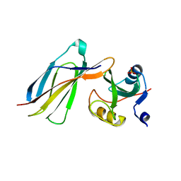
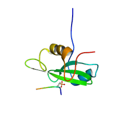
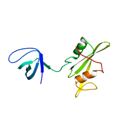
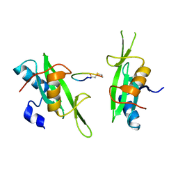
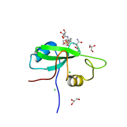
Crystal structure of the Nedd4 C2/Grb10 SH2 complex
Descriptor: E3 ubiquitin-protein ligase NEDD4, Growth factor receptor-bound protein 10
Authors:Huang, Q, Szebenyi, M.
Deposit date:2010-03-16
Release date:2010-10-27
Last modified:2023-09-06
Method:X-RAY DIFFRACTION (2 Å)
Cite:Structural Basis for the Interaction between the Growth Factor-binding Protein GRB10 and the E3 Ubiquitin Ligase NEDD4.
J.Biol.Chem., 285, 2010
3MAZ
DownloadVisualize
BU of 3maz by Molmil
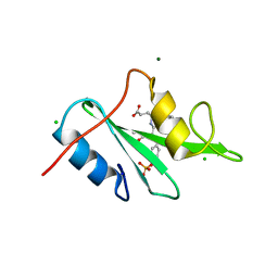
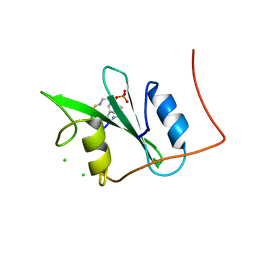
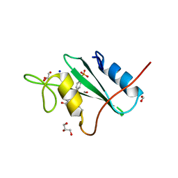
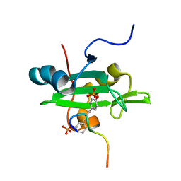
Crystal Structure of the Human BRDG1/STAP-1 SH2 Domain in Complex with the NTAL pTyr136 Peptide
Descriptor: CheD family protein, MALONATE ION, Signal-transducing adaptor protein 1
Authors:Kaneko, T, Huang, H, Zhao, B, Li, L, Liu, H, Voss, C.K, Wu, C, Schiller, M.R, Li, S.S.
Deposit date:2010-03-24
Release date:2010-05-12
Last modified:2023-11-22
Method:X-RAY DIFFRACTION (1.9 Å)
Cite:Loops govern SH2 domain specificity by controlling access to binding pockets.
Sci.Signal., 3, 2010
3MXC
DownloadVisualize
BU of 3mxc by Molmil
Structures of Grb2-SH2 Domain and AICD peptide Complexes Reveal a Conformational Switch and Their Functional Implications.
Descriptor: AICD peptide, Growth factor receptor-bound protein 2
Authors:Sen, U, Das, S.
Deposit date:2010-05-07
Release date:2011-05-11
Last modified:2023-11-22
Method:X-RAY DIFFRACTION (2 Å)
Cite:Functional Implications of the Conformational Switch in AICD Peptide upon Binding to Grb2-SH2 Domain.
J.Mol.Biol., 414, 2011
3MXY
DownloadVisualize
BU of 3mxy by Molmil
Structures of Grb2-SH2 Domain and AICD peptide Complexes Reveal a Conformational Switch and Their Functional Implications.
Descriptor: AICD peptide E683V variant, Growth factor receptor-bound protein 2
Authors:Sen, U, Das, S.
Deposit date:2010-05-08
Release date:2011-05-11
Last modified:2023-11-22
Method:X-RAY DIFFRACTION (2.3 Å)
Cite:Functional Implications of the Conformational Switch in AICD Peptide upon Binding to Grb2-SH2 Domain.
J.Mol.Biol., 414, 2011
3N7Y
DownloadVisualize
BU of 3n7y by Molmil
Crystal Structure of the Grb2 SH2 Domain in Complex with a 20-Membered Macrocyclic Ligand Having the Sequence pYVNV
Descriptor: 20-membered peptide-like macrocyclic ligand, Growth factor receptor-bound protein 2
Authors:Whiddon, B.B, Clements, J.H, Martin, S.F.
Deposit date:2010-05-27
Release date:2011-01-12
Last modified:2023-11-15
Method:X-RAY DIFFRACTION (2.02 Å)
Cite:Thermodynamic and Structural Effects of Macrocyclization as a Constraining Method in Protein-Ligand Interactions.
ACS MED.CHEM.LETT., 1, 2010
3N84
DownloadVisualize
BU of 3n84 by Molmil
Crystal Structure of the Grb2 SH2 Domain in Complex with a 23-Membered Macrocyclic Ligand Having the Sequence pYVNVP
Descriptor: 23-membered peptide-like macrocyclic ligand, CHLORIDE ION, GLYCEROL, ...
Authors:Clements, J.H, Martin, S.F.
Deposit date:2010-05-27
Release date:2011-01-12
Last modified:2023-11-15
Method:X-RAY DIFFRACTION (2 Å)
Cite:Thermodynamic and Structural Effects of Macrocyclization as a Constraining Method in Protein-Ligand Interactions.
ACS MED.CHEM.LETT., 1, 2010
3N8M
DownloadVisualize
BU of 3n8m by Molmil
Crystal Structure of the Grb2 SH2 Domain in Complex with An Acyclic Ligand Having the Sequence pYVNVP
Descriptor: GLYCEROL, Growth factor receptor-bound protein 2, PEPTIDE
Authors:Whiddon, B.B, Clements, J.H, Martin, S.F.
Deposit date:2010-05-28
Release date:2010-12-15
Last modified:2023-11-22
Method:X-RAY DIFFRACTION (2 Å)
Cite:Thermodynamic and Structural Effects of Macrocyclization as a Constraining Method in Protein-Ligand Interactions.
ACS MED.CHEM.LETT., 1, 2010
3NHN
DownloadVisualize
BU of 3nhn by Molmil
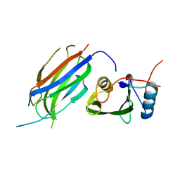
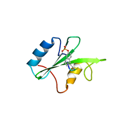
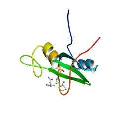
Crystal structure of the SRC-family kinase HCK SH3-SH2-linker regulatory region
Descriptor: Tyrosine-protein kinase HCK
Authors:Alvarado, J.J, Betts, L, Moroco, J.A, Smithgall, T.E, Yeh, J.I.
Deposit date:2010-06-14
Release date:2010-09-01
Last modified:2023-09-06
Method:X-RAY DIFFRACTION (2.61 Å)
Cite:Crystal Structure of the Src Family Kinase Hck SH3-SH2 Linker Regulatory Region Supports an SH3-dominant Activation Mechanism.
J.Biol.Chem., 285, 2010
3OV1
DownloadVisualize
BU of 3ov1 by Molmil
Crystal Structure of the Grb2 SH2 Domain in Complex with a pYXN-Derived Tripeptide
Descriptor: FORMIC ACID, GLYCEROL, Growth factor receptor-bound protein 2, ...
Authors:Clements, J.H, Martin, S.F.
Deposit date:2010-09-15
Release date:2011-11-02
Last modified:2023-12-06
Method:X-RAY DIFFRACTION (1.6 Å)
Cite:Protein-ligand interactions: thermodynamic effects associated with increasing nonpolar surface area.
J.Am.Chem.Soc., 133, 2011
3OVE
DownloadVisualize
BU of 3ove by Molmil
Crystal Structure of the Grb2 SH2 Domain in Complex with a pYXN-Derived Tripeptide
Descriptor: CHLORIDE ION, Growth factor receptor-bound protein 2, PYAC7CN
Authors:Clements, J.H, Martin, S.F.
Deposit date:2010-09-16
Release date:2011-11-02
Last modified:2023-12-06
Method:X-RAY DIFFRACTION (1.82 Å)
Cite:Protein-ligand interactions: thermodynamic effects associated with increasing nonpolar surface area.
J.Am.Chem.Soc., 133, 2011
2L3T
DownloadVisualize
BU of 2l3t by Molmil
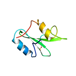
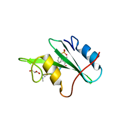
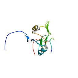
Solution structure of tandem SH2 domain from Spt6
Descriptor: Transcription elongation factor SPT6
Authors:Liu, J, Zhang, J, Wu, J, Shi, Y.
Deposit date:2010-09-22
Release date:2011-06-15
Last modified:2024-05-01
Method:SOLUTION NMR
Cite:Solution structure of the tandem SH2 domains from Spt6 and their binding to the phosphorylated RNA polymerase II C-terminal domain
To be Published
2L4K
DownloadVisualize
BU of 2l4k by Molmil
Water refined solution structure of the human Grb7-SH2 domain in complex with the 10 amino acid peptide pY1139
Descriptor: Growth factor receptor-bound protein 7, Receptor tyrosine-protein kinase erbB-2
Authors:Pias, S.C, Ivancic, M, Brescia, P.J, Johnson, D.L, Smith, D.E, Daly, R.J, Lyons, B.A.
Deposit date:2010-10-07
Release date:2010-11-17
Last modified:2012-10-03
Method:SOLUTION NMR
Cite:Water-Refined Solution Structure of the Human Grb7-SH2 Domain in Complex with the erbB2 Receptor Peptide pY1139.
Protein Pept.Lett., 19, 2012
2L6K
DownloadVisualize
BU of 2l6k by Molmil
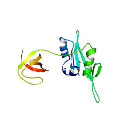
Solution Structure of a Nonphosphorylated Peptide Recognizing Domain
Descriptor: Tensin-like C1 domain-containing phosphatase
Authors:Dai, K, Liao, S, Zhang, J, Zhang, X, Tu, X.
Deposit date:2010-11-22
Release date:2011-10-12
Last modified:2024-05-15
Method:SOLUTION NMR
Cite:Solution structure of Tensin2 SH2 domain and its phosphotyrosine-independent interaction with DLC-1
Plos One, 6, 2011
3PQZ
DownloadVisualize
BU of 3pqz by Molmil
Grb7 SH2 with peptide
Descriptor: Growth factor receptor-bound protein 7, cyclic peptide
Authors:Wilce, J.A.
Deposit date:2010-11-29
Release date:2011-07-20
Last modified:2011-09-21
Method:X-RAY DIFFRACTION (2.413 Å)
Cite:Structural basis of binding by cyclic nonphosphorylated Peptide antagonists of grb7 implicated in breast cancer progression
J.Mol.Biol., 412, 2011
3PS5
DownloadVisualize
BU of 3ps5 by Molmil
Crystal structure of the full-length Human Protein Tyrosine Phosphatase SHP-1
Descriptor: SULFATE ION, Tyrosine-protein phosphatase non-receptor type 6
Authors:Wang, W, Liu, L, Song, X, Mo, Y, Komma, C, Bellamy, H.D, Zhao, Z.J, Zhou, G.W.
Deposit date:2010-11-30
Release date:2011-04-20
Last modified:2023-09-06
Method:X-RAY DIFFRACTION (3.1 Å)
Cite:Crystal structure of human protein tyrosine phosphatase SHP-1 in the open conformation.
J.Cell.Biochem., 112, 2011
3PSI
DownloadVisualize
BU of 3psi by Molmil
Crystal Structure of the Spt6 core domain from Saccharomyces cerevisiae, Form Spt6(239-1451)
Descriptor: Transcription elongation factor SPT6
Authors:Close, D, Hill, C.P, Johnson, S.J.
Deposit date:2010-12-01
Release date:2011-03-30
Last modified:2011-08-03
Method:X-RAY DIFFRACTION (3.3 Å)
Cite:Crystal structures of the S. cerevisiae Spt6 core and C-terminal tandem SH2 domain.
J.Mol.Biol., 408, 2011
3PSJ
DownloadVisualize
BU of 3psj by Molmil
Crystal Structure of the Spt6 Tandem SH2 Domain from Saccharomyces cerevisiae, Form Se-Spt6 (1247-1451)
Descriptor: SULFATE ION, Transcription elongation factor SPT6
Authors:Close, D, Hill, C.P.
Deposit date:2010-12-01
Release date:2011-03-30
Last modified:2017-11-08
Method:X-RAY DIFFRACTION (2.702 Å)
Cite:Crystal structures of the S. cerevisiae Spt6 core and C-terminal tandem SH2 domain.
J.Mol.Biol., 408, 2011
3PSK
DownloadVisualize
BU of 3psk by Molmil
Crystal Structure of the Spt6 Tandem SH2 Domain from Saccharomyces cerevisiae, Form Native Spt6 (1247-1451)
Descriptor: SULFATE ION, Transcription elongation factor SPT6
Authors:Close, D, Hill, C.P.
Deposit date:2010-12-01
Release date:2011-03-30
Last modified:2023-09-06
Method:X-RAY DIFFRACTION (2.1 Å)
Cite:Crystal structures of the S. cerevisiae Spt6 core and C-terminal tandem SH2 domain.
J.Mol.Biol., 408, 2011
2Y3A
DownloadVisualize
BU of 2y3a by Molmil
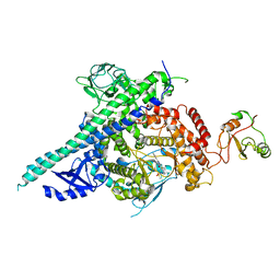
Crystal structure of p110beta in complex with icSH2 of p85beta and the drug GDC-0941
Descriptor: 2-(1H-indazol-4-yl)-6-{[4-(methylsulfonyl)piperazin-1-yl]methyl}-4-morpholin-4-yl-thieno[3,2-d]pyrimidine, PHOSPHATIDYLINOSITOL 3-KINASE REGULATORY SUBUNIT BETA, PHOSPHATIDYLINOSITOL-4,5-BISPHOSPHATE 3-KINASE CATALYTIC SUBUNIT BETA ISOFORM
Authors:Zhang, X, Vadas, O, Perisic, O, Williams, R.L.
Deposit date:2010-12-20
Release date:2011-03-16
Last modified:2023-12-20
Method:X-RAY DIFFRACTION (3.3 Å)
Cite:Structure of Lipid Kinase P110Beta-P85Beta Elucidates an Unusual Sh2-Domain-Mediated Inhibitory Mechanism.
Mol.Cell, 41, 2011
3QWX
DownloadVisualize
BU of 3qwx by Molmil
CED-2 1-174
Descriptor: Cell death abnormality protein 2, SULFATE ION
Authors:Kang, Y, Sun, J, Liu, Y.
Deposit date:2011-02-28
Release date:2011-06-08
Last modified:2024-02-21
Method:X-RAY DIFFRACTION (2.01 Å)
Cite:Crystal structure of the cell corpse engulfment protein CED-2 in Caenorhabditis elegans.
Biochem.Biophys.Res.Commun., 410, 2011
3QWY
DownloadVisualize
BU of 3qwy by Molmil
CED-2
Descriptor: Cell death abnormality protein 2, GLYCEROL, SULFATE ION
Authors:Kang, Y, Sun, J, Liu, Y, Sun, D, Hu, Y, Liu, Y.F.
Deposit date:2011-02-28
Release date:2011-06-08
Last modified:2024-02-21
Method:X-RAY DIFFRACTION (2.52 Å)
Cite:Crystal structure of the cell corpse engulfment protein CED-2 in Caenorhabditis elegans.
Biochem.Biophys.Res.Commun., 410, 2011
2LCT
DownloadVisualize
BU of 2lct by Molmil
Solution structure of the Vav1 SH2 domain complexed with a Syk-derived doubly phosphorylated peptide
Descriptor: Proto-oncogene vav, Tyrosine-protein kinase SYK
Authors:Chen, C, Gorenstein, N, Post, C.
Deposit date:2011-05-09
Release date:2011-06-01
Last modified:2023-06-14
Method:SOLUTION NMR
Cite:Two closely spaced tyrosines regulate NFAT signaling in B cells via Syk association with Vav.
Mol.Cell.Biol., 31, 2011
3S8N
DownloadVisualize
BU of 3s8n by Molmil
Crystal Structure of the Grb2 SH2 Domain in Complex with a pYXN-Derived Tripeptide
Descriptor: CHLORIDE ION, GLYCEROL, Growth factor receptor-bound protein 2, ...
Authors:Clements, J.H, Stephen, F.M.
Deposit date:2011-05-29
Release date:2011-11-02
Last modified:2023-12-06
Method:X-RAY DIFFRACTION (1.71 Å)
Cite:Protein-ligand interactions: thermodynamic effects associated with increasing nonpolar surface area.
J.Am.Chem.Soc., 133, 2011

221051

PDB entries from 2024-06-12

PDB statisticsPDBj update infoContact PDBjnumon