Loading
PDBj
MenuPDBj@FacebookPDBj@TwitterPDBj@YouTubewwPDB FoundationwwPDB
RCSB PDBPDBeBMRBAdv. SearchSearch help
PDB: 20 results

4TZA
DownloadVisualize
BU of 4tza by Molmil
TGP, an extremely thermostable green fluorescent protein created by structure-guided surface engineering
Descriptor: Fluorescent Protein
Authors:Close, D.W, Bradbury, A.R.M.
Deposit date:2014-07-09
Release date:2014-10-22
Last modified:2023-11-15
Method:X-RAY DIFFRACTION (1.9 Å)
Cite:Thermal green protein, an extremely stable, nonaggregating fluorescent protein created by structure-guided surface engineering.
Proteins, 83, 2015
4TZG
DownloadVisualize
BU of 4tzg by Molmil
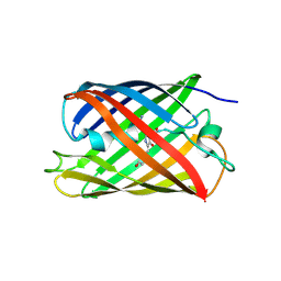
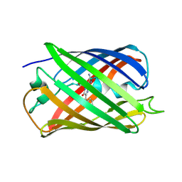
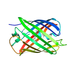
Crystal structure of eCGP123, an extremely thermostable green fluorescent protein
Descriptor: Fluorescent Protein
Authors:Close, D.W, Don Paul, C, Traore, D.A.K, Wilce, M.C.J, Prescott, M, Bradbury, A.R.M.
Deposit date:2014-07-10
Release date:2014-10-22
Last modified:2023-11-15
Method:X-RAY DIFFRACTION (2.1 Å)
Cite:Thermal green protein, an extremely stable, nonaggregating fluorescent protein created by structure-guided surface engineering.
Proteins, 83, 2015
6WZN
DownloadVisualize
BU of 6wzn by Molmil
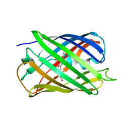
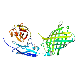
Crystal Structure of a Fluorescent Single Chain Fv Chimera
Descriptor: Fluorescent Single Chain Fv Chimera, GLYCEROL
Authors:Close, D, Velappan, N, Hung, L.W, Naranjo, L, Hemez, C, DeVore, N, McCullough, D, Lillo, A.M, Waldo, G, Bradbury, A.R.M.
Deposit date:2020-05-14
Release date:2021-01-27
Last modified:2023-11-15
Method:X-RAY DIFFRACTION (2.5 Å)
Cite:Construction, characterization and crystal structure of a fluorescent single-chain Fv chimera.
Protein Eng.Des.Sel., 34, 2021
3PSK
DownloadVisualize
BU of 3psk by Molmil
Crystal Structure of the Spt6 Tandem SH2 Domain from Saccharomyces cerevisiae, Form Native Spt6 (1247-1451)
Descriptor: SULFATE ION, Transcription elongation factor SPT6
Authors:Close, D, Hill, C.P.
Deposit date:2010-12-01
Release date:2011-03-30
Last modified:2023-09-06
Method:X-RAY DIFFRACTION (2.1 Å)
Cite:Crystal structures of the S. cerevisiae Spt6 core and C-terminal tandem SH2 domain.
J.Mol.Biol., 408, 2011
4PHB
DownloadVisualize
BU of 4phb by Molmil
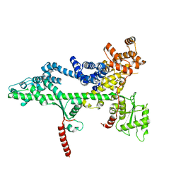
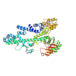
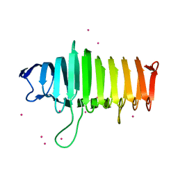
Structure of the polysaccharide lyase-like protein Cthe_2159 from C. thermocellum, Gadolinium derivative
Descriptor: GADOLINIUM ATOM, Uncharacterized protein
Authors:Close, D.W, D'Angelo, S, Bradbury, A.R.M.
Deposit date:2014-05-06
Release date:2014-08-27
Last modified:2023-12-27
Method:X-RAY DIFFRACTION (2.1781 Å)
Cite:A new family of beta-helix proteins with similarities to the polysaccharide lyases.
Acta Crystallogr.,Sect.D, 70, 2014
4PEU
DownloadVisualize
BU of 4peu by Molmil
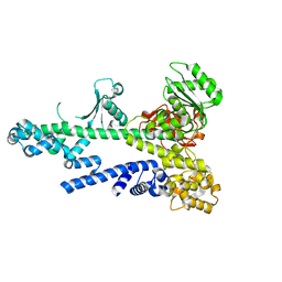
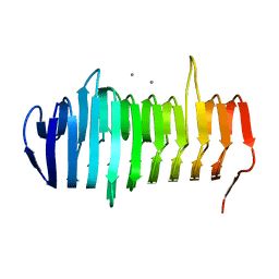
Structure of the polysaccharide lyase-like protein Cthe_2159 from C. thermocellum, native form with Calcium bound
Descriptor: CALCIUM ION, Uncharacterized protein
Authors:Close, D.W, D'Angelo, S, Bradbury, A.R.M.
Deposit date:2014-04-24
Release date:2014-10-01
Last modified:2023-12-27
Method:X-RAY DIFFRACTION (1.8001 Å)
Cite:A new family of beta-helix proteins with similarities to the polysaccharide lyases.
Acta Crystallogr.,Sect.D, 70, 2014
5EB7
DownloadVisualize
BU of 5eb7 by Molmil
Crystal Structure of the Reversibly photoswitching chromoprotein Dathail, Metastable State
Descriptor: Reversibly photoswitching chromoprotein Dathail
Authors:Close, D.W, Langan, P.S, Bradbury, A.R.M.
Deposit date:2015-10-18
Release date:2016-04-06
Last modified:2023-11-15
Method:X-RAY DIFFRACTION (1.65 Å)
Cite:Evolution and characterization of a new reversibly photoswitching chromogenic protein, Dathail.
J.Mol.Biol., 428, 2016
5EB6
DownloadVisualize
BU of 5eb6 by Molmil
Crystal Structure of the Reversibly photoswitching chromoprotein Dathail, Ground State
Descriptor: Reversible photoswitching chromoprotein Dathail
Authors:Close, D.W, Langan, P.S, Bradbury, A.R.M.
Deposit date:2015-10-18
Release date:2016-04-06
Last modified:2023-11-15
Method:X-RAY DIFFRACTION (1.65008247 Å)
Cite:Evolution and characterization of a new reversibly photoswitching chromogenic protein, Dathail.
J.Mol.Biol., 428, 2016
5EJU
DownloadVisualize
BU of 5eju by Molmil
Ensemble refinement of the Crystal Structure of the Reversibly photoswitching chromoprotein Dathail, Ground State
Descriptor: Reversibly photoswitching protein Dathail
Authors:Close, D.W, Langan, P.S, Bradbury, A.R.M.
Deposit date:2015-11-02
Release date:2016-04-06
Last modified:2023-11-15
Method:X-RAY DIFFRACTION (1.65 Å)
Cite:Evolution and characterization of a new reversibly photoswitching chromogenic protein, Dathail.
J.Mol.Biol., 428, 2016
5EXU
DownloadVisualize
BU of 5exu by Molmil
Reversibly photoswitching protein Dathail, Ensemble refinement
Descriptor: Reversibly photoswitching protein Dathail
Authors:Close, D.W, Langan, P.S, Bradbury, A.R.M.
Deposit date:2015-11-24
Release date:2016-04-06
Last modified:2023-11-15
Method:X-RAY DIFFRACTION (1.65 Å)
Cite:Evolution and characterization of a new reversibly photoswitching chromogenic protein, Dathail.
J.Mol.Biol., 428, 2016
3PSJ
DownloadVisualize
BU of 3psj by Molmil
Crystal Structure of the Spt6 Tandem SH2 Domain from Saccharomyces cerevisiae, Form Se-Spt6 (1247-1451)
Descriptor: SULFATE ION, Transcription elongation factor SPT6
Authors:Close, D, Hill, C.P.
Deposit date:2010-12-01
Release date:2011-03-30
Last modified:2017-11-08
Method:X-RAY DIFFRACTION (2.702 Å)
Cite:Crystal structures of the S. cerevisiae Spt6 core and C-terminal tandem SH2 domain.
J.Mol.Biol., 408, 2011
3PSI
DownloadVisualize
BU of 3psi by Molmil
Crystal Structure of the Spt6 core domain from Saccharomyces cerevisiae, Form Spt6(239-1451)
Descriptor: Transcription elongation factor SPT6
Authors:Close, D, Hill, C.P, Johnson, S.J.
Deposit date:2010-12-01
Release date:2011-03-30
Last modified:2011-08-03
Method:X-RAY DIFFRACTION (3.3 Å)
Cite:Crystal structures of the S. cerevisiae Spt6 core and C-terminal tandem SH2 domain.
J.Mol.Biol., 408, 2011
3PSF
DownloadVisualize
BU of 3psf by Molmil
Crystal Structure of the Spt6 core domain from Saccharomyces cerevisiae, Form Spt6(236-1259)
Descriptor: Transcription elongation factor SPT6
Authors:Close, D, Hill, C.P.
Deposit date:2010-12-01
Release date:2011-03-30
Last modified:2024-02-21
Method:X-RAY DIFFRACTION (2.59 Å)
Cite:Crystal structures of the S. cerevisiae Spt6 core and C-terminal tandem SH2 domain.
J.Mol.Biol., 408, 2011
4PPK
DownloadVisualize
BU of 4ppk by Molmil
Crystal structure of eCGP123 T69V variant at pH 7.5
Descriptor: Monomeric Azami Green
Authors:Don Paul, C, Traore, D.A.K, Devenish, R.J, Close, D, Bell, T, Bradbury, A, Wilce, M.C.J, Prescott, M.
Deposit date:2014-02-27
Release date:2015-04-08
Last modified:2024-04-03
Method:X-RAY DIFFRACTION (2 Å)
Cite:X-Ray Crystal Structure and Properties of Phanta, a Weakly Fluorescent Photochromic GFP-Like Protein.
Plos One, 10, 2015
4PPJ
DownloadVisualize
BU of 4ppj by Molmil
Crystal structure of Phanta, a weakly fluorescent photochromic GFP-like protein. ON state
Descriptor: Monomeric Azami Green
Authors:Don Paul, C, Traore, D.A.K, Devenish, R.J, Close, D, Bell, T, Bradbury, A, Wilce, M.C.J, Prescott, M.
Deposit date:2014-02-27
Release date:2015-04-08
Last modified:2024-04-03
Method:X-RAY DIFFRACTION (2.3 Å)
Cite:X-Ray Crystal Structure and Properties of Phanta, a Weakly Fluorescent Photochromic GFP-Like Protein.
Plos One, 10, 2015
4PPL
DownloadVisualize
BU of 4ppl by Molmil
Crystal structure of eCGP123 H193Q variant at pH 7.5
Descriptor: Monomeric Azami Green
Authors:Don Paul, C, Traore, D.A.K, Devenish, R.J, Close, D, Bell, T, Bradbury, A, Wilce, M.C.J, Prescott, M.
Deposit date:2014-02-27
Release date:2015-04-08
Last modified:2024-04-03
Method:X-RAY DIFFRACTION (2.2 Å)
Cite:X-Ray Crystal Structure and Properties of Phanta, a Weakly Fluorescent Photochromic GFP-Like Protein.
Plos One, 10, 2015
3OAK
DownloadVisualize
BU of 3oak by Molmil
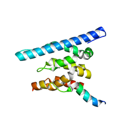
Crystal structure of a Spn1 (Iws1)-Spt6 complex
Descriptor: Transcription elongation factor SPT6, Transcription factor IWS1
Authors:McDonald, S.M, Close, D, Hill, C.P.
Deposit date:2010-08-05
Release date:2010-11-24
Last modified:2024-02-21
Method:X-RAY DIFFRACTION (2.15 Å)
Cite:Structure and biological importance of the spn1-spt6 interaction, and its regulatory role in nucleosome binding.
Mol.Cell, 40, 2010
3BZC
DownloadVisualize
BU of 3bzc by Molmil
Crystal Structure of the Tex protein from Pseudomonas aeruginosa, crystal form I
Descriptor: Tex
Authors:Johnson, S.J, Close, D, Hill, C.P.
Deposit date:2008-01-17
Release date:2008-04-08
Last modified:2024-02-21
Method:X-RAY DIFFRACTION (2.27 Å)
Cite:Crystal structure and RNA binding of the Tex protein from Pseudomonas aeruginosa.
J.Mol.Biol., 377, 2008
3BZK
DownloadVisualize
BU of 3bzk by Molmil
Crystal Structure of the Tex protein from Pseudomonas aeruginosa, crystal form 2
Descriptor: Tex
Authors:Johnson, S.J, Close, D, Hill, C.P.
Deposit date:2008-01-18
Release date:2008-04-08
Last modified:2023-08-30
Method:X-RAY DIFFRACTION (2.3 Å)
Cite:Crystal structure and RNA binding of the Tex protein from Pseudomonas aeruginosa.
J.Mol.Biol., 377, 2008
3O8Z
DownloadVisualize
BU of 3o8z by Molmil
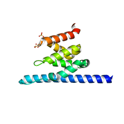
Crystal structure of Spn1 (Iws1) core domain
Descriptor: SULFATE ION, Transcription factor IWS1
Authors:McDonald, S.M, Close, D, Hill, C.P.
Deposit date:2010-08-03
Release date:2010-11-24
Last modified:2024-02-21
Method:X-RAY DIFFRACTION (2.15 Å)
Cite:Structure and biological importance of the spn1-spt6 interaction, and its regulatory role in nucleosome binding.
Mol.Cell, 40, 2010

218853

PDB entries from 2024-04-24

PDB statisticsPDBj update infoContact PDBjnumon