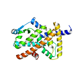
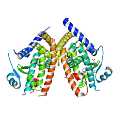
5HT6
 
 | | Crystal structure of the SET domain of the human MLL5 methyltransferase | | 分子名称: | Histone-lysine N-methyltransferase 2E | | 著者 | le Maire, A, Mas-y-Mas, S, Dumas, C, Lebedev, A. | | 登録日 | 2016-01-26 | | 公開日 | 2016-11-16 | | 最終更新日 | 2024-05-08 | | 実験手法 | X-RAY DIFFRACTION (2.093 Å) | | 主引用文献 | The Human Mixed Lineage Leukemia 5 (MLL5), a Sequentially and Structurally Divergent SET Domain-Containing Protein with No Intrinsic Catalytic Activity.
Plos One, 11, 2016
|
|
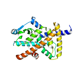
2CKK
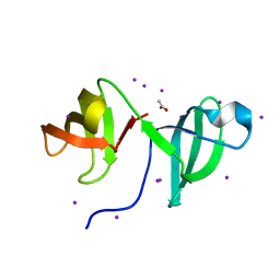 | | High resolution crystal structure of the human kin17 C-terminal domain containing a kow motif | | 分子名称: | ACETATE ION, IODIDE ION, KIN17 | | 著者 | le Maire, A, Schiltz, M, Pinon-Lataillade, G, Stura, E, Couprie, J, Gondry, M, Angulo-Mora, J, Zinn-Justin, S. | | 登録日 | 2006-04-20 | | 公開日 | 2006-10-04 | | 最終更新日 | 2024-05-08 | | 実験手法 | X-RAY DIFFRACTION (1.45 Å) | | 主引用文献 | A Tandem of SH3-Like Domains Participates in RNA Binding in Kin17, a Human Protein Activated in Response to Genotoxics.
J.Mol.Biol., 364, 2006
|
|
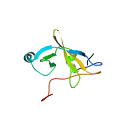
1S1N
 | | SH3 domain of human nephrocystin | | 分子名称: | Nephrocystin 1 | | 著者 | Le Maire, A, Weber, T, Saunier, S, Antignac, C, Ducruix, A, Dardel, F. | | 登録日 | 2004-01-07 | | 公開日 | 2005-01-18 | | 最終更新日 | 2024-05-01 | | 実験手法 | SOLUTION NMR | | 主引用文献 | Solution NMR structure of the SH3 domain of human nephrocystin and analysis of a mutation-causing juvenile nephronophthisis.
Proteins, 59, 2005
|
|
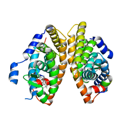
6SSQ
 | | Crystal structure of RARbeta LBD in complex with LG 100754 | | 分子名称: | (2E,4E,6Z)-3-methyl-7-(5,5,8,8-tetramethyl-3-propoxy-5,6,7,8-tetrahydronaphthalen-2-yl)octa-2,4,6-trienoic acid, CITRATE ANION, GLYCEROL, ... | | 著者 | le Maire, A, Teyssier, C, Germain, P, Bourguet, W. | | 登録日 | 2019-09-09 | | 公開日 | 2019-11-20 | | 最終更新日 | 2024-01-24 | | 実験手法 | X-RAY DIFFRACTION (2.3 Å) | | 主引用文献 | Regulation of RXR-RAR Heterodimers by RXR- and RAR-Specific Ligands and Their Combinations.
Cells, 8, 2019
|
|
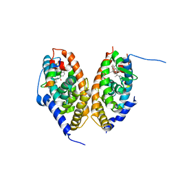
6STI
 | | Crystal structure of RXRalpha LBD in complex with LG 100754 and a coactivator peptide | | 分子名称: | (2E,4E,6Z)-3-methyl-7-(5,5,8,8-tetramethyl-3-propoxy-5,6,7,8-tetrahydronaphthalen-2-yl)octa-2,4,6-trienoic acid, ACETATE ION, Nuclear receptor coactivator 2, ... | | 著者 | le Maire, A, Teyssier, C, Germain, P, Bourguet, W. | | 登録日 | 2019-09-10 | | 公開日 | 2019-11-20 | | 最終更新日 | 2024-01-31 | | 実験手法 | X-RAY DIFFRACTION (1.892 Å) | | 主引用文献 | Regulation of RXR-RAR Heterodimers by RXR- and RAR-Specific Ligands and Their Combinations.
Cells, 8, 2019
|
|
3KWY
 | |
7PDQ
 | | Crystal structure of a mutated form of RXRalpha ligand binding domain in complex with LG100268 and a coactivator fragment | | 分子名称: | 6-[1-(3,5,5,8,8-PENTAMETHYL-5,6,7,8-TETRAHYDRONAPHTHALEN-2-YL)CYCLOPROPYL]PYRIDINE-3-CARBOXYLIC ACID, Nuclear receptor coactivator 2, Retinoic acid receptor RXR-alpha | | 著者 | le Maire, A, Bourguet, W, Guee, L. | | 登録日 | 2021-08-06 | | 公開日 | 2022-08-03 | | 最終更新日 | 2024-01-31 | | 実験手法 | X-RAY DIFFRACTION (1.58 Å) | | 主引用文献 | Design and in vitro characterization of RXR variants as tools to investigate the biological role of endogenous rexinoids.
J.Mol.Endocrinol., 69, 2022
|
|
7PDT
 | | Crystal structure of a mutated form of RXRalpha ligand binding domain in complex with BMS649 and a coactivator fragment | | 分子名称: | 4-[2-(5,5,8,8-TETRAMETHYL-5,6,7,8-TETRAHYDRO-NAPHTHALEN-2-YL)-[1,3]DIOXOLAN-2-YL]-BENZOIC ACID, Nuclear receptor coactivator 2, Retinoic acid receptor RXR-alpha | | 著者 | le Maire, A, Bourguet, W, Guee, L. | | 登録日 | 2021-08-07 | | 公開日 | 2022-08-03 | | 最終更新日 | 2024-01-31 | | 実験手法 | X-RAY DIFFRACTION (3.3 Å) | | 主引用文献 | Design and in vitro characterization of RXR variants as tools to investigate the biological role of endogenous rexinoids.
J.Mol.Endocrinol., 69, 2022
|
|
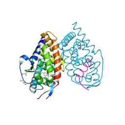
7QAA
 | | Crystal structure of RARalpha/RXRalpha ligand binding domain heterodimer in complex with BMS614 and oleic acid | | 分子名称: | 4-[(4,4-DIMETHYL-1,2,3,4-TETRAHYDRO-[1,2']BINAPTHALENYL-7-CARBONYL)-AMINO]-BENZOIC ACID, Isoform Alpha-1-deltaBC of Retinoic acid receptor alpha, OLEIC ACID, ... | | 著者 | le Maire, A, Vivat, V, Guee, L, Blanc, P, Malosse, C, Chamot-Rooke, J, Germain, P, Bourguet, w. | | 登録日 | 2021-11-16 | | 公開日 | 2022-10-05 | | 最終更新日 | 2024-01-31 | | 実験手法 | X-RAY DIFFRACTION (2.76 Å) | | 主引用文献 | Design and in vitro characterization of RXR variants as tools to investigate the biological role of endogenous rexinoids.
J.Mol.Endocrinol., 69, 2022
|
|
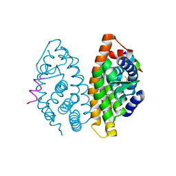
3OSW
 | | Crystal structure of PPARgamma ligand binding domain in complex with tetrabromo-bisphenol A (TBBPA) | | 分子名称: | 4,4'-propane-2,2-diylbis(2,6-dibromophenol), Peroxisome proliferator-activated receptor gamma, S-1,2-PROPANEDIOL | | 著者 | le Maire, A, Bourguet, W. | | 登録日 | 2010-09-10 | | 公開日 | 2011-05-25 | | 最終更新日 | 2024-02-21 | | 実験手法 | X-RAY DIFFRACTION (2.55 Å) | | 主引用文献 | Peroxisome proliferator-activated receptor Gamma is a target for halogenated analogs of bisphenol A.
Environ.Health Perspect., 119, 2011
|
|
3OSI
 | | Crystal structure of PPARgamma ligand binding domain in complex with tetrachloro-bisphenol A (TCBPA) | | 分子名称: | 4,4'-propane-2,2-diylbis(2,6-dichlorophenol), Peroxisome proliferator-activated receptor gamma, S-1,2-PROPANEDIOL | | 著者 | le Maire, A, Bourguet, W. | | 登録日 | 2010-09-09 | | 公開日 | 2011-05-25 | | 最終更新日 | 2024-02-21 | | 実験手法 | X-RAY DIFFRACTION (2.7 Å) | | 主引用文献 | Peroxisome proliferator-activated receptor Gamma is a target for halogenated analogs of bisphenol A.
Environ.Health Perspect., 119, 2011
|
|
7APO
 | | Crystal structure of RARalpha ligand binding domain in complex with a fragment of the TIF2 coactivator | | 分子名称: | 4-{[(5,5,8,8-tetramethyl-5,6,7,8-tetrahydronaphthalen-2-yl)carbonyl]amino}benzoic acid, GLYCEROL, Nuclear receptor coactivator 2, ... | | 著者 | le Maire, A, Guee, L, Bourguet, W. | | 登録日 | 2020-10-19 | | 公開日 | 2021-08-04 | | 最終更新日 | 2024-01-31 | | 実験手法 | X-RAY DIFFRACTION (2.4 Å) | | 主引用文献 | Structural Insights into the Interaction of the Intrinsically Disordered Co-activator TIF2 with Retinoic Acid Receptor Heterodimer (RXR/RAR).
J.Mol.Biol., 433, 2021
|
|
3PBA
 | | Crystal structure of PPARgamma ligand binding domain in complex with monosulfate tetrabromo-bisphenol A (MonoTBBPA) | | 分子名称: | 2,6-dibromo-4-[2-(3,5-dibromo-4-hydroxyphenyl)propan-2-yl]phenyl hydrogen sulfate, Peroxisome proliferator-activated receptor gamma, S-1,2-PROPANEDIOL | | 著者 | le Maire, A, Bourguet, W. | | 登録日 | 2010-10-20 | | 公開日 | 2011-06-08 | | 最終更新日 | 2024-02-21 | | 実験手法 | X-RAY DIFFRACTION (2.3 Å) | | 主引用文献 | Characterization of Novel Ligands of ER{alpha}, Er{beta}, and PPAR{gamma}: The Case of Halogenated Bisphenol A and Their Conjugated Metabolites.
Toxicol Sci, 122, 2011
|
|
7BK4
 | | Crystal structure of RXRalpha ligand binding domain in complex with a fragment of the TIF2 coactivator | | 分子名称: | 6-[1-(3,5,5,8,8-PENTAMETHYL-5,6,7,8-TETRAHYDRONAPHTHALEN-2-YL)CYCLOPROPYL]PYRIDINE-3-CARBOXYLIC ACID, Nuclear receptor coactivator 2, Retinoic acid receptor RXR-alpha | | 著者 | le Maire, A, Bourguet, W. | | 登録日 | 2021-01-15 | | 公開日 | 2021-08-04 | | 最終更新日 | 2024-01-31 | | 実験手法 | X-RAY DIFFRACTION (2.8 Å) | | 主引用文献 | Structural Insights into the Interaction of the Intrinsically Disordered Co-activator TIF2 with Retinoic Acid Receptor Heterodimer (RXR/RAR).
J.Mol.Biol., 433, 2021
|
|
7AOS
 | | crystal structure of the RARalpha/RXRalpha ligand binding domain heterodimer in complex with a fragment of SRC1 coactivator | | 分子名称: | 4-{[(5,5,8,8-tetramethyl-5,6,7,8-tetrahydronaphthalen-2-yl)carbonyl]amino}benzoic acid, 6-[1-(3,5,5,8,8-PENTAMETHYL-5,6,7,8-TETRAHYDRONAPHTHALEN-2-YL)CYCLOPROPYL]PYRIDINE-3-CARBOXYLIC ACID, GLYCEROL, ... | | 著者 | le Maire, A, Guee, L, Bourguet, W. | | 登録日 | 2020-10-15 | | 公開日 | 2021-08-04 | | 最終更新日 | 2024-01-31 | | 実験手法 | X-RAY DIFFRACTION (2.55 Å) | | 主引用文献 | Structural Insights into the Interaction of the Intrinsically Disordered Co-activator TIF2 with Retinoic Acid Receptor Heterodimer (RXR/RAR).
J.Mol.Biol., 433, 2021
|
|
3PO9
 | |
6AN1
 | | Crystal structure of the complex between PPARgamma LBD and the ligand AM-879 | | 分子名称: | 4-({2-[(1,3-dioxo-1,3-dihydro-2H-inden-2-ylidene)methyl]phenoxy}methyl)benzoic acid, Peroxisome proliferator-activated receptor gamma | | 著者 | Veras, H, Figueira, A.C, le Maire, A. | | 登録日 | 2017-08-11 | | 公開日 | 2018-02-14 | | 最終更新日 | 2024-03-13 | | 実験手法 | X-RAY DIFFRACTION (2.687 Å) | | 主引用文献 | Screening for PPAR Non-Agonist Ligands Followed by Characterization of a Hit, AM-879, with Additional No-Adipogenic and cdk5-Mediated Phosphorylation Inhibition Properties.
Front Endocrinol (Lausanne), 9, 2018
|
|
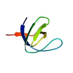
2V1N
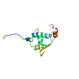 | | SOLUTION STRUCTURE OF THE REGION 51-160 OF HUMAN KIN17 REVEALS A WINGED HELIX FOLD | | 分子名称: | PROTEIN KIN HOMOLOG | | 著者 | Carlier, L, Le Maire, A, Gondry, M, Guilhaudis, L, Milazzo, I, Davoust, D, Couprie, J, Gilquin, B, Zinn-Justin, S. | | 登録日 | 2007-05-27 | | 公開日 | 2007-11-27 | | 最終更新日 | 2024-05-15 | | 実験手法 | SOLUTION NMR | | 主引用文献 | Solution Structure of the Region 51-160 of Human Kin17 Reveals an Atypical Winged Helix Domain
Protein Sci., 16, 2007
|
|
3E94
 | |
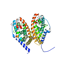
3KMZ
 | | Crystal structure of RARalpha ligand binding domain in complex with the inverse agonist BMS493 and a corepressor fragment | | 分子名称: | 4-{(E)-2-[5,5-dimethyl-8-(phenylethynyl)-5,6-dihydronaphthalen-2-yl]ethenyl}benzoic acid, GLYCEROL, Nuclear receptor corepressor 1, ... | | 著者 | Bourguet, W, le Maire, A. | | 登録日 | 2009-11-11 | | 公開日 | 2010-06-02 | | 最終更新日 | 2023-11-22 | | 実験手法 | X-RAY DIFFRACTION (2.1 Å) | | 主引用文献 | A unique secondary-structure switch controls constitutive gene repression by retinoic acid receptor.
Nat.Struct.Mol.Biol., 17, 2010
|
|


