8G05
 
 | | Cryo-EM structure of an orphan GPCR-Gi protein signaling complex | | 分子名称: | 6-(octylamino)pyrimidine-2,4(3H,5H)-dione, CHOLESTEROL, G-protein coupled receptor 84, ... | | 著者 | Zhang, X, Wang, Y.J, Li, X, Liu, G.B, Gong, W.M, Zhang, C. | | 登録日 | 2023-01-31 | | 公開日 | 2023-11-01 | | 実験手法 | ELECTRON MICROSCOPY (3 Å) | | 主引用文献 | Pro-phagocytic function and structural basis of GPR84 signaling.
Nat Commun, 14, 2023
|
|
4J0U
 | | Crystal structure of IFIT5/ISG58 | | 分子名称: | Interferon-induced protein with tetratricopeptide repeats 5 | | 著者 | Liu, Y, Liang, H, Feng, F, Yuan, L, Wang, Y.E, Crowley, C, Lv, Z, Li, J, Zeng, S, Cheng, G. | | 登録日 | 2013-01-31 | | 公開日 | 2013-02-13 | | 実験手法 | X-RAY DIFFRACTION (1.969 Å) | | 主引用文献 | Crystal Structure of IFIT5
To be Published
|
|
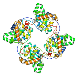
1KGX
 | | Three Dimensional Structure Analysis of the R195Q Variant of Human Pancreatic Alpha Amylase | | 分子名称: | 2-acetamido-2-deoxy-beta-D-glucopyranose, ALPHA-AMYLASE, PANCREATIC, ... | | 著者 | Numao, S, Maurus, R, Sidhu, G, Wang, Y, Overall, C.M, Brayer, G.D, Withers, S.G. | | 登録日 | 2001-11-28 | | 公開日 | 2002-01-16 | | 最終更新日 | 2023-08-16 | | 実験手法 | X-RAY DIFFRACTION (2 Å) | | 主引用文献 | Probing the role of the chloride ion in the mechanism of human pancreatic alpha-amylase.
Biochemistry, 41, 2002
|
|
6JE8
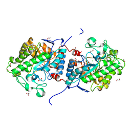
 | | crystal structure of a beta-N-acetylhexosaminidase | | 分子名称: | Beta-N-acetylhexosaminidase, FORMIC ACID, GLYCEROL, ... | | 著者 | Chen, X, Wang, J.C, Liu, M.J, Yang, W.Y, Wang, Y.Z, Tang, R.P, Zhang, M. | | 登録日 | 2019-02-04 | | 公開日 | 2019-03-13 | | 最終更新日 | 2023-11-22 | | 実験手法 | X-RAY DIFFRACTION (1.798 Å) | | 主引用文献 | Crystallographic evidence for substrate-assisted catalysis of beta-N-acetylhexosaminidas from Akkermansia muciniphila.
Biochem. Biophys. Res. Commun., 511, 2019
|
|
4ZHY
 | | Crystal structure of a bacterial signalling complex | | 分子名称: | FORMIC ACID, SULFATE ION, YfiB, ... | | 著者 | Li, S, Li, T, Wang, Y, Bartlam, M. | | 登録日 | 2015-04-27 | | 公開日 | 2016-04-27 | | 実験手法 | X-RAY DIFFRACTION (1.969 Å) | | 主引用文献 | Structural insights into YfiR sequestering by YfiB in Pseudomonas aeruginosa PAO1
Sci Rep, 5, 2015
|
|
3KMY
 | | Structure of BACE bound to SCH12472 | | 分子名称: | 3-[2-(3-chlorophenyl)ethyl]pyridin-2-amine, Beta-secretase 1 | | 著者 | Strickland, C, Wang, Y. | | 登録日 | 2009-11-11 | | 公開日 | 2010-01-19 | | 最終更新日 | 2023-09-06 | | 実験手法 | X-RAY DIFFRACTION (1.9 Å) | | 主引用文献 | Application of Fragment-Based NMR Screening, X-ray Crystallography, Structure-Based Design, and Focused Chemical Library Design to Identify Novel muM Leads for the Development of nM BACE-1 (beta-Site APP Cleaving Enzyme 1) Inhibitors.
J.Med.Chem., 53, 2010
|
|
6JEB
 | | crystal structure of a beta-N-acetylhexosaminidase | | 分子名称: | ACETAMIDE, Beta-N-acetylhexosaminidase, ZINC ION | | 著者 | Chen, X, Wang, J.C, Liu, M.J, Yang, W.Y, Wang, Y.Z, Tang, R.P, Zhang, M. | | 登録日 | 2019-02-05 | | 公開日 | 2019-03-13 | | 最終更新日 | 2023-11-22 | | 実験手法 | X-RAY DIFFRACTION (1.498 Å) | | 主引用文献 | Crystallographic evidence for substrate-assisted catalysis of beta-N-acetylhexosaminidas from Akkermansia muciniphila.
Biochem. Biophys. Res. Commun., 511, 2019
|
|
4J1G
 | | Leanyer orthobunyavirus nucleoprotein-ssRNA complex | | 分子名称: | Nucleocapsid, RNA (45-MER) | | 著者 | Niu, F, Shaw, N, Wang, Y, Jiao, L, Ding, W, Li, X, Zhu, P, Upur, H, Ouyang, S, Cheng, G, Liu, Z.J. | | 登録日 | 2013-02-01 | | 公開日 | 2013-04-10 | | 最終更新日 | 2024-02-28 | | 実験手法 | X-RAY DIFFRACTION (2.789 Å) | | 主引用文献 | Structure of the Leanyer orthobunyavirus nucleoprotein-RNA complex reveals unique architecture for RNA encapsidation.
Proc.Natl.Acad.Sci.USA, 110, 2013
|
|
7C8U
 | | The crystal structure of COVID-19 main protease in complex with GC376 | | 分子名称: | (1S,2S)-2-({N-[(benzyloxy)carbonyl]-L-leucyl}amino)-1-hydroxy-3-[(3S)-2-oxopyrrolidin-3-yl]propane-1-sulfonic acid, 3C-like proteinase | | 著者 | Luan, X, Shang, W, Wang, Y, Yin, W, Jiang, Y, Feng, S, Wang, Y, Liu, M, Zhou, R, Zhang, Z, Wang, F, Cheng, W, Gao, M, Wang, H, Wu, W, Tian, R, Tian, Z, Jin, Y, Jiang, H.W, Zhang, L, Xu, H.E, Zhang, S. | | 登録日 | 2020-06-03 | | 公開日 | 2020-06-24 | | 最終更新日 | 2023-11-29 | | 実験手法 | X-RAY DIFFRACTION (2.35 Å) | | 主引用文献 | The crystal structure of COVID-19 main protease in complex with GC376
To Be Published
|
|
3S45
 | | wild-type HIV-2 protease with antiviral drug amprenavir | | 分子名称: | CHLORIDE ION, IMIDAZOLE, Protease, ... | | 著者 | Tie, Y.-F, Wang, Y.-F, Weber, I.T. | | 登録日 | 2011-05-18 | | 公開日 | 2012-03-21 | | 最終更新日 | 2023-09-13 | | 実験手法 | X-RAY DIFFRACTION (1.51 Å) | | 主引用文献 | Critical differences in HIV-1 and HIV-2 protease specificity for clinical inhibitors.
Protein Sci., 21, 2012
|
|
4ZHV
 | | Crystal structure of a bacterial signalling protein | | 分子名称: | SULFATE ION, YfiB | | 著者 | Li, S, Li, T, Wang, Y, Bartlam, M. | | 登録日 | 2015-04-27 | | 公開日 | 2016-04-27 | | 最終更新日 | 2024-03-20 | | 実験手法 | X-RAY DIFFRACTION (1.585 Å) | | 主引用文献 | Structural insights into YfiR sequestering by YfiB in Pseudomonas aeruginosa PAO1
Sci Rep, 5, 2015
|
|
8J59
 | |
7C3V
 | | Structure of a thermostable Alcohol dehydrogenase from Kluyveromyces polyspora(KpADH) | | 分子名称: | Alcohol dehydrogenase, NADP NICOTINAMIDE-ADENINE-DINUCLEOTIDE PHOSPHATE | | 著者 | Dai, W, Ni, Y, Xu, G, Liu, Y, Wang, Y, Zhou, J. | | 登録日 | 2020-05-14 | | 公開日 | 2021-05-19 | | 最終更新日 | 2023-11-29 | | 実験手法 | X-RAY DIFFRACTION (2.20042944 Å) | | 主引用文献 | Structure of a thermostable Alcohol dehydrogenase from Kluyveromyces polyspora(KpADH)
To Be Published
|
|
5WQN
 | |
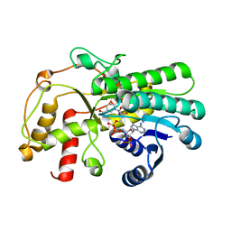
4BGG
 | | Crystal structure of the ACVR1 kinase in complex with LDN-213844 | | 分子名称: | 1,2-ETHANEDIOL, 1-{4-[5-(3,4,5-trimethoxyphenyl)pyridin-3-yl]phenyl}piperazine, ACTIVIN RECEPTOR TYPE-1, ... | | 著者 | Sanvitale, C, Canning, P, Cooper, C, Wang, Y, Mohedas, A.H, Choi, S, Yu, P.B, Cuny, G.D, Nowak, R, Coutandin, D, Vollmar, M, von Delft, F, Arrowsmith, C.H, Edwards, A.M, Bountra, C, Bullock, A, Structural Genomics Consortium (SGC) | | 登録日 | 2013-03-26 | | 公開日 | 2013-04-10 | | 最終更新日 | 2023-12-20 | | 実験手法 | X-RAY DIFFRACTION (2.56 Å) | | 主引用文献 | Structure-Activity Relationship of 3,5-Diaryl-2-Aminopyridine Alk2 Inhibitors Reveals Unaltered Binding Affinity for Fibrodysplasia Ossificans Progressiva Causing Mutants.
J.Med.Chem., 57, 2014
|
|
5WQO
 | | Crystal structure of a carbonyl reductase from Pseudomonas aeruginosa PAO1 in complex with NADP (condition I) | | 分子名称: | 1,2-ETHANEDIOL, NADP NICOTINAMIDE-ADENINE-DINUCLEOTIDE PHOSPHATE, Probable dehydrogenase, ... | | 著者 | Li, S, Wang, Y, Bartlam, M. | | 登録日 | 2016-11-27 | | 公開日 | 2017-10-04 | | 最終更新日 | 2023-11-08 | | 実験手法 | X-RAY DIFFRACTION (1.78 Å) | | 主引用文献 | Structure and characterization of a NAD(P)H-dependent carbonyl reductase from Pseudomonas aeruginosa PAO1.
FEBS Lett., 591, 2017
|
|
5WQM
 | |
6KHX
 | | Crystal structure of Prx from Akkermansia muciniphila | | 分子名称: | CALCIUM ION, Peroxiredoxin | | 著者 | Li, M, Wang, J, Xu, W, Wang, Y, Zhang, M, Wang, M. | | 登録日 | 2019-07-16 | | 公開日 | 2020-02-19 | | 最終更新日 | 2023-11-22 | | 実験手法 | X-RAY DIFFRACTION (2.58 Å) | | 主引用文献 | Crystal structure of Akkermansia muciniphila peroxiredoxin reveals a novel regulatory mechanism of typical 2-Cys Prxs by a distinct loop.
Febs Lett., 594, 2020
|
|
3PMR
 | | Crystal Structure of E2 domain of Human Amyloid Precursor-Like Protein 1 | | 分子名称: | Amyloid-like protein 1, PHOSPHATE ION | | 著者 | Lee, S, Xue, Y, Hu, J, Wang, Y, Liu, X, Demeler, B, Ha, Y. | | 登録日 | 2010-11-17 | | 公開日 | 2011-06-01 | | 最終更新日 | 2024-05-22 | | 実験手法 | X-RAY DIFFRACTION (2.11 Å) | | 主引用文献 | The E2 Domains of APP and APLP1 Share a Conserved Mode of Dimerization.
Biochemistry, 50, 2011
|
|
6KZC
 | | crystal structure of TRKc in complex with 3-(imidazo[1,2-a]pyrazin-3-ylethynyl)-2-methyl-N-(3-((4- methylpiperazin-1-yl)methyl)-5- (trifluoromethyl)phenyl)benzamide | | 分子名称: | 3-(2-imidazo[1,2-a]pyrazin-3-ylethynyl)-2-methyl-~{N}-[3-(4-methylpiperazin-1-yl)-5-(trifluoromethyl)phenyl]benzamide, NT-3 growth factor receptor | | 著者 | Zhang, Z.M, Wang, Y. | | 登録日 | 2019-09-23 | | 公開日 | 2019-10-09 | | 最終更新日 | 2024-03-27 | | 実験手法 | X-RAY DIFFRACTION (2 Å) | | 主引用文献 | Design, synthesis and biological evaluation of 3-(imidazo[1,2-a]pyrazin-3-ylethynyl)-2-methylbenzamides as potent and selective pan-tropomyosin receptor kinase (TRK) inhibitors.
Eur.J.Med.Chem., 179, 2019
|
|
3QG9
 | | crystal structure of FBF-2/gld-1 FBEa A7U mutant complex | | 分子名称: | 1,2-ETHANEDIOL, 5'-R(*UP*GP*UP*GP*CP*CP*UP*UP*A)-3', Fem-3 mRNA-binding factor 2 | | 著者 | Koh, Y.Y, Wang, Y, Qiu, C, Opperman, L, Gross, L, Hall, T.M.T, Wickens, M. | | 登録日 | 2011-01-24 | | 公開日 | 2011-03-23 | | 最終更新日 | 2018-01-24 | | 実験手法 | X-RAY DIFFRACTION (2.25 Å) | | 主引用文献 | Stacking interactions in PUF-RNA complexes.
Rna, 17, 2011
|
|
4ZHU
 | | Crystal structure of a bacterial repressor protein | | 分子名称: | SULFATE ION, YfiR | | 著者 | Li, S, Li, T, Wang, Y, Bartlam, M. | | 登録日 | 2015-04-27 | | 公開日 | 2016-04-27 | | 実験手法 | X-RAY DIFFRACTION (2.3968 Å) | | 主引用文献 | Structural insights into YfiR sequestering by YfiB in Pseudomonas aeruginosa PAO1
Sci Rep, 5, 2015
|
|
5IY4
 | |
8KDE
 | | Cryo-EM structure of an intermediate-state complex during the process of photosystem II repair | | 分子名称: | 1,2-DI-O-ACYL-3-O-[6-DEOXY-6-SULFO-ALPHA-D-GLUCOPYRANOSYL]-SN-GLYCEROL, 1,2-DIPALMITOYL-PHOSPHATIDYL-GLYCEROLE, 1,2-DISTEAROYL-MONOGALACTOSYL-DIGLYCERIDE, ... | | 著者 | Li, A, Wang, Y, Liu, Z. | | 登録日 | 2023-08-09 | | 公開日 | 2024-06-19 | | 最終更新日 | 2024-06-26 | | 実験手法 | ELECTRON MICROSCOPY (2.6 Å) | | 主引用文献 | Structural basis for an early stage of the photosystem II repair cycle in Chlamydomonas reinhardtii.
Nat Commun, 15, 2024
|
|
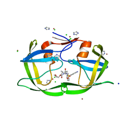
1FHS
 | | THE THREE-DIMENSIONAL SOLUTION STRUCTURE OF THE SRC HOMOLOGY DOMAIN-2 OF THE GROWTH FACTOR RECEPTOR BOUND PROTEIN-2, NMR, 18 STRUCTURES | | 分子名称: | GROWTH FACTOR RECEPTOR BOUND PROTEIN-2 | | 著者 | Senior, M.M, Frederick, A.F, Black, S, Perkins, L.M, Wilson, O, Snow, M.E, Wang, Y.-S. | | 登録日 | 1997-06-12 | | 公開日 | 1998-06-17 | | 最終更新日 | 2024-05-22 | | 実験手法 | SOLUTION NMR | | 主引用文献 | The three-dimensional solution structure of the Src homology domain-2 of the growth factor receptor-bound protein-2.
J.Biomol.NMR, 11, 1998
|
|


