Loading
PDBj
メニューPDBj@FacebookPDBj@TwitterPDBj@YouTubewwPDB FoundationwwPDB
RCSB PDBPDBeBMRBAdv. SearchSearch help
PDB: 373 件

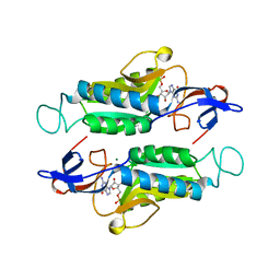
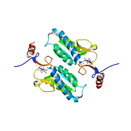
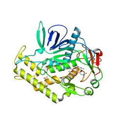
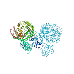
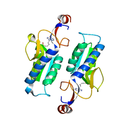
5KAM
DownloadVisualize
BU of 5kam by Molmil
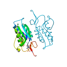
Trypanosome brucei Hypoxanthine-guanine phosphoribosyltranferase in complex with Inosine 5' monophosphate
分子名称: Hypoxanthine-guanine phosphoribosyltransferase, INOSINIC ACID, MAGNESIUM ION, ...
著者Teran, D, Guddat, L.
登録日2016-06-01
公開日2016-11-09
最終更新日2023-09-27
実験手法X-RAY DIFFRACTION (2.481 Å)
主引用文献Crystal structures and inhibition of Trypanosoma brucei hypoxanthine-guanine phosphoribosyltransferase.
Sci Rep, 6, 2016
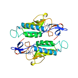
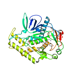
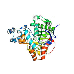
5K51
DownloadVisualize
BU of 5k51 by Molmil
Trypanosome brucei Hypoxanthine-guanine phosphoribosyltranferase in complex with a 9-[5-(phosphonoheptyl]hypoxanthine
分子名称: 5-(6-oxidanylidene-3~{H}-purin-9-yl)pentylphosphonic acid, Hypoxanthine-guanine phosphoribosyltransferase
著者Teran, D, Guddat, L.
登録日2016-05-22
公開日2016-11-09
最終更新日2023-09-27
実験手法X-RAY DIFFRACTION (2.956 Å)
主引用文献Crystal structures and inhibition of Trypanosoma brucei hypoxanthine-guanine phosphoribosyltransferase.
Sci Rep, 6, 2016
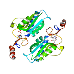
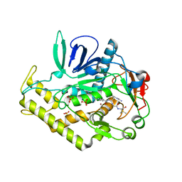
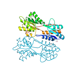
6MXC
DownloadVisualize
BU of 6mxc by Molmil
Crystal structure of Trypanosoma brucei hypoxanthine-guanine-xanthine phosphoribosyltranferase in complex with GMP
分子名称: GUANOSINE-5'-MONOPHOSPHATE, Hypoxanthine-guanine phosphoribosyltransferase, MAGNESIUM ION
著者Teran, D, Guddat, L.
登録日2018-10-30
公開日2019-07-24
最終更新日2023-10-11
実験手法X-RAY DIFFRACTION (1.993 Å)
主引用文献Crystal structures of Trypanosoma brucei hypoxanthine - guanine - xanthine phosphoribosyltransferase in complex with IMP, GMP and XMP.
Febs J., 286, 2019
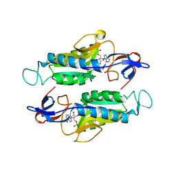
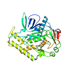
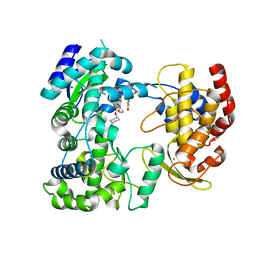
6MXB
DownloadVisualize
BU of 6mxb by Molmil
Crystal structure of Trypanosoma brucei hypoxanthine-guanine-xanthine phosphoribosyltranferase in complex with XMP
分子名称: Hypoxanthine-guanine phosphoribosyltransferase, putative, MAGNESIUM ION, ...
著者Teran, D, Guddat, L.
登録日2018-10-30
公開日2019-07-24
最終更新日2023-10-11
実験手法X-RAY DIFFRACTION (2.192 Å)
主引用文献Crystal structures of Trypanosoma brucei hypoxanthine - guanine - xanthine phosphoribosyltransferase in complex with IMP, GMP and XMP.
Febs J., 286, 2019
6MXG
DownloadVisualize
BU of 6mxg by Molmil
Crystal structure of Trypanosoma brucei hypoxanthine-guanine phosphoribosyltranferase in complex with XMP
分子名称: Hypoxanthine-guanine phosphoribosyltransferase, MAGNESIUM ION, SULFATE ION, ...
著者Teran, D, Guddat, L.
登録日2018-10-30
公開日2019-07-24
最終更新日2023-10-11
実験手法X-RAY DIFFRACTION (2.392 Å)
主引用文献Crystal structures of Trypanosoma brucei hypoxanthine - guanine - xanthine phosphoribosyltransferase in complex with IMP, GMP and XMP.
Febs J., 286, 2019
6MXD
DownloadVisualize
BU of 6mxd by Molmil
Crystal structure of Trypanosoma brucei hypoxanthine-guanine-xanthine phosphoribosyltranferase in complex with IMP
分子名称: Hypoxanthine-guanine phosphoribosyltransferase, putative, INOSINIC ACID, ...
著者Teran, D, Guddat, L.
登録日2018-10-30
公開日2019-07-24
最終更新日2023-10-11
実験手法X-RAY DIFFRACTION (2.96 Å)
主引用文献Crystal structures of Trypanosoma brucei hypoxanthine - guanine - xanthine phosphoribosyltransferase in complex with IMP, GMP and XMP.
Febs J., 286, 2019
5JV5
DownloadVisualize
BU of 5jv5 by Molmil
Trypanosome brucei Hypoxanthine-guanine phosphoribosyltranferase in complex with Guanosine 5' monophosphate
分子名称: GUANOSINE-5'-MONOPHOSPHATE, Hypoxanthine-guanine phosphoribosyltransferase, MAGNESIUM ION, ...
著者Teran, D, Guddat, L.
登録日2016-05-10
公開日2016-11-09
最終更新日2023-09-27
実験手法X-RAY DIFFRACTION (2.73 Å)
主引用文献Crystal structures and inhibition of Trypanosoma brucei hypoxanthine-guanine phosphoribosyltransferase.
Sci Rep, 6, 2016
6HVC
DownloadVisualize
BU of 6hvc by Molmil
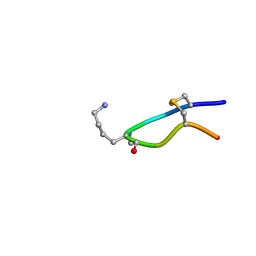
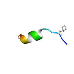
NMR structure of Urotensin Peptide Asp-c[Cys-Phe-Trp-(N-Me)Lys-Tyr-Cys]-Val in SDS solution
分子名称: Urotensin-2
著者Brancaccio, D, Carotenuto, A, Merlino, F, Billard, E, Yousif, A.M, Di Maro, S, Abate, L, Bellavita, R, D'Emmanuele di Villa Bianca, R, Santicioli, P, Marinelli, L, Novellino, E, Hebert, T.E, Lubell, W.D, Chatenet, D, Grieco, P.
登録日2018-10-10
公開日2019-01-16
最終更新日2019-04-24
実験手法SOLUTION NMR
主引用文献Functional Selectivity Revealed by N-Methylation Scanning of Human Urotensin II and Related Peptides.
J.Med.Chem., 62, 2019
3N52
DownloadVisualize
BU of 3n52 by Molmil
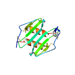
crystal Structure analysis of MIP2
分子名称: C-X-C motif chemokine 2
著者Rajasekaran, D.
登録日2010-05-24
公開日2011-06-08
最終更新日2017-11-08
実験手法X-RAY DIFFRACTION (1.9 Å)
主引用文献A Model of GAG/MIP-2/CXCR2 Interfaces and Its Functional Effects.
Biochemistry, 51, 2012
6QXB
DownloadVisualize
BU of 6qxb by Molmil
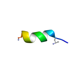
NMR structure of peptide 7, characterized by a cis-4-amino-Pro residue, with a significant lower MIC on E. coli
分子名称: PHE-VAL-CAP-TRP-PHE-SER-LYS-PHE-LEU-GLY-ARG-ILE-LEU-NH2
著者Brancaccio, D, Carotenuto, A, Merlino, F, Grieco, P, Novellino, E.
登録日2019-03-07
公開日2019-05-29
最終更新日2023-06-14
実験手法SOLUTION NMR
主引用文献The Outcomes of Decorated Prolines in the Discovery of Antimicrobial Peptides from Temporin-L.
Chemmedchem, 14, 2019
6QXC
DownloadVisualize
BU of 6qxc by Molmil
NMR structure of peptide 8, characterized by a trans-4-cyclohexyl-Pro, with a dramatic reduction in activity on E. coli ATCC and lost effect on P. aeruginosa.
分子名称: PHE-VAL-TCP-TRP-PHE-SER-LYS-PHE-LEU-GLY-ARG-ILE-LEU-NH2
著者Brancaccio, D, Carotenuto, A, Merlino, F, Grieco, P, Novellino, E.
登録日2019-03-07
公開日2019-05-29
最終更新日2023-06-14
実験手法SOLUTION NMR
主引用文献The Outcomes of Decorated Prolines in the Discovery of Antimicrobial Peptides from Temporin-L.
Chemmedchem, 14, 2019
3BPQ
DownloadVisualize
BU of 3bpq by Molmil
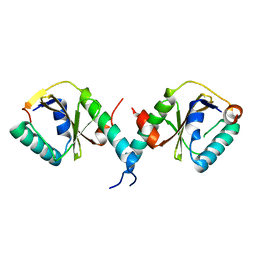
Crystal Structure of RelB-RelE antitoxin-toxin complex from Methanococcus jannaschii
分子名称: Antitoxin RelB3, Toxin RelE3
著者Francuski, D, Saenger, W.
登録日2007-12-19
公開日2008-12-23
最終更新日2024-03-13
実験手法X-RAY DIFFRACTION (2.2 Å)
主引用文献Crystal structure of the antitoxin-toxin protein complex RelB-RelE from Methanococcus jannaschii
J.Mol.Biol., 393, 2009
3C8A
DownloadVisualize
BU of 3c8a by Molmil
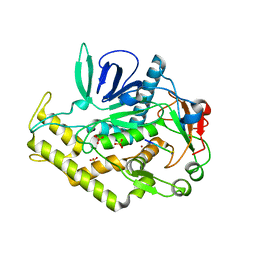
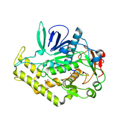
Crystal structure of the catalytic domain of botulinum neurotoxin serotype A with inhibitory peptide RRGL
分子名称: Botulinum neurotoxin A light chain, Inhibitor peptide RRGL, SULFATE ION, ...
著者Kumaran, D, Swaminathan, S.
登録日2008-02-11
公開日2008-04-22
最終更新日2024-10-30
実験手法X-RAY DIFFRACTION (1.52 Å)
主引用文献Structure- and Substrate-based Inhibitor Design for Clostridium botulinum Neurotoxin Serotype A
J.Biol.Chem., 283, 2008
3BWI
DownloadVisualize
BU of 3bwi by Molmil
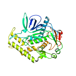
Crystal structure of the catalytic domain of botulinum neurotoxin serotype A with an acetate ion bound at the active site
分子名称: ACETATE ION, Botulinum neurotoxin A light chain, SULFATE ION, ...
著者Kumaran, D, Rawat, R, Swaminathan, S.
登録日2008-01-09
公開日2008-04-22
最終更新日2023-08-30
実験手法X-RAY DIFFRACTION (1.7 Å)
主引用文献Structure- and Substrate-based Inhibitor Design for Clostridium botulinum Neurotoxin Serotype A
J.Biol.Chem., 283, 2008
3C88
DownloadVisualize
BU of 3c88 by Molmil
Crystal structure of the catalytic domain of botulinum neurotoxin serotype A with inhibitory peptide RRGC
分子名称: Botulinum neurotoxin A light chain, Inhibitor peptide RRGC, SODIUM ION, ...
著者Kumaran, D, Swaminathan, S.
登録日2008-02-11
公開日2008-04-22
最終更新日2023-08-30
実験手法X-RAY DIFFRACTION (1.6 Å)
主引用文献Structure- and Substrate-based Inhibitor Design for Clostridium botulinum Neurotoxin Serotype A
J.Biol.Chem., 283, 2008
3C8B
DownloadVisualize
BU of 3c8b by Molmil
Crystal structure of the catalytic domain of botulinum neurotoxin serotype A with inhibitory peptide RRGI
分子名称: Botulinum neurotoxin A light chain, Inhibitor peptide RRGI, SULFATE ION, ...
著者Kumaran, D, Swaminathan, S.
登録日2008-02-11
公開日2008-04-22
最終更新日2024-10-30
実験手法X-RAY DIFFRACTION (1.47 Å)
主引用文献Structure- and Substrate-based Inhibitor Design for Clostridium botulinum Neurotoxin Serotype A
J.Biol.Chem., 283, 2008
3C89
DownloadVisualize
BU of 3c89 by Molmil
Crystal structure of the catalytic domain of botulinum neurotoxin serotype A with inhibitory peptide RRGM
分子名称: Botulinum neurotoxin A light chain, Inhibitor peptide RRGM, SULFATE ION, ...
著者Kumaran, D, Swaminathan, S.
登録日2008-02-11
公開日2008-04-22
最終更新日2023-08-30
実験手法X-RAY DIFFRACTION (1.58 Å)
主引用文献Structure- and Substrate-based Inhibitor Design for Clostridium botulinum Neurotoxin Serotype A
J.Biol.Chem., 283, 2008
3DDB
DownloadVisualize
BU of 3ddb by Molmil
Crystal structure of the catalytic domain of Botulinum neurotoxin serotype a with a substrate analog peptide
分子名称: Botulinum neurotoxin A light chain, SULFATE ION, Synaptosomal-associated protein 25, ...
著者Kumaran, D, Swaminathan, S.
登録日2008-06-05
公開日2008-09-16
最終更新日2023-08-30
実験手法X-RAY DIFFRACTION (1.6 Å)
主引用文献Substrate binding mode and its implication on drug design for botulinum neurotoxin A
Plos Pathog., 4, 2008
3CIH
DownloadVisualize
BU of 3cih by Molmil
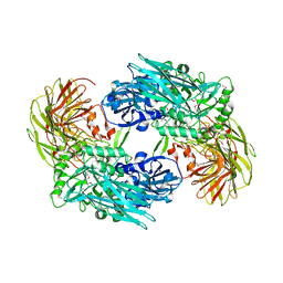
Crystal structure of a putative alpha-rhamnosidase from Bacteroides thetaiotaomicron
分子名称: 1,2-ETHANEDIOL, 2-AMINO-2-HYDROXYMETHYL-PROPANE-1,3-DIOL, Putative alpha-rhamnosidase
著者Kumaran, D, Burley, S.K, Swaminathan, S, New York SGX Research Center for Structural Genomics (NYSGXRC)
登録日2008-03-11
公開日2008-04-01
最終更新日2024-10-30
実験手法X-RAY DIFFRACTION (2.33 Å)
主引用文献Crystal structure of a putative alpha-rhamnosidase from Bacteroides thetaiotaomicron.
To be Published
3K2G
DownloadVisualize
BU of 3k2g by Molmil
Crystal structure of a Resiniferatoxin-binding protein from Rhodobacter sphaeroides
分子名称: (2S,3S)-1,4-DIMERCAPTOBUTANE-2,3-DIOL, MAGNESIUM ION, Resiniferatoxin-binding, ...
著者Kumaran, D, Burley, S.K, Swaminathan, S, New York SGX Research Center for Structural Genomics (NYSGXRC)
登録日2009-09-30
公開日2009-10-13
最終更新日2021-02-10
実験手法X-RAY DIFFRACTION (1.8 Å)
主引用文献Crystal structure of a Resiniferatoxin-binding protein from Rhodobacter sphaeroides
To be Published
3BBL
DownloadVisualize
BU of 3bbl by Molmil
Crystal structure of a regulatory protein of LacI family from Chloroflexus aggregans
分子名称: 1,2-ETHANEDIOL, Regulatory protein of LacI family
著者Kumaran, D, Burley, S.K, Swaminathan, S, New York SGX Research Center for Structural Genomics (NYSGXRC)
登録日2007-11-09
公開日2007-11-27
最終更新日2021-02-03
実験手法X-RAY DIFFRACTION (2.35 Å)
主引用文献Crystal structure of a regulatory protein of LacI family from the Chloroflexus aggregans.
To be Published
5IQ6
DownloadVisualize
BU of 5iq6 by Molmil
Crystal structure of Dengue virus serotype 3 RNA dependent RNA polymerase bound to HeE1-2Tyr, a new pyridobenzothizole inhibitor
分子名称: N-[8-(cyclohexyloxy)-1-oxo-2-phenyl-1H-pyrido[2,1-b][1,3]benzothiazole-4-carbonyl]-L-tyrosine, RNA dependent RNA polymerase, ZINC ION
著者Tarantino, D, Mastrangelo, E, Milani, M.
登録日2016-03-10
公開日2016-10-05
最終更新日2024-05-08
実験手法X-RAY DIFFRACTION (3 Å)
主引用文献Targeting flavivirus RNA dependent RNA polymerase through a pyridobenzothiazole inhibitor.
Antiviral Res., 134, 2016
3BGA
DownloadVisualize
BU of 3bga by Molmil
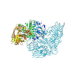
Crystal structure of beta-galactosidase from Bacteroides thetaiotaomicron VPI-5482
分子名称: Beta-galactosidase, CHLORIDE ION, MAGNESIUM ION, ...
著者Kumaran, D, Burley, S.K, Swaminathan, S, New York SGX Research Center for Structural Genomics (NYSGXRC)
登録日2007-11-26
公開日2007-12-11
最終更新日2024-10-30
実験手法X-RAY DIFFRACTION (2.1 Å)
主引用文献Crystal structure analysis of beta-galactosidase from Bacteroides thetaiotaomicron VPI-5482.
To be Published
3DEC
DownloadVisualize
BU of 3dec by Molmil
Crystal structure of a glycosyl hydrolases family 2 protein from Bacteroides thetaiotaomicron
分子名称: Beta-galactosidase, POTASSIUM ION
著者Kumaran, D, Bonanno, J, Romero, R, Burley, S.K, Swaminathan, S, New York SGX Research Center for Structural Genomics (NYSGXRC)
登録日2008-06-09
公開日2008-06-17
最終更新日2024-11-06
実験手法X-RAY DIFFRACTION (2.8 Å)
主引用文献Crystal Structure of a Glycosyl Hydrolases Family 2 protein from Bacteroides thetaiotaomicron.
To be Published
3DDA
DownloadVisualize
BU of 3dda by Molmil
Crystal structure of the catalytic domain of Botulinum neurotoxin serotype a with a snap-25 peptide
分子名称: Botulinum neurotoxin A light chain, SULFATE ION, Synaptosomal-associated protein 25, ...
著者Kumaran, D, Swaminathan, S.
登録日2008-06-05
公開日2008-09-16
最終更新日2024-10-30
実験手法X-RAY DIFFRACTION (1.5 Å)
主引用文献Substrate binding mode and its implication on drug design for botulinum neurotoxin A
Plos Pathog., 4, 2008

227111

件を2024-11-06に公開中

PDB statisticsPDBj update infoContact PDBjnumon