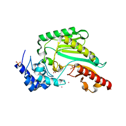
6N45
 
 | |
5YK7
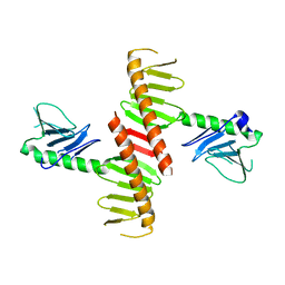
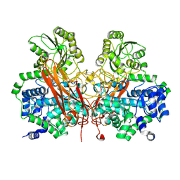
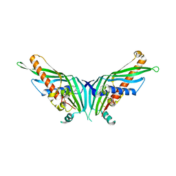
 | | Crystal Structure of Mdm12-Mmm1 complex | | 分子名称: | Maintenance of mitochondrial morphology protein 1, Mitochondrial distribution and morphology protein 12, PHOSPHATE ION | | 著者 | Jeong, H, Park, J, Lee, C. | | 登録日 | 2017-10-12 | | 公開日 | 2018-01-10 | | 最終更新日 | 2023-11-22 | | 実験手法 | X-RAY DIFFRACTION (3.799 Å) | | 主引用文献 | Crystal structures of Mmm1 and Mdm12-Mmm1 reveal mechanistic insight into phospholipid trafficking at ER-mitochondria contact sites.
Proc. Natl. Acad. Sci. U.S.A., 114, 2017
|
|
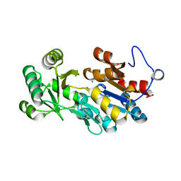
4PVY
 | | Crystal structure of human FPPS in complex with [({5-[4-(propan-2-yloxy)phenyl]pyridin-3-yl}amino)methanediyl]bis(phosphonic acid) | | 分子名称: | Farnesyl pyrophosphate synthase, GLYCEROL, MAGNESIUM ION, ... | | 著者 | Rodionov, D, Park, J, De Schutter, J.W, Tsantrizos, Y.S, Berghuis, A.M. | | 登録日 | 2014-03-18 | | 公開日 | 2015-04-15 | | 最終更新日 | 2023-09-20 | | 実験手法 | X-RAY DIFFRACTION (2.05 Å) | | 主引用文献 | Crystallographic and thermodynamic characterization of phenylaminopyridine bisphosphonates binding to human farnesyl pyrophosphate synthase.
PLoS ONE, 12, 2017
|
|
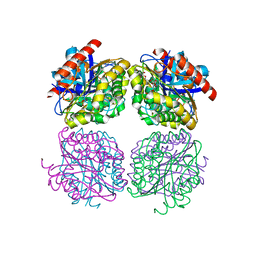
3U4A
 | | From soil to structure: a novel dimeric family 3-beta-glucosidase isolated from compost using metagenomic analysis | | 分子名称: | CALCIUM ION, JMB19063, beta-D-glucopyranose, ... | | 著者 | McAndrew, R.P, Park, J.I, Reindl, W, Friedland, G.D, D'haeseleer, P, Northen, T, Sale, K.L, Simmons, B.A, Adams, P.D. | | 登録日 | 2011-10-07 | | 公開日 | 2013-04-10 | | 最終更新日 | 2024-02-28 | | 実験手法 | X-RAY DIFFRACTION (2.196 Å) | | 主引用文献 | From soil to structure: a novel dimeric family 3-beta--glucosidase isolated from compost using metagenomic analysis
To be Published
|
|
3U48
 | | From soil to structure: a novel dimeric family 3-beta-glucosidase isolated from compost using metagenomic analysis | | 分子名称: | CALCIUM ION, JMB19063, beta-D-glucopyranose | | 著者 | McAndrew, R.P, Park, J.I, Reindl, W, Friedland, G.D, D'haeseleer, P, Northen, T, Sale, K.L, Simmons, B.A, Adams, P.D. | | 登録日 | 2011-10-07 | | 公開日 | 2013-04-10 | | 最終更新日 | 2024-02-28 | | 実験手法 | X-RAY DIFFRACTION (2.2 Å) | | 主引用文献 | From soil to structure: a novel dimeric family 3-beta-glucosidase isolated from compost using metagenomic analysis
To be Published
|
|
4PVX
 | | Crystal structure of human FPPS in complex with [({4-[4-(cyclopropyloxy)phenyl]pyridin-2-yl}amino)methanediyl]bis(phosphonic acid) | | 分子名称: | Farnesyl pyrophosphate synthase, GLYCEROL, MAGNESIUM ION, ... | | 著者 | Rodionov, D, Park, J, Lin, Y.-S, Tsantrizos, Y.S, Berghuis, A.M. | | 登録日 | 2014-03-18 | | 公開日 | 2015-04-15 | | 最終更新日 | 2023-09-20 | | 実験手法 | X-RAY DIFFRACTION (2.18 Å) | | 主引用文献 | Crystallographic and thermodynamic characterization of phenylaminopyridine bisphosphonates binding to human farnesyl pyrophosphate synthase.
PLoS ONE, 12, 2017
|
|
6OCP
 | | Crystal structure of a human GABAB receptor peptide bound to KCTD16 T1 | | 分子名称: | BTB/POZ domain-containing protein KCTD16, Gamma-aminobutyric acid type B receptor subunit 2 | | 著者 | Zuo, H, Glaaser, I, Zhao, Y, Kurinov, I, Mosyak, L, Wang, H, Liu, J, Park, J, Frangaj, A, Sturchler, E, Zhou, M, McDonald, P, Geng, Y, Slesinger, P.A, Fan, Q.R. | | 登録日 | 2019-03-25 | | 公開日 | 2019-04-10 | | 最終更新日 | 2023-10-11 | | 実験手法 | X-RAY DIFFRACTION (2.35 Å) | | 主引用文献 | Structural basis for auxiliary subunit KCTD16 regulation of the GABABreceptor.
Proc.Natl.Acad.Sci.USA, 116, 2019
|
|
7E42
 | |
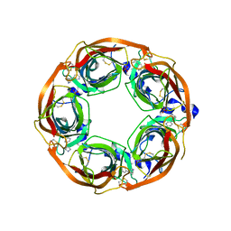
5GYD
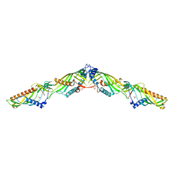
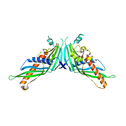
 | | Crystal Structure of Mdm12 | | 分子名称: | 1,2-dioleoyl-sn-glycero-3-phosphoethanolamine, Mitochondrial distribution and morphology protein 12 | | 著者 | Jeong, H, Park, J, Lee, C. | | 登録日 | 2016-09-22 | | 公開日 | 2016-11-16 | | 最終更新日 | 2024-03-20 | | 実験手法 | X-RAY DIFFRACTION (3.106 Å) | | 主引用文献 | Crystal structure of Mdm12 reveals the architecture and dynamic organization of the ERMES complex
EMBO Rep., 17, 2016
|
|
5GYK
 | | Crystal Structure of Mdm12-deletion mutant | | 分子名称: | 1,2-dioleoyl-sn-glycero-3-phosphoethanolamine, Mitochondrial distribution and morphology protein 12 | | 著者 | Jeong, H, Park, J, Lee, C. | | 登録日 | 2016-09-22 | | 公開日 | 2016-11-16 | | 最終更新日 | 2023-11-08 | | 実験手法 | X-RAY DIFFRACTION (3.596 Å) | | 主引用文献 | Crystal structure of Mdm12 reveals the architecture and dynamic organization of the ERMES complex
EMBO Rep., 17, 2016
|
|
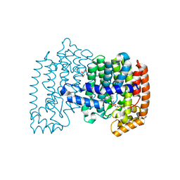
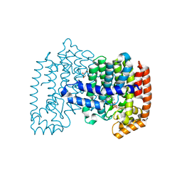
6JG0
 | | Crystal structure of the N-terminal domain single mutant (S92E) of the human mitochondrial calcium uniporter fused with T4 lysozyme | | 分子名称: | Endolysin,Calcium uniporter protein, SULFATE ION | | 著者 | Lee, Y, Park, J, Min, C.K, Kang, J.Y, Kim, T.G, Yamamoto, T, Kim, D.H, Eom, S.H. | | 登録日 | 2019-02-13 | | 公開日 | 2020-02-19 | | 最終更新日 | 2023-11-22 | | 実験手法 | X-RAY DIFFRACTION (2.5 Å) | | 主引用文献 | Crystal structure of calcium channel domain
To Be Published
|
|
5YK6
 | | Crystal Structure of Mmm1 | | 分子名称: | 1,2-DIACYL-GLYCEROL-3-SN-PHOSPHATE, Maintenance of mitochondrial morphology protein 1 | | 著者 | Jeong, H, Park, J, Lee, C. | | 登録日 | 2017-10-12 | | 公開日 | 2018-01-10 | | 最終更新日 | 2024-03-27 | | 実験手法 | X-RAY DIFFRACTION (2.8 Å) | | 主引用文献 | Crystal structures of Mmm1 and Mdm12-Mmm1 reveal mechanistic insight into phospholipid trafficking at ER-mitochondria contact sites.
Proc. Natl. Acad. Sci. U.S.A., 114, 2017
|
|
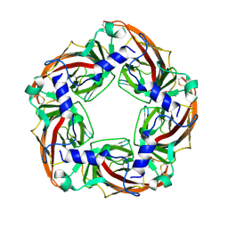
6NAF
 | | De novo designed homo-trimeric amantadine-binding protein | | 分子名称: | (3S,5S,7S)-tricyclo[3.3.1.1~3,7~]decan-1-amine, SODIUM ION, amantadine-binding protein | | 著者 | Selvaraj, B, Park, J, Cuneo, M.J, Myles, D.A.A, Baker, D. | | 登録日 | 2018-12-05 | | 公開日 | 2019-12-18 | | 最終更新日 | 2023-10-25 | | 実験手法 | NEUTRON DIFFRACTION (1.923 Å), X-RAY DIFFRACTION | | 主引用文献 | De novo design of a homo-trimeric amantadine-binding protein.
Elife, 8, 2019
|
|
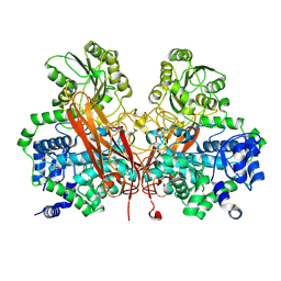
6KY3
 | | Structure of arginine kinase H284A mutant | | 分子名称: | ARGININE, Arginine kinase, PHOSPHATE ION, ... | | 著者 | Rao, Z, Park, J.H, Kim, S.Y, Kim, D.S. | | 登録日 | 2019-09-16 | | 公開日 | 2020-09-16 | | 最終更新日 | 2023-11-22 | | 実験手法 | X-RAY DIFFRACTION (1.34 Å) | | 主引用文献 | Insight into Structural Aspects of Histidine 284 of Daphnia magna Arginine Kinase.
Mol.Cells, 43, 2020
|
|
4QJB
 | |
4ZJS
 | | Crystal structure of a chimeric acetylcholine binding protein from Aplysia Californica (Ac-AChBP) containing the main immunogenic region (MIR) from the human alpha 1 subunit of the muscle nicotinic acetylcholine receptor in complex with anatoxin-A. | | 分子名称: | 1-[(1R,6R)-9-azabicyclo[4.2.1]non-2-en-2-yl]ethanone, Acetylcholine receptor subunit alpha,Soluble acetylcholine receptor,Acetylcholine receptor subunit alpha,Soluble acetylcholine receptor | | 著者 | Talley, T.T, Bobango, J, Wu, J, Park, J.F, Luo, J, Lindsatrom, J, Taylor, P. | | 登録日 | 2015-04-29 | | 公開日 | 2015-05-13 | | 最終更新日 | 2023-09-27 | | 実験手法 | X-RAY DIFFRACTION (2.2301 Å) | | 主引用文献 | Main immunogenic region structure promotes binding of conformation-dependent myasthenia gravis autoantibodies, nicotinic acetylcholine receptor conformation maturation, and agonist sensitivity.
J. Neurosci., 29, 2009
|
|
6AK2
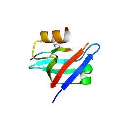 | | Crystal structure of the syntenin PDZ1 domain in complex with the peptide inhibitor KSL-128018 | | 分子名称: | Syntenin-1, peptide inhibitor KSL-128018 | | 著者 | Jin, Z.Y, Park, J.H, Yun, J.H, Haugaard-Kedstrom, L.M, Lee, W.T. | | 登録日 | 2018-08-29 | | 公開日 | 2019-09-04 | | 最終更新日 | 2023-11-22 | | 実験手法 | X-RAY DIFFRACTION (1.868 Å) | | 主引用文献 | A High-Affinity Peptide Ligand Targeting Syntenin Inhibits Glioblastoma.
J.Med.Chem., 64, 2021
|
|
4ZRU
 | | X-ray crystal structure of Lymnaea stagnalis acetylcholine binding protein (Ls-AChBP) in complex with 3-[2-[(2S)-pyrrolidin-2-yl]ethynyl]pyridine (TI-5180) | | 分子名称: | 3-[(2S)-pyrrolidin-2-ylethynyl]pyridine, Acetylcholine-binding protein, PHOSPHATE ION | | 著者 | Bobango, J, Sankaran, B, Park, J.F, Wu, J, Talley, T.T. | | 登録日 | 2015-05-12 | | 公開日 | 2015-05-27 | | 最終更新日 | 2023-09-27 | | 実験手法 | X-RAY DIFFRACTION (1.9 Å) | | 主引用文献 | Comparisons of Binding Affinities for Neuronal Nicotinic Receptors (NNRs) and AChBPs, and Structural Features of a High-Affinity, Non-selective NNR Ligand-AChBP Co-crystal Structure
To be Published
|
|
5GHG
 | | Transaminase W58L with SMBA | | 分子名称: | 4'-DEOXY-4'-AMINOPYRIDOXAL-5'-PHOSPHATE, Aminotransferase class-III | | 著者 | Kim, J, Park, J. | | 登録日 | 2016-06-20 | | 公開日 | 2017-05-24 | | 最終更新日 | 2024-03-20 | | 実験手法 | X-RAY DIFFRACTION (2 Å) | | 主引用文献 | Active Site Engineering of omega-Transaminase Guided by Docking Orientation Analysis and Virtual Activity Screening
Acs Catalysis, 7, 2017
|
|
5GHF
 | | Transaminase with L-ala | | 分子名称: | 4'-DEOXY-4'-AMINOPYRIDOXAL-5'-PHOSPHATE, Aminotransferase class-III | | 著者 | Kim, J, Park, J. | | 登録日 | 2016-06-19 | | 公開日 | 2017-05-24 | | 最終更新日 | 2024-03-20 | | 実験手法 | X-RAY DIFFRACTION (2 Å) | | 主引用文献 | Active Site Engineering of omega-Transaminase Guided by Docking Orientation Analysis and Virtual Activity Screening
Acs Catalysis, 7, 2017
|
|
7W7Q
 | |
5HOE
 | | Crystal structrue of Est24, a carbohydrate acetylesterase from Sinorhizobium meliloti | | 分子名称: | HEXAETHYLENE GLYCOL, Hydrolase, PHOSPHATE ION | | 著者 | Oh, C, Ryu, B.H, An, D.R, Nguyen, D.D, Yoo, W, Kim, T, Ngo, D.T, Kim, H.S, Park, J.S, Kim, K.K, Kim, T.D. | | 登録日 | 2016-01-19 | | 公開日 | 2016-04-27 | | 最終更新日 | 2023-11-08 | | 実験手法 | X-RAY DIFFRACTION (1.45 Å) | | 主引用文献 | Structural and Biochemical Characterization of an Octameric Carbohydrate Acetylesterase from Sinorhizobium meliloti.
Febs Lett., 590, 2016
|
|
6LFK
 | |
7C3M
 | | Structure of FERM protein | | 分子名称: | Fermitin family homolog 3,Fermitin family homolog 3,Fermitin family homolog 3 | | 著者 | Bu, W, Loh, Z.Y, Jin, S, Basu, S, Ero, R, Park, J.E, Yan, X, Wang, M, Sze, S.K, Tan, S.M, Gao, Y.G. | | 登録日 | 2020-05-13 | | 公開日 | 2020-06-03 | | 最終更新日 | 2024-03-27 | | 実験手法 | X-RAY DIFFRACTION (3.6 Å) | | 主引用文献 | Structural basis of human full-length kindlin-3 homotrimer in an auto-inhibited state.
Plos Biol., 18, 2020
|
|


