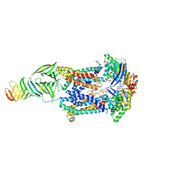
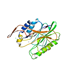
5JQH
 
 | | Structure of beta2 adrenoceptor bound to carazolol and inactive-state stabilizing nanobody, Nb60 | | 分子名称: | (2S)-1-(9H-Carbazol-4-yloxy)-3-(isopropylamino)propan-2-ol, CHOLESTEROL, Endolysin,Beta-2 adrenergic receptor, ... | | 著者 | Staus, D.P, Strachan, R.T, Manglik, A, Pani, B, Kahsai, A.W, Kim, T.H, Wingler, L.M, Ahn, S, Chatterjee, A, Masoudi, A, Kruse, A.C, Pardon, E, Steyaert, J, Weis, W.I, Prosser, R.S, Kobilka, B.K, Costa, T, Lefkowitz, R.J. | | 登録日 | 2016-05-05 | | 公開日 | 2016-07-13 | | 最終更新日 | 2023-09-27 | | 実験手法 | X-RAY DIFFRACTION (3.2 Å) | | 主引用文献 | Allosteric nanobodies reveal the dynamic range and diverse mechanisms of G-protein-coupled receptor activation.
Nature, 535, 2016
|
|
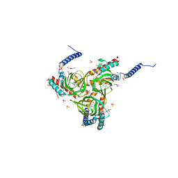
6MJP
 | | LptB(E163Q)FGC from Vibrio cholerae | | 分子名称: | 2-(2-ETHOXYETHOXY)ETHANOL, 6-cyclohexylhexyl beta-D-glucopyranoside, ABC transporter ATP-binding protein, ... | | 著者 | Owens, T.W, Kahne, D, Kruse, A.C. | | 登録日 | 2018-09-21 | | 公開日 | 2019-03-27 | | 最終更新日 | 2023-10-11 | | 実験手法 | X-RAY DIFFRACTION (2.85 Å) | | 主引用文献 | Structural basis of unidirectional export of lipopolysaccharide to the cell surface.
Nature, 567, 2019
|
|
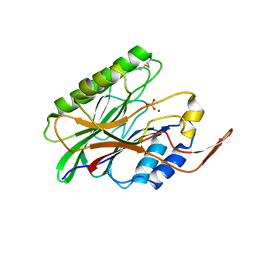
5IUS
 | | Crystal structure of human PD-L1 in complex with high affinity PD-1 mutant | | 分子名称: | CHLORIDE ION, Programmed cell death 1 ligand 1, Programmed cell death protein 1 | | 著者 | Pascolutti, R, Sun, X, Kao, J, Maute, R, Ring, A.M, Bowman, G.R, Kruse, A.C. | | 登録日 | 2016-03-18 | | 公開日 | 2016-09-28 | | 最終更新日 | 2023-09-27 | | 実験手法 | X-RAY DIFFRACTION (2.889 Å) | | 主引用文献 | Structure and Dynamics of PD-L1 and an Ultra-High-Affinity PD-1 Receptor Mutant.
Structure, 24, 2016
|
|
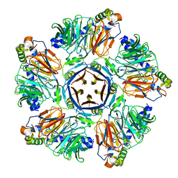
6M8R
 | |
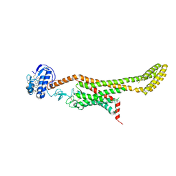
6MIT
 | | LptBFGC from Enterobacter cloacae | | 分子名称: | CHLORIDE ION, LPS export ABC transporter permease LptF, Lipopolysaccharide export system ATP-binding protein, ... | | 著者 | Owens, T.W, Kahne, D, Kruse, A.C. | | 登録日 | 2018-09-20 | | 公開日 | 2019-03-27 | | 最終更新日 | 2023-10-11 | | 実験手法 | X-RAY DIFFRACTION (3.2 Å) | | 主引用文献 | Structural basis of unidirectional export of lipopolysaccharide to the cell surface.
Nature, 567, 2019
|
|
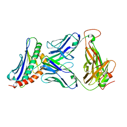
6M8S
 | |
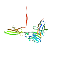
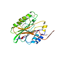
3TJH
 | | 42F3-p3A1/H2-Ld complex | | 分子名称: | 42F3 alpha, 42F3 beta, H2-Ld SBM2, ... | | 著者 | Adams, J.J, Kruse, A, Kranz, D.M, Garcia, K.C. | | 登録日 | 2011-08-24 | | 公開日 | 2011-12-07 | | 最終更新日 | 2023-09-13 | | 実験手法 | X-RAY DIFFRACTION (2.12 Å) | | 主引用文献 | T cell receptor signaling is limited by docking geometry to peptide-major histocompatibility complex.
Immunity, 35, 2011
|
|
4QKX
 | | Structure of beta2 adrenoceptor bound to a covalent agonist and an engineered nanobody | | 分子名称: | 4-[(1R)-1-hydroxy-2-({2-[3-methoxy-4-(2-sulfanylethoxy)phenyl]ethyl}amino)ethyl]benzene-1,2-diol, Beta-2 adrenergic receptor, R9 protein, ... | | 著者 | Weichert, D, Kruse, A.C, Manglik, A, Hiller, C, Zhang, C, Huebner, H, Kobilka, B.K, Gmeiner, P. | | 登録日 | 2014-06-10 | | 公開日 | 2014-07-23 | | 最終更新日 | 2017-06-28 | | 実験手法 | X-RAY DIFFRACTION (3.3 Å) | | 主引用文献 | Covalent agonists for studying G protein-coupled receptor activation.
Proc.Natl.Acad.Sci.USA, 111, 2014
|
|
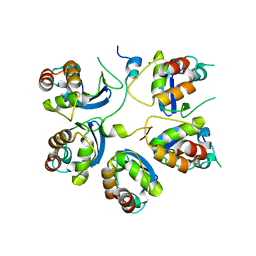
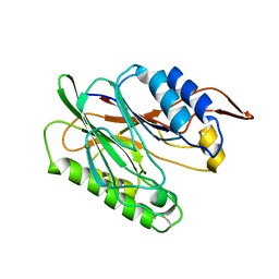
3HK1
 | | Identification and Characterization of a Small Molecule Inhibitor of Fatty Acid Binding Proteins | | 分子名称: | 4-{[2-(methoxycarbonyl)-5-(2-thienyl)-3-thienyl]amino}-4-oxo-2-butenoic acid, Fatty acid-binding protein, adipocyte | | 著者 | Hertzel, A.V, Hellberg, K, Reynolds, J.M, Kruse, A.C, Juhlmann, B.E, Smith, A.J, Sanders, M.A, Ohlendorf, D.H, Suttles, J, Bernlohr, D.A. | | 登録日 | 2009-05-22 | | 公開日 | 2009-09-22 | | 最終更新日 | 2023-09-06 | | 実験手法 | X-RAY DIFFRACTION (1.7 Å) | | 主引用文献 | Identification and characterization of a small molecule inhibitor of Fatty Acid binding proteins.
J.Med.Chem., 52, 2009
|
|
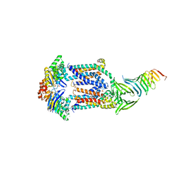
3SN6
 | | Crystal structure of the beta2 adrenergic receptor-Gs protein complex | | 分子名称: | 8-[(1R)-2-{[1,1-dimethyl-2-(2-methylphenyl)ethyl]amino}-1-hydroxyethyl]-5-hydroxy-2H-1,4-benzoxazin-3(4H)-one, Camelid antibody VHH fragment, Endolysin,Beta-2 adrenergic receptor, ... | | 著者 | Rasmussen, S.G.F, DeVree, B.T, Zou, Y, Kruse, A.C, Chung, K.Y, Kobilka, T.S, Thian, F.S, Chae, P.S, Pardon, E, Calinski, D, Mathiesen, J.M, Shah, S.T.A, Lyons, J.A, Caffrey, M, Gellman, S.H, Steyaert, J, Skiniotis, G, Weis, W.I, Sunahara, R.K, Kobilka, B.K. | | 登録日 | 2011-06-28 | | 公開日 | 2011-07-20 | | 最終更新日 | 2023-09-13 | | 実験手法 | X-RAY DIFFRACTION (3.2 Å) | | 主引用文献 | Crystal structure of the beta2 adrenergic receptor-Gs protein complex
Nature, 477, 2011
|
|
6DK0
 | | Human sigma-1 receptor bound to NE-100 | | 分子名称: | (2R)-2,3-dihydroxypropyl (9Z)-octadec-9-enoate, GLYCEROL, N-{2-[4-methoxy-3-(2-phenylethoxy)phenyl]ethyl}-N-propylpropan-1-amine, ... | | 著者 | Schmidt, H.R, Kruse, A.C. | | 登録日 | 2018-05-28 | | 公開日 | 2018-10-17 | | 最終更新日 | 2023-10-11 | | 実験手法 | X-RAY DIFFRACTION (2.9 Å) | | 主引用文献 | Structural basis for sigma1receptor ligand recognition.
Nat. Struct. Mol. Biol., 25, 2018
|
|
6DJZ
 | | Human sigma-1 receptor bound to haloperidol | | 分子名称: | (2R)-2,3-dihydroxypropyl (9Z)-octadec-9-enoate, 4-[4-(4-chlorophenyl)-4-hydroxypiperidin-1-yl]-1-(4-fluorophenyl)butan-1-one, GLYCEROL, ... | | 著者 | Schmidt, H.R, Kruse, A.C. | | 登録日 | 2018-05-28 | | 公開日 | 2018-10-17 | | 最終更新日 | 2023-10-11 | | 実験手法 | X-RAY DIFFRACTION (3.084 Å) | | 主引用文献 | Structural basis for sigma1receptor ligand recognition.
Nat. Struct. Mol. Biol., 25, 2018
|
|
3I46
 | | Crystal structure of beta toxin from Staphylococcus aureus F277A, P278A mutant with bound calcium ions | | 分子名称: | Beta-hemolysin, CALCIUM ION, CHLORIDE ION | | 著者 | Huseby, M, Shi, K, Kruse, A.C, Ohlendorf, D.H. | | 登録日 | 2009-07-01 | | 公開日 | 2010-07-14 | | 最終更新日 | 2024-04-03 | | 実験手法 | X-RAY DIFFRACTION (2.6 Å) | | 主引用文献 | Structure and biological functions of beta toxin from Staphylococcus aureus: Role of the hydrophobic beta hairpin in virulence
To be Published
|
|
6DK1
 | | Human sigma-1 receptor bound to (+)-pentazocine | | 分子名称: | (2R)-2,3-dihydroxypropyl (9Z)-octadec-9-enoate, (2S,6S,11S)-6,11-dimethyl-3-(3-methylbut-2-en-1-yl)-1,2,3,4,5,6-hexahydro-2,6-methano-3-benzazocin-8-ol, GLYCEROL, ... | | 著者 | Schmidt, H.R, Kruse, A.C. | | 登録日 | 2018-05-28 | | 公開日 | 2018-10-17 | | 最終更新日 | 2023-10-11 | | 実験手法 | X-RAY DIFFRACTION (3.12 Å) | | 主引用文献 | Structural basis for sigma1receptor ligand recognition.
Nat. Struct. Mol. Biol., 25, 2018
|
|
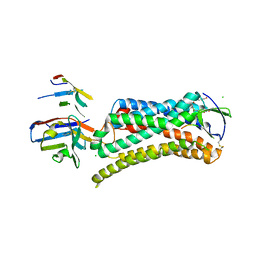
6D35
 | | Crystal structure of Xenopus Smoothened in complex with cholesterol | | 分子名称: | CHOLESTEROL, Smoothened,Soluble cytochrome b562,Smoothened | | 著者 | Huang, P, Zheng, S, Kim, Y, Kruse, A.C, Salic, A. | | 登録日 | 2018-04-14 | | 公開日 | 2018-05-23 | | 最終更新日 | 2023-10-04 | | 実験手法 | X-RAY DIFFRACTION (3.9 Å) | | 主引用文献 | Structural Basis of Smoothened Activation in Hedgehog Signaling.
Cell, 174, 2018
|
|
6D32
 | | Crystal structure of Xenopus Smoothened in complex with cyclopamine | | 分子名称: | Cyclopamine, Smoothened,Soluble cytochrome b562,Smoothened | | 著者 | Huang, P, Zheng, S, Kim, Y, Kruse, A.C, Salic, A. | | 登録日 | 2018-04-14 | | 公開日 | 2018-05-23 | | 最終更新日 | 2023-10-04 | | 実験手法 | X-RAY DIFFRACTION (3.751 Å) | | 主引用文献 | Structural Basis of Smoothened Activation in Hedgehog Signaling.
Cell, 174, 2018
|
|
3I48
 | | Crystal structure of beta toxin from Staphylococcus aureus F277A, P278A mutant with bound magnesium ions | | 分子名称: | Beta-hemolysin, MAGNESIUM ION, PHOSPHATE ION | | 著者 | Huseby, M, Shi, K, Kruse, A.C, Ohlendorf, D.H. | | 登録日 | 2009-07-01 | | 公開日 | 2010-07-14 | | 最終更新日 | 2023-09-06 | | 実験手法 | X-RAY DIFFRACTION (1.8 Å) | | 主引用文献 | Structure and biological functions of beta toxin from Staphylococcus aureus: Role of the hydrophobic beta hairpin in virulence
to be published, 2009
|
|
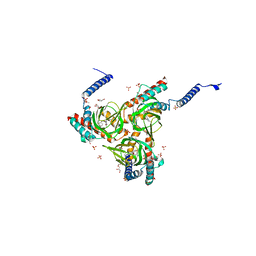
3I5V
 | |
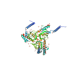
3I41
 | |
6DO1
 | | Structure of nanobody-stabilized angiotensin II type 1 receptor bound to S1I8 | | 分子名称: | (2R)-2,3-dihydroxypropyl (9Z)-octadec-9-enoate, 2-acetamido-2-deoxy-beta-D-glucopyranose, Angiotensin-like peptide S1I8, ... | | 著者 | Wingler, L.M, McMahon, C, Staus, D.P, Lefkowitz, R.J, Kruse, A.C. | | 登録日 | 2018-06-08 | | 公開日 | 2019-01-30 | | 最終更新日 | 2023-10-11 | | 実験手法 | X-RAY DIFFRACTION (2.901 Å) | | 主引用文献 | Distinctive Activation Mechanism for Angiotensin Receptor Revealed by a Synthetic Nanobody.
Cell, 176, 2019
|
|
3UON
 | | Structure of the human M2 muscarinic acetylcholine receptor bound to an antagonist | | 分子名称: | (3R)-1-azabicyclo[2.2.2]oct-3-yl hydroxy(diphenyl)acetate, CHLORIDE ION, Human M2 muscarinic acetylcholine, ... | | 著者 | Haga, K, Kruse, A.C, Asada, H, Yurugi-Kobayashi, T, Shiroishi, M, Zhang, C, Weis, W.I, Okada, T, Kobilka, B.K, Haga, T, Kobayashi, T. | | 登録日 | 2011-11-16 | | 公開日 | 2012-02-01 | | 最終更新日 | 2023-09-13 | | 実験手法 | X-RAY DIFFRACTION (3 Å) | | 主引用文献 | Structure of the human M2 muscarinic acetylcholine receptor bound to an antagonist.
Nature, 482, 2012
|
|


