7VM4
 
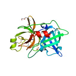
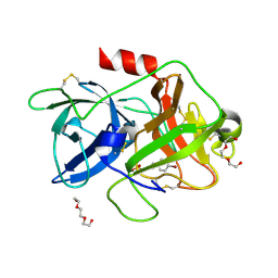
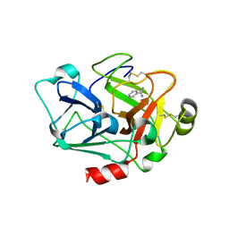
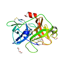 | | Crystal structure of uPA in complex with nafamostat | | 分子名称: | 4-carbamimidamidobenzoic acid, TRIETHYLENE GLYCOL, Urokinase-type plasminogen activator | | 著者 | Jiang, L.G, Huang, M.D. | | 登録日 | 2021-10-07 | | 公開日 | 2022-10-12 | | 最終更新日 | 2023-11-29 | | 実験手法 | X-RAY DIFFRACTION (2.01 Å) | | 主引用文献 | Structural study of the uPA-nafamostat complex reveals a covalent inhibitory mechanism of nafamostat.
Biophys.J., 121, 2022
|
|
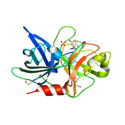
7VM5
 | |
7VM6
 | |
3TBA
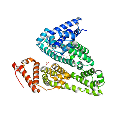
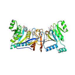
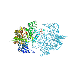 | | Structure of Yeast Ribonucleotide Reductase 1 Q288A with dGTP and ADP | | 分子名称: | 2'-DEOXYGUANOSINE-5'-TRIPHOSPHATE, ADENOSINE-5'-DIPHOSPHATE, MAGNESIUM ION, ... | | 著者 | Ahmad, M.F, Kaushal, P.S, Wan, Q, Wijeratna, S.R, Huang, M, Dealwis, C. | | 登録日 | 2011-08-05 | | 公開日 | 2012-04-04 | | 最終更新日 | 2024-02-28 | | 実験手法 | X-RAY DIFFRACTION (2.8 Å) | | 主引用文献 | Role of Arginine 293 and Glutamine 288 in Communication between Catalytic and Allosteric Sites in Yeast Ribonucleotide Reductase.
J.Mol.Biol., 419, 2012
|
|
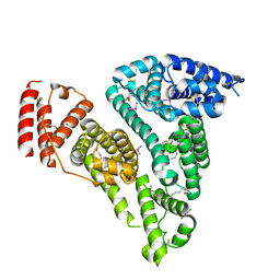
3TB9
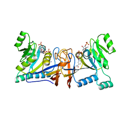
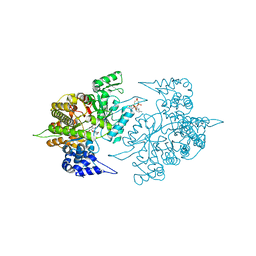 | | Structure of Yeast Ribonucleotide Reductase 1 Q288A with AMPPNP and CDP | | 分子名称: | CYTIDINE-5'-DIPHOSPHATE, MAGNESIUM ION, PHOSPHOAMINOPHOSPHONIC ACID-ADENYLATE ESTER, ... | | 著者 | Ahmad, M.F, Kaushal, P.S, Wan, Q, Wijeratna, S.R, Huang, M, Dealwis, C.D. | | 登録日 | 2011-08-05 | | 公開日 | 2012-04-04 | | 最終更新日 | 2023-09-13 | | 実験手法 | X-RAY DIFFRACTION (2.53 Å) | | 主引用文献 | Role of Arginine 293 and Glutamine 288 in Communication between Catalytic and Allosteric Sites in Yeast Ribonucleotide Reductase.
J.Mol.Biol., 419, 2012
|
|
6A8O
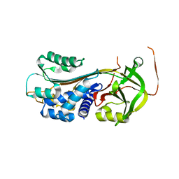
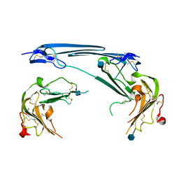
 | | Crystal structures of the serine protease domain of murine plasma kallikrein with peptide inhibitor mupain-1-16 | | 分子名称: | Plasma kallikrein, alpha-D-mannopyranose, peptide inhibitor,, ... | | 著者 | Xu, M, Jiang, L, Huang, M. | | 登録日 | 2018-07-09 | | 公開日 | 2019-07-10 | | 最終更新日 | 2023-11-22 | | 実験手法 | X-RAY DIFFRACTION (2.77 Å) | | 主引用文献 | Crystal structure of plasma kallikrein reveals the unusual flexibility of the S1 pocket triggered by Glu217.
Febs Lett., 592, 2018
|
|
2NWN
 | | New Pharmacophore for Serine Protease Inhibition Revealed by Crystal Structure of Human Urokinase-type Plasminogen Activator Complexed with a Cyclic Peptidyl Inhibitor, upain-1 | | 分子名称: | Plasminogen activator, urokinase, upain-1 | | 著者 | Zhao, G, Yuan, C, Wind, T, Andreasen, P.A, Huang, Z, Huang, M, Structural Genomics Consortium (SGC) | | 登録日 | 2006-11-16 | | 公開日 | 2007-10-16 | | 最終更新日 | 2023-10-25 | | 実験手法 | X-RAY DIFFRACTION (2.15 Å) | | 主引用文献 | Structural basis of specificity of a peptidyl urokinase inhibitor, upain-1
J.Struct.Biol., 160, 2007
|
|
2O8U
 | | Crystal Structure and Binding Epitopes of Urokinase-type Plasminogen Activator (C122A/N145Q/S195A) in complex with Inhibitors | | 分子名称: | BENZAMIDINE, DI(HYDROXYETHYL)ETHER, SULFATE ION, ... | | 著者 | Zhao, G, Yuan, C, Jiang, L, Huang, Z, Huang, M. | | 登録日 | 2006-12-12 | | 公開日 | 2007-12-25 | | 最終更新日 | 2023-12-27 | | 実験手法 | X-RAY DIFFRACTION (1.7 Å) | | 主引用文献 | Crystal Structure and Binding Epitopes of Urokinase-type Plasminogen Activator (C122A/N145Q/S195A) in complex with Inhibitors
To be Published
|
|
2O8T
 | | Crystal Structure and Binding Epitopes of Urokinase-type Plasminogen Activator (C122A/N145Q) in complex with Inhibitors | | 分子名称: | DI(HYDROXYETHYL)ETHER, PENTAETHYLENE GLYCOL, SULFATE ION, ... | | 著者 | Zhao, G, Yuan, C, Jiang, L, Huang, Z, Huang, M. | | 登録日 | 2006-12-12 | | 公開日 | 2007-12-25 | | 最終更新日 | 2023-12-27 | | 実験手法 | X-RAY DIFFRACTION (1.45 Å) | | 主引用文献 | Crystal Structure and Binding Epitopes of Urokinase-type Plasminogen Activator (C122A/N145Q/S195A) in complex with Inhibitors
To be Published
|
|
2O8W
 | | Crystal Structure and Binding Epitopes of Urokinase-type Plasminogen Activator (C122A/N145Q/S195A) in complex with Inhibitors | | 分子名称: | 1-phenylguanidine, SULFATE ION, TETRAETHYLENE GLYCOL, ... | | 著者 | Zhao, G, Yuan, C, Jiang, L, Huang, Z, Huang, M. | | 登録日 | 2006-12-12 | | 公開日 | 2007-12-25 | | 最終更新日 | 2023-12-27 | | 実験手法 | X-RAY DIFFRACTION (1.86 Å) | | 主引用文献 | Crystal Structure and Binding Epitopes of Urokinase-type Plasminogen Activator (C122A/N145Q/S195A) in complex with Inhibitors
To be Published
|
|
7WOK
 | | Crystal structure of HSA soaked with cisplatin for one week | | 分子名称: | Albumin, Cisplatin, PHOSPHATE ION | | 著者 | Chen, S.L, Yuan, C, Jiang, L.G, Luo, Z.P, Huang, M.D. | | 登録日 | 2022-01-21 | | 公開日 | 2022-07-20 | | 最終更新日 | 2023-11-29 | | 実験手法 | X-RAY DIFFRACTION (2.9 Å) | | 主引用文献 | Crystallographic analysis of interaction between cisplatin and human serum albumin: Effect of fatty acid.
Int.J.Biol.Macromol., 216, 2022
|
|
7WOJ
 | | Crystal structure of HSA-Myr complex soaked with cisplatin for one week | | 分子名称: | Albumin, Cisplatin, MYRISTIC ACID | | 著者 | Chen, S.L, Yuan, C, Jiang, L.G, Luo, Z.P, Huang, M.D. | | 登録日 | 2022-01-21 | | 公開日 | 2022-07-20 | | 最終更新日 | 2023-11-29 | | 実験手法 | X-RAY DIFFRACTION (2.89 Å) | | 主引用文献 | Crystallographic analysis of interaction between cisplatin and human serum albumin: Effect of fatty acid.
Int.J.Biol.Macromol., 216, 2022
|
|
5Z9Y
 | | Crystal structure of Mycobacterium tuberculosis thiazole synthase (ThiG) complexed with DXP | | 分子名称: | 1-DEOXY-D-XYLULOSE-5-PHOSPHATE, Thiazole synthase | | 著者 | Zhang, J, Zhang, B, Zhao, Y, Yang, X, Huang, M, Cui, P, Zhang, W, Li, J, Zhang, Y. | | 登録日 | 2018-02-05 | | 公開日 | 2018-04-11 | | 実験手法 | X-RAY DIFFRACTION (1.48 Å) | | 主引用文献 | Snapshots of catalysis: Structure of covalently bound substrate trapped in Mycobacterium tuberculosis thiazole synthase (ThiG).
Biochem. Biophys. Res. Commun., 497, 2018
|
|
3UT3
 | | A novel PAI-I inhibitor and its structural mechanism | | 分子名称: | 2,5-dihydroxy-3-undecylcyclohexa-2,5-diene-1,4-dione, Plasminogen activator inhibitor 1 | | 著者 | Lin, Z.H, Hong, Z.B, Shi, X.L, Hu, L.H, Andreasen, P.A, Huang, M.D. | | 登録日 | 2011-11-25 | | 公開日 | 2013-02-06 | | 最終更新日 | 2023-11-08 | | 実験手法 | X-RAY DIFFRACTION (2.42 Å) | | 主引用文献 | A novel PAI-I inhibitor and its structural mechanism
To be Published
|
|
5YC7
 | |
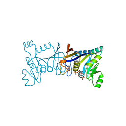
5ZLZ
 | | Structure of tPA and PAI-1 | | 分子名称: | GLYCEROL, Plasminogen activator inhibitor 1, Tissue-type plasminogen activator | | 著者 | Min, L, Huang, M. | | 登録日 | 2018-03-31 | | 公開日 | 2019-04-03 | | 最終更新日 | 2023-11-22 | | 実験手法 | X-RAY DIFFRACTION (3.581 Å) | | 主引用文献 | Development of a PAI-1 trapping agent (PAItrap2) based on inactivated tPA-SPD and the crystal structure of PAItrap2 in complex with PAI-1
To Be Published
|
|
5Z1C
 | | The crystal structure of uPA in complex with 4-Iodobenzylamine at pH7.4 | | 分子名称: | 1-(4-iodophenyl)methanamine, Urokinase-type plasminogen activator | | 著者 | Jiang, L.G, Zhang, X, Luo, Z.P, Huang, M.D. | | 登録日 | 2017-12-25 | | 公開日 | 2018-12-26 | | 最終更新日 | 2023-11-22 | | 実験手法 | X-RAY DIFFRACTION (1.45 Å) | | 主引用文献 | Halogen bonding for the design of inhibitors by targeting the S1 pocket of serine proteases
Rsc Adv, 49, 2018
|
|
5YUP
 | | Crystal Structure of the Fab fragment of FVIIa antibody mAb4F5 | | 分子名称: | the heavy chain of the Fab fragment of FVIIa antibody mAb4F5, the light chain of the Fab fragment of FVIIa antibody mAb4F5 | | 著者 | Jiang, L.G, Persson, E, Huang, M.D. | | 登録日 | 2017-11-22 | | 公開日 | 2019-06-19 | | 最終更新日 | 2023-11-22 | | 実験手法 | X-RAY DIFFRACTION (1.81 Å) | | 主引用文献 | Crystal structure, epitope, and functional impact of an antibody against a superactive FVIIa provide insights into allosteric mechanism.
Res Pract Thromb Haemost, 3, 2019
|
|
5YC6
 | | The crystal structure of uPA in complex with 4-Bromobenzylamirne at pH4.6 | | 分子名称: | 1-(4-BROMOPHENYL)METHANAMINE, SULFATE ION, TRIETHYLENE GLYCOL, ... | | 著者 | Jiang, L.G, Zhang, X, Huang, M.D. | | 登録日 | 2017-09-06 | | 公開日 | 2018-10-03 | | 最終更新日 | 2023-11-22 | | 実験手法 | X-RAY DIFFRACTION (1.18 Å) | | 主引用文献 | Halogen bonding for the design of inhibitors by targeting the S1 pocket of serine proteases
Rsc Adv, 8, 2018
|
|
5XUX
 | | Crystal structure of Rib7 from Methanosarcina mazei | | 分子名称: | Conserved protein, NADP NICOTINAMIDE-ADENINE-DINUCLEOTIDE PHOSPHATE | | 著者 | Yeh, T.M, Chen, S.C, Chang, T.H, Huang, M.F, Liaw, S.H. | | 登録日 | 2017-06-26 | | 公開日 | 2018-07-04 | | 最終更新日 | 2023-11-22 | | 実験手法 | X-RAY DIFFRACTION (2.27 Å) | | 主引用文献 | Evolution of archaeal Rib7 and eubacterial RibG reductases in riboflavin biosynthesis: Substrate specificity and cofactor preference.
Biochem. Biophys. Res. Commun., 503, 2018
|
|
5XV0
 | | Crystal structure of Rib7 mutant D33N from Methanosarcina mazei | | 分子名称: | Conserved protein, NADP NICOTINAMIDE-ADENINE-DINUCLEOTIDE PHOSPHATE | | 著者 | Yeh, T.M, Chen, S.C, Chang, T.H, Huang, M.F, Liaw, S.H. | | 登録日 | 2017-06-26 | | 公開日 | 2018-07-04 | | 最終更新日 | 2023-11-22 | | 実験手法 | X-RAY DIFFRACTION (1.95 Å) | | 主引用文献 | Evolution of archaeal Rib7 and eubacterial RibG reductases in riboflavin biosynthesis: Substrate specificity and cofactor preference.
Biochem. Biophys. Res. Commun., 503, 2018
|
|
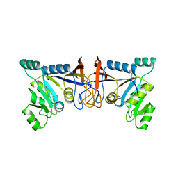
7V63
 | | Structure of dimeric uPAR at low pH | | 分子名称: | 2-acetamido-2-deoxy-beta-D-glucopyranose, Urokinase plasminogen activator surface receptor | | 著者 | Yuan, C, Huang, M. | | 登録日 | 2021-08-19 | | 公開日 | 2021-12-22 | | 最終更新日 | 2022-04-06 | | 実験手法 | X-RAY DIFFRACTION (2.906 Å) | | 主引用文献 | Crystal structure and cellular functions of uPAR dimer
Nat Commun, 13, 2022
|
|
5XV5
 | | Crystal structure of Rib7 mutant S88E from Methanosarcina mazei | | 分子名称: | Conserved protein | | 著者 | Yeh, T.M, Chen, S.C, Chang, T.H, Huang, M.F, Liaw, S.H. | | 登録日 | 2017-06-26 | | 公開日 | 2018-07-04 | | 最終更新日 | 2023-11-22 | | 実験手法 | X-RAY DIFFRACTION (2.5 Å) | | 主引用文献 | Evolution of archaeal Rib7 and eubacterial RibG reductases in riboflavin biosynthesis: Substrate specificity and cofactor preference.
Biochem. Biophys. Res. Commun., 503, 2018
|
|
5XV2
 | | Crystal structure of Rib7 mutant D33A from Methanosarcina mazei | | 分子名称: | Conserved protein, NADP NICOTINAMIDE-ADENINE-DINUCLEOTIDE PHOSPHATE | | 著者 | Yeh, T.M, Chen, S.C, Chang, T.H, Huang, M.F, Liaw, S.H. | | 登録日 | 2017-06-26 | | 公開日 | 2018-07-04 | | 最終更新日 | 2023-11-22 | | 実験手法 | X-RAY DIFFRACTION (2 Å) | | 主引用文献 | Evolution of archaeal Rib7 and eubacterial RibG reductases in riboflavin biosynthesis: Substrate specificity and cofactor preference.
Biochem. Biophys. Res. Commun., 503, 2018
|
|
7CJ1
 | |


