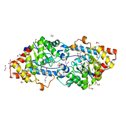
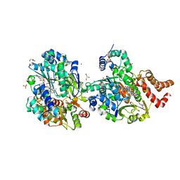
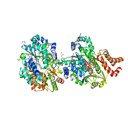
2V0P
 
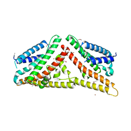 | | The Structure of Tap42 Alpha4 Subunit | | 分子名称: | TYPE 2A PHOSPHATASE-ASSOCIATED PROTEIN 42, ZINC ION | | 著者 | Roe, S.M, Yang, J, Barford, D. | | 登録日 | 2007-05-15 | | 公開日 | 2007-07-17 | | 最終更新日 | 2019-05-15 | | 実験手法 | X-RAY DIFFRACTION (1.8 Å) | | 主引用文献 | The Structure of Tap42/Alpha4 Reveals a Tetratricopeptide Repeat-Like Fold and Provides Insights Into Pp2A Regulation.
Biochemistry, 46, 2007
|
|
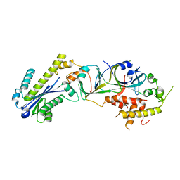
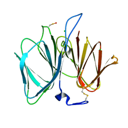
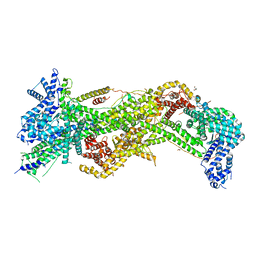
2V5S
 | | Structural basis for Dscam isoform specificity | | 分子名称: | 2-acetamido-2-deoxy-beta-D-glucopyranose-(1-4)-2-acetamido-2-deoxy-beta-D-glucopyranose, DSCAM | | 著者 | Meijers, R, Puettmann-Holgado, R, Skiniotis, G, Liu, J.-H, Walz, T, Schmucker, D, Wang, J.-H. | | 登録日 | 2007-07-09 | | 公開日 | 2007-09-11 | | 最終更新日 | 2023-12-13 | | 実験手法 | X-RAY DIFFRACTION (2.3 Å) | | 主引用文献 | Structural Basis of Dscam Isoform Specificity
Nature, 449, 2007
|
|
3P5J
 | |
2V4B
 | | Crystal Structure of Human ADAMTS-1 catalytic Domain and Cysteine- Rich Domain (apo-form) | | 分子名称: | ADAMTS-1, CADMIUM ION, MAGNESIUM ION, ... | | 著者 | Gerhardt, S, Hassall, G, Hawtin, P, McCall, E, Flavell, L, Minshull, C, Hargreaves, D, Ting, A, Pauptit, R.A, Parker, A.E, Abbott, W.M. | | 登録日 | 2007-06-28 | | 公開日 | 2008-01-15 | | 最終更新日 | 2019-04-24 | | 実験手法 | X-RAY DIFFRACTION (2 Å) | | 主引用文献 | Crystal Structures of Human Adamts-1 Reveal a Conserved Catalytic Domain and a Disintegrin-Like Domain with a Fold Homologous to Cysteine-Rich Domains.
J.Mol.Biol., 373, 2007
|
|
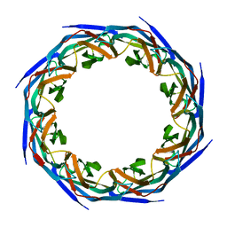
1MFR
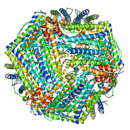 | | CRYSTAL STRUCTURE OF M FERRITIN | | 分子名称: | M FERRITIN, MAGNESIUM ION | | 著者 | Ha, Y, Shi, D, Allewell, N.M. | | 登録日 | 1998-06-18 | | 公開日 | 1999-06-22 | | 最終更新日 | 2024-05-22 | | 実験手法 | X-RAY DIFFRACTION (2.8 Å) | | 主引用文献 | Crystal structure of bullfrog M ferritin at 2.8 A resolution: analysis of subunit interactions and the binuclear metal center
J.Biol.Inorg.Chem., 4, 1999
|
|
3P8T
 | |
8QGW
 | |
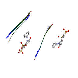
2VIP
 | | Fragment-Based Discovery of Mexiletine Derivatives as Orally Bioavailable Inhibitors of Urokinase-Type Plasminogen Activator | | 分子名称: | 4-(2-AMINOETHOXY)-3,5-DICHLORO-N-[3-(1-METHYLETHOXY)PHENYL]BENZAMIDE, ACETATE ION, SULFATE ION, ... | | 著者 | Frederickson, M, Callaghan, O, Chessari, G, Congreve, M, Cowan, S.R, Matthews, J.E, McMenamin, R, Smith, D, Vinkovic, M, Wallis, N.G. | | 登録日 | 2007-12-05 | | 公開日 | 2008-01-22 | | 最終更新日 | 2017-07-05 | | 実験手法 | X-RAY DIFFRACTION (1.72 Å) | | 主引用文献 | Fragment-Based Discovery of Mexiletine Derivatives as Orally Bioavailable Inhibitors of Urokinase-Type Plasminogen Activator
J.Med.Chem., 51, 2008
|
|
4D8A
 | | Crystal structure of B. anthracis DHPS with compound 21 | | 分子名称: | Dihydropteroate synthase, LYSINE, SULFATE ION, ... | | 著者 | Hammoudeh, D, Lee, R.E, White, S.W. | | 登録日 | 2012-01-10 | | 公開日 | 2012-04-04 | | 最終更新日 | 2023-09-13 | | 実験手法 | X-RAY DIFFRACTION (2.183 Å) | | 主引用文献 | Structure-Based Design of Novel Pyrimido[4,5-c]pyridazine Derivatives as Dihydropteroate Synthase Inhibitors with Increased Affinity.
Chemmedchem, 7, 2012
|
|
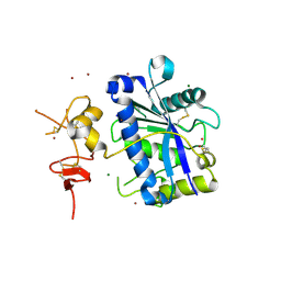
3P1X
 | | Crystal structure of DRBM 2 domain of Interleukin enhancer-binding factor 3 from Homo sapiens, Northeast Structural Genomics Consortium Target HR4527E | | 分子名称: | Interleukin enhancer-binding factor 3 | | 著者 | Seetharaman, J, Neely, H, Wang, D, Janjua, H, Cunningham, K, Owens, L, Xiao, R, Liu, J, Baran, M.C, Acton, T.B, Montelione, G.T, Tong, L, Hunt, J.F, Northeast Structural Genomics Consortium (NESG) | | 登録日 | 2010-09-30 | | 公開日 | 2010-12-29 | | 最終更新日 | 2012-02-22 | | 実験手法 | X-RAY DIFFRACTION (1.9 Å) | | 主引用文献 | Crystal structure of DRBM 2 domain of Interleukin enhancer-binding factor 3 from Homo sapiens, Northeast Structural Genomics Consortium Target HR4527E
To be Published
|
|
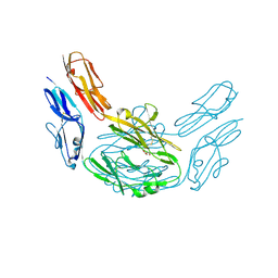
8QZR
 | | SARS-CoV-2 delta RBD complexed with BA.4/5-9 Fab | | 分子名称: | 2-acetamido-2-deoxy-beta-D-glucopyranose, BA.4/5-9 heavy chain, BA.4/5-9 light chain, ... | | 著者 | Zhou, D, Ren, J, Stuart, D.I. | | 登録日 | 2023-10-29 | | 公開日 | 2024-04-03 | | 最終更新日 | 2024-05-08 | | 実験手法 | X-RAY DIFFRACTION (3.77 Å) | | 主引用文献 | Emerging variants develop total escape from potent monoclonal antibodies induced by BA.4/5 infection.
Nat Commun, 15, 2024
|
|
8QH4
 | | Crystal structure of reduced respiratory Complex I subunits NuoEF from Aquifex aeolicus bound to oxidized 3-acetylpyridine adenine dinucleotide | | 分子名称: | 1-DEOXY-1-(7,8-DIMETHYL-2,4-DIOXO-3,4-DIHYDRO-2H-BENZO[G]PTERIDIN-1-ID-10(5H)-YL)-5-O-PHOSPHONATO-D-RIBITOL, 3-ACETYLPYRIDINE ADENINE DINUCLEOTIDE, FE2/S2 (INORGANIC) CLUSTER, ... | | 著者 | Wohlwend, D, Friedrich, T. | | 登録日 | 2023-09-06 | | 公開日 | 2024-04-03 | | 最終更新日 | 2024-06-19 | | 実験手法 | X-RAY DIFFRACTION (1.96 Å) | | 主引用文献 | Structures of 3-acetylpyridine adenine dinucleotide and ADP-ribose bound to the electron input module of respiratory complex I.
Structure, 32, 2024
|
|
8QG1
 | |
2V5M
 | | Structural basis for Dscam isoform specificity | | 分子名称: | 2-acetamido-2-deoxy-beta-D-glucopyranose-(1-4)-2-acetamido-2-deoxy-beta-D-glucopyranose, DSCAM, GLYCEROL | | 著者 | Meijers, R, Puettmann-Holgado, R, Skiniotis, G, Liu, J.-H, Walz, T, Schmucker, D, Wang, J.-H. | | 登録日 | 2007-07-06 | | 公開日 | 2007-09-11 | | 最終更新日 | 2020-07-29 | | 実験手法 | X-RAY DIFFRACTION (1.95 Å) | | 主引用文献 | Structural Basis of Dscam Isoform Specificity
Nature, 449, 2007
|
|
8RTY
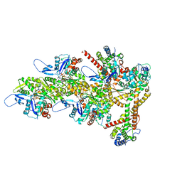 | | Structure of the F-actin barbed end bound by Cdc12 and profilin (ring complex) at a resolution of 6.3 Angstrom | | 分子名称: | ADENOSINE-5'-DIPHOSPHATE, Actin, cytoplasmic 1, ... | | 著者 | Oosterheert, W, Boiero Sanders, M, Funk, J, Prumbaum, D, Raunser, S, Bieling, P. | | 登録日 | 2024-01-29 | | 公開日 | 2024-04-10 | | 最終更新日 | 2024-04-24 | | 実験手法 | ELECTRON MICROSCOPY (6.25 Å) | | 主引用文献 | Molecular mechanism of actin filament elongation by formins.
Science, 384, 2024
|
|
2V97
 | | Structure of the unphotolysed complex of TcAChE with 1-(2- nitrophenyl)-2,2,2-trifluoroethyl-arsenocholine after a 9 seconds annealing to room temperature | | 分子名称: | 1-(2-nitrophenyl)-2,2,2-trifluoroethyl]-arsenocholine, 2-acetamido-2-deoxy-beta-D-glucopyranose, ACETYLCHOLINESTERASE, ... | | 著者 | Colletier, J.-P, Sanson, B, Royant, A, Specht, A, Nachon, F, Masson, P, Zaccai, G, Sussman, J.L, Goeldner, M, Silman, I, Bourgeois, D, Weik, M. | | 登録日 | 2007-08-22 | | 公開日 | 2007-11-20 | | 最終更新日 | 2023-12-13 | | 実験手法 | X-RAY DIFFRACTION (2.4 Å) | | 主引用文献 | Use of a 'Caged' Analog to Study Traffic of Choline within Acetylcholinesterase by Kinetic Crystallography
Acta Crystallogr.,Sect.D, 63, 2007
|
|
2V5J
 | | Apo Class II aldolase HpcH | | 分子名称: | 2,4-DIHYDROXYHEPT-2-ENE-1,7-DIOIC ACID ALDOLASE, GLYCEROL, PHOSPHATE ION | | 著者 | Rea, D, Fulop, V, Bugg, T.D.H, Roper, D.I. | | 登録日 | 2007-07-05 | | 公開日 | 2007-10-02 | | 最終更新日 | 2023-12-13 | | 実験手法 | X-RAY DIFFRACTION (1.6 Å) | | 主引用文献 | Structure and Mechanism of Hpch: A Metal Ion Dependent Class II Aldolase from the Homoprotocatechuate Degradation Pathway of Escherichia Coli.
J.Mol.Biol., 373, 2007
|
|
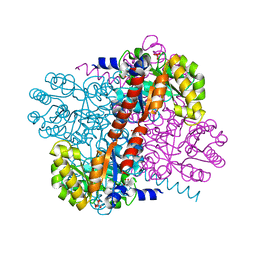
2VC7
 | | Structural basis for natural lactonase and promiscuous phosphotriesterase activities | | 分子名称: | (4S)-4-(decanoylamino)-5-hydroxy-3,4-dihydro-2H-thiophenium, 1,2-ETHANEDIOL, ARYLDIALKYLPHOSPHATASE, ... | | 著者 | Elias, M, Dupuy, J, Merone, L, Mandrich, L, Moniot, S, Rochu, D, Lecomte, C, Rossi, M, Masson, P, Manco, G, Chabriere, E. | | 登録日 | 2007-09-19 | | 公開日 | 2008-04-15 | | 最終更新日 | 2023-12-13 | | 実験手法 | X-RAY DIFFRACTION (2.05 Å) | | 主引用文献 | Structural Basis for Natural Lactonase and Promiscuous Phosphotriesterase Activities.
J.Mol.Biol., 379, 2008
|
|
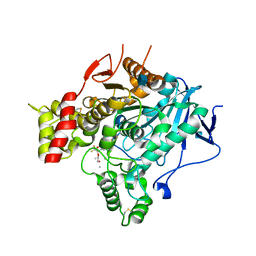
2VEC
 | | The crystal structure of the protein YhaK from Escherichia coli | | 分子名称: | CHLORIDE ION, PIRIN-LIKE PROTEIN YHAK | | 著者 | Gurmu, D, Lu, J, Johnson, K.A, Nordlund, P, Holmgren, A, Erlandsen, H. | | 登録日 | 2007-10-18 | | 公開日 | 2008-07-01 | | 最終更新日 | 2011-07-13 | | 実験手法 | X-RAY DIFFRACTION (1.85 Å) | | 主引用文献 | The Crystal Structure of the Protein Yhak from Escherichia Coli Reveals a New Subclass of Redox Sensitive Enterobacterial Bicupins.
Proteins, 74, 2008
|
|
8RHZ
 | | Structure of CUL9-RBX1 ubiquitin E3 ligase complex in unneddylated conformation - symmetry expanded unneddylated dimer | | 分子名称: | Cullin-9, E3 ubiquitin-protein ligase RBX1, ZINC ION | | 著者 | Hopf, L.V.M, Horn-Ghetko, D, Prabu, J.R, Schulman, B.A. | | 登録日 | 2023-12-17 | | 公開日 | 2024-04-17 | | 最終更新日 | 2024-07-31 | | 実験手法 | ELECTRON MICROSCOPY (3.37 Å) | | 主引用文献 | Noncanonical assembly, neddylation and chimeric cullin-RING/RBR ubiquitylation by the 1.8 MDa CUL9 E3 ligase complex.
Nat.Struct.Mol.Biol., 31, 2024
|
|
2V9U
 | | Rim domain of main porin from Mycobacteria smegmatis | | 分子名称: | MSPA | | 著者 | Grueninger, D, Ziegler, M.O.P, Koetter, J.W.A, Treiber, N, Schulze, M.-S, Schulz, G.E. | | 登録日 | 2007-08-27 | | 公開日 | 2008-01-15 | | 最終更新日 | 2024-05-08 | | 実験手法 | X-RAY DIFFRACTION (2.59 Å) | | 主引用文献 | Designed Protein-Protein Association.
Science, 319, 2008
|
|
8QH7
 | | Crystal structure of respiratory Complex I subunits NuoEF from Aquifex aeolicus bound to reduced 3-acetylpyridine adenine dinucleotide (without reducing agent) | | 分子名称: | 1-DEOXY-1-(7,8-DIMETHYL-2,4-DIOXO-3,4-DIHYDRO-2H-BENZO[G]PTERIDIN-1-ID-10(5H)-YL)-5-O-PHOSPHONATO-D-RIBITOL, 2-[BIS-(2-HYDROXY-ETHYL)-AMINO]-2-HYDROXYMETHYL-PROPANE-1,3-DIOL, ACETYL PYRIDINE ADENINE DINUCLEOTIDE, ... | | 著者 | Wohlwend, D, Friedrich, T. | | 登録日 | 2023-09-06 | | 公開日 | 2024-04-03 | | 最終更新日 | 2024-06-19 | | 実験手法 | X-RAY DIFFRACTION (1.85 Å) | | 主引用文献 | Structures of 3-acetylpyridine adenine dinucleotide and ADP-ribose bound to the electron input module of respiratory complex I.
Structure, 32, 2024
|
|
3P8C
 | | Structure and Control of the Actin Regulatory WAVE Complex | | 分子名称: | 2-AMINO-2-HYDROXYMETHYL-PROPANE-1,3-DIOL, Abl interactor 2, CHLORIDE ION, ... | | 著者 | Chen, Z, Borek, D, Padrick, S.B, Gomez, T.S, Metlagel, Z, Ismail, A.M, Umetani, J, Billadeau, D.D, Otwinowski, Z, Rosen, M.K. | | 登録日 | 2010-10-13 | | 公開日 | 2010-12-01 | | 最終更新日 | 2024-02-21 | | 実験手法 | X-RAY DIFFRACTION (2.29 Å) | | 主引用文献 | Structure and control of the actin regulatory WAVE complex.
Nature, 468, 2010
|
|
3OVJ
 | |
8QHK
 | | Crystal structure of reduced respiratory Complex I subunits NuoEF from Aquifex aeolicus bound to reduced 3-acetylpyridine adenine dinucleotide | | 分子名称: | 1-DEOXY-1-(7,8-DIMETHYL-2,4-DIOXO-3,4-DIHYDRO-2H-BENZO[G]PTERIDIN-1-ID-10(5H)-YL)-5-O-PHOSPHONATO-D-RIBITOL, ACETYL PYRIDINE ADENINE DINUCLEOTIDE, REDUCED, ... | | 著者 | Wohlwend, D, Friedrich, T, Bucka, S. | | 登録日 | 2023-09-08 | | 公開日 | 2024-04-03 | | 最終更新日 | 2024-06-19 | | 実験手法 | X-RAY DIFFRACTION (1.95 Å) | | 主引用文献 | Structures of 3-acetylpyridine adenine dinucleotide and ADP-ribose bound to the electron input module of respiratory complex I.
Structure, 32, 2024
|
|


