Loading
PDBj
MenuPDBj@FacebookPDBj@TwitterPDBj@YouTubewwPDB FoundationwwPDB
RCSB PDBPDBeBMRBAdv. SearchSearch help
Search by PDB author
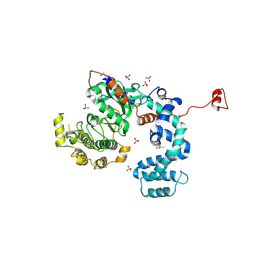
3NYN
DownloadVisualize
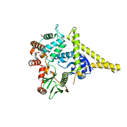
BU of 3nyn by Molmil
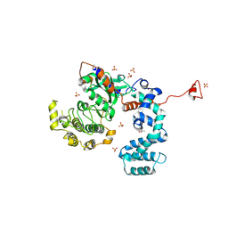
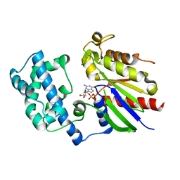
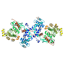
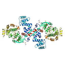
Crystal Structure of G Protein-Coupled Receptor Kinase 6 in Complex with Sangivamycin
Descriptor: (R,R)-2,3-BUTANEDIOL, G protein-coupled receptor kinase 6, SANGIVAMYCIN, ...
Authors:Tesmer, J.J.G, Singh, P.
Deposit date:2010-07-15
Release date:2010-09-22
Last modified:2024-02-21
Method:X-RAY DIFFRACTION (2.72 Å)
Cite:Molecular basis for activation of G protein-coupled receptor kinases.
Embo J., 29, 2010
3NYO
DownloadVisualize
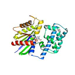
BU of 3nyo by Molmil
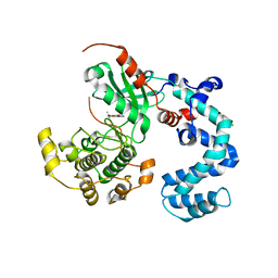
Crystal Structure of G Protein-Coupled Receptor Kinase 6 in Complex with AMP
Descriptor: (R,R)-2,3-BUTANEDIOL, ADENOSINE MONOPHOSPHATE, G protein-coupled receptor kinase 6, ...
Authors:Tesmer, J.J.G, Singh, P.
Deposit date:2010-07-15
Release date:2010-09-22
Last modified:2024-02-21
Method:X-RAY DIFFRACTION (2.92 Å)
Cite:Molecular basis for activation of G protein-coupled receptor kinases.
Embo J., 29, 2010
2BCJ
DownloadVisualize
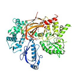
BU of 2bcj by Molmil
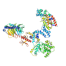
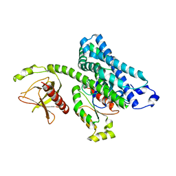
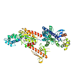
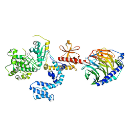
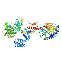
Crystal Structure of G Protein-Coupled Receptor Kinase 2 in Complex with Galpha-q and Gbetagamma Subunits
Descriptor: G-protein-coupled receptor kinase 2, GUANOSINE-5'-DIPHOSPHATE, Guanine nucleotide-binding protein G(I)/G(S)/G(O) subunit gamma-2, ...
Authors:Tesmer, J.J.G.
Deposit date:2005-10-19
Release date:2005-12-20
Last modified:2023-08-23
Method:X-RAY DIFFRACTION (3.061 Å)
Cite:Snapshot of Activated G Proteins at the Membrane: The Gq-GRK2-G Complex
Science, 310, 2005
1TXD
DownloadVisualize
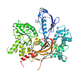
BU of 1txd by Molmil
Crystal Structure of the DH/PH domains of Leukemia-associated RhoGEF
Descriptor: Rho guanine nucleotide exchange factor 12
Authors:Kristelly, R, Gao, G, Tesmer, J.J.
Deposit date:2004-07-03
Release date:2004-09-21
Last modified:2024-02-14
Method:X-RAY DIFFRACTION (2.13 Å)
Cite:Structural determinants of RhoA binding and nucleotide exchange in leukemia-associated Rho guanine-nucleotide exchange factor.
J.Biol.Chem., 279, 2004
4WBO
DownloadVisualize
BU of 4wbo by Molmil
Bovine G Protein Coupled Receptor Kinase 1 in Complex with Amlexanox
Descriptor: 2-amino-7-(1-methylethyl)-5-oxo-5H-chromeno[2,3-b]pyridine-3-carboxylic acid, CHLORIDE ION, Rhodopsin kinase
Authors:Homan, K.T, Tesmer, J.J.G.
Deposit date:2014-09-03
Release date:2014-11-05
Last modified:2023-12-27
Method:X-RAY DIFFRACTION (2.81 Å)
Cite:Identification and characterization of amlexanox as a g protein-coupled receptor kinase 5 inhibitor.
Molecules, 19, 2014
9ABP
DownloadVisualize
BU of 9abp by Molmil
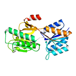
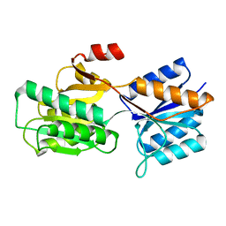
A PRO TO GLY MUTATION IN THE HINGE OF THE ARABINOSE-BINDING PROTEIN ENHANCES BINDING AND ALTERS SPECIFICITY: SUGAR-BINDING AND CRYSTALLOGRAPHIC STUDIES
Descriptor: L-ARABINOSE-BINDING PROTEIN, alpha-D-galactopyranose, beta-D-galactopyranose
Authors:Vermersch, P.S, Tesmer, J.J.G, Quiocho, F.A.
Deposit date:1991-11-15
Release date:1992-01-15
Last modified:2024-02-14
Method:X-RAY DIFFRACTION (1.97 Å)
Cite:A Pro to Gly mutation in the hinge of the arabinose-binding protein enhances binding and alters specificity. Sugar-binding and crystallographic studies.
J.Biol.Chem., 265, 1990
8ABP
DownloadVisualize
BU of 8abp by Molmil
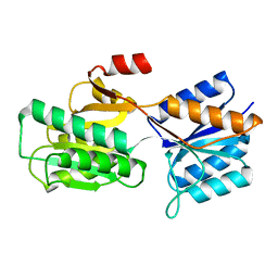
SUGAR-BINDING AND CRYSTALLOGRAPHIC STUDIES OF AN ARABINOSE-BINDING PROTEIN MUTANT (MET108LEU) WHICH EXHIBITS ENHANCED AFFINITY AND ALTERED SPECIFICITY
Descriptor: L-ARABINOSE-BINDING PROTEIN, alpha-D-galactopyranose, beta-D-galactopyranose
Authors:Vermersch, P.S, Tesmer, J.J.G, Quiocho, F.A.
Deposit date:1991-04-25
Release date:1991-10-15
Last modified:2024-02-14
Method:X-RAY DIFFRACTION (1.49 Å)
Cite:Sugar-binding and crystallographic studies of an arabinose-binding protein mutant (Met108Leu) that exhibits enhanced affinity and altered specificity.
Biochemistry, 30, 1991
1BAP
DownloadVisualize
BU of 1bap by Molmil
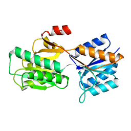
A PRO TO GLY MUTATION IN THE HINGE OF THE ARABINOSE-BINDING PROTEIN ENHANCES BINDING AND ALTERS SPECIFICITY: SUGAR-BINDING AND CRYSTALLOGRAPHIC STUDIES
Descriptor: L-ARABINOSE-BINDING PROTEIN, alpha-L-arabinopyranose, beta-L-arabinopyranose
Authors:Vermersch, P.S, Tesmer, J.J.G, Quiocho, F.A.
Deposit date:1991-11-15
Release date:1992-01-15
Last modified:2024-02-07
Method:X-RAY DIFFRACTION (1.75 Å)
Cite:A Pro to Gly mutation in the hinge of the arabinose-binding protein enhances binding and alters specificity. Sugar-binding and crystallographic studies.
J.Biol.Chem., 265, 1990
1X86
DownloadVisualize
BU of 1x86 by Molmil
Crystal Structure of the DH/PH domains of Leukemia-associated RhoGEF in complex with RhoA
Descriptor: PHOSPHATE ION, Rho guanine nucleotide exchange factor 12, Transforming protein RhoA
Authors:Kristelly, R, Gao, G, Tesmer, J.J.
Deposit date:2004-08-17
Release date:2004-09-21
Last modified:2024-02-14
Method:X-RAY DIFFRACTION (3.22 Å)
Cite:Structural determinants of RhoA binding and nucleotide exchange in leukemia-associated Rho guanine-nucleotide exchange factor.
J.Biol.Chem., 279, 2004
1ZCB
DownloadVisualize
BU of 1zcb by Molmil
Crystal structure of G alpha 13 in complex with GDP
Descriptor: G alpha i/13, GUANOSINE-5'-DIPHOSPHATE
Authors:Nance, M.R, Tesmer, J.J.G.
Deposit date:2005-04-11
Release date:2005-11-15
Last modified:2023-08-23
Method:X-RAY DIFFRACTION (2 Å)
Cite:A new approach to producing functional G alpha subunits yields the activated and deactivated structures of G alpha(12/13) proteins.
Biochemistry, 45, 2006
2RGN
DownloadVisualize
BU of 2rgn by Molmil
Crystal Structure of p63RhoGEF complex with Galpha-q and RhoA
Descriptor: GUANOSINE-5'-DIPHOSPHATE, Guanine nucleotide-binding protein G(i) subunit alpha-1,Guanine nucleotide-binding protein G(q) subunit alpha, MAGNESIUM ION, ...
Authors:Shankaranarayanan, A, Nance, M.R, Tesmer, J.J.G.
Deposit date:2007-10-04
Release date:2008-01-15
Last modified:2023-08-30
Method:X-RAY DIFFRACTION (3.5 Å)
Cite:Structure of Galphaq-p63RhoGEF-RhoA complex reveals a pathway for the activation of RhoA by GPCRs.
Science, 318, 2007
7ABP
DownloadVisualize
BU of 7abp by Molmil
SUGAR-BINDING AND CRYSTALLOGRAPHIC STUDIES OF AN ARABINOSE-BINDING PROTEIN MUTANT (MET108LEU) WHICH EXHIBITS ENHANCED AFFINITY AND ALTERED SPECIFICITY
Descriptor: L-ARABINOSE-BINDING PROTEIN, alpha-D-fucopyranose, beta-D-fucopyranose
Authors:Vermersch, P.S, Tesmer, J.J.G, Quiocho, F.A.
Deposit date:1991-04-25
Release date:1991-10-15
Last modified:2024-03-06
Method:X-RAY DIFFRACTION (1.67 Å)
Cite:Sugar-binding and crystallographic studies of an arabinose-binding protein mutant (Met108Leu) that exhibits enhanced affinity and altered specificity.
Biochemistry, 30, 1991
6MVD
DownloadVisualize
BU of 6mvd by Molmil
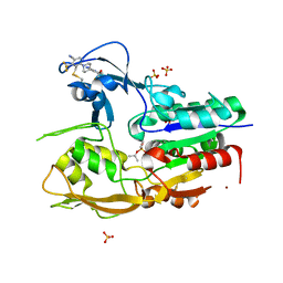
Crystal structure of Lecithin:cholesterol acyltransferase (LCAT) in complex with isopropyl dodec-11-enylfluorophosphonate (IDFP) and a small molecule activator
Descriptor: 2-acetamido-2-deoxy-beta-D-glucopyranose, 6-{4-[(4R)-4-hydroxy-6-oxo-4-(trifluoromethyl)-4,5,6,7-tetrahydro-2H-pyrazolo[3,4-b]pyridin-3-yl]piperidin-1-yl}-4-(trifluoromethyl)pyridine-3-carbonitrile, NICKEL (II) ION, ...
Authors:Manthei, K.A, Chang, L, Tesmer, J.J.G.
Deposit date:2018-10-25
Release date:2018-12-05
Last modified:2023-10-11
Method:X-RAY DIFFRACTION (3.1 Å)
Cite:Molecular basis for activation of lecithin:cholesterol acyltransferase by a compound that increases HDL cholesterol.
Elife, 7, 2018
4L9I
DownloadVisualize
BU of 4l9i by Molmil
Bovine G Protein Coupled Receptor Kinase 1 in Complex with Paroxetine
Descriptor: 2-(N-MORPHOLINO)-ETHANESULFONIC ACID, CHLORIDE ION, Paroxetine, ...
Authors:Homan, K.T, Tesmer, J.J.G.
Deposit date:2013-06-18
Release date:2014-01-22
Last modified:2023-09-20
Method:X-RAY DIFFRACTION (2.32 Å)
Cite:Structural and functional analysis of g protein-coupled receptor kinase inhibition by paroxetine and a rationally designed analog.
Mol.Pharmacol., 85, 2014
4MK0
DownloadVisualize
BU of 4mk0 by Molmil
Crystal structure of G protein-coupled receptor kinase 2 in complex with a a rationally designed paroxetine derivative
Descriptor: 5-{[(3S,4R)-4-(4-fluorophenyl)piperidin-3-yl]methoxy}-1H-isoindol-1-one, Beta-adrenergic receptor kinase 1, GLYCEROL, ...
Authors:Homan, K.T, Tesmer, J.J.G.
Deposit date:2013-09-04
Release date:2014-01-22
Last modified:2024-02-28
Method:X-RAY DIFFRACTION (2.4 Å)
Cite:Structural and functional analysis of g protein-coupled receptor kinase inhibition by paroxetine and a rationally designed analog.
Mol.Pharmacol., 85, 2014
4GNK
DownloadVisualize
BU of 4gnk by Molmil
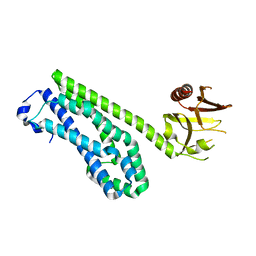
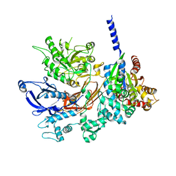
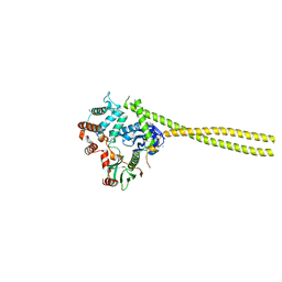
Crystal structure of Galphaq in complex with full-length human PLCbeta3
Descriptor: 1-phosphatidylinositol 4,5-bisphosphate phosphodiesterase beta-3, CALCIUM ION, GUANOSINE-5'-DIPHOSPHATE, ...
Authors:Lyon, A.M, Tesmer, J.J.G.
Deposit date:2012-08-17
Release date:2013-02-06
Last modified:2023-09-13
Method:X-RAY DIFFRACTION (4 Å)
Cite:Full-length G alpha (q)-phospholipase C-beta 3 structure reveals interfaces of the C-terminal coiled-coil domain.
Nat.Struct.Mol.Biol., 20, 2013
2I1K
DownloadVisualize
BU of 2i1k by Molmil
Moesin from Spodoptera frugiperda reveals the coiled-coil domain at 3.0 angstrom resolution
Descriptor: CHLORIDE ION, GLYCEROL, Moesin, ...
Authors:Nance, M.R, Tesmer, J.J.G.
Deposit date:2006-08-14
Release date:2006-12-19
Last modified:2024-02-21
Method:X-RAY DIFFRACTION (3 Å)
Cite:Self-masking in an Intact ERM-merlin Protein: An Active Role for the Central alpha-Helical Domain.
J.Mol.Biol., 365, 2007
2I1J
DownloadVisualize
BU of 2i1j by Molmil
Moesin from Spodoptera frugiperda at 2.1 angstroms resolution
Descriptor: CHLORIDE ION, GLYCEROL, Moesin, ...
Authors:Li, Q, Nance, M.R, Tesmer, J.J.G.
Deposit date:2006-08-14
Release date:2006-12-19
Last modified:2024-02-21
Method:X-RAY DIFFRACTION (2.1 Å)
Cite:Self-masking in an Intact ERM-merlin Protein: An Active Role for the Central alpha-Helical Domain.
J.Mol.Biol., 365, 2007
1ZCA
DownloadVisualize
BU of 1zca by Molmil
Crystal structure of G alpha 12 in complex with GDP, Mg2+ and AlF4-
Descriptor: G alpha i/12, GUANOSINE-5'-DIPHOSPHATE, MAGNESIUM ION, ...
Authors:Nance, M.R, Tesmer, J.J.G.
Deposit date:2005-04-11
Release date:2005-11-15
Last modified:2023-08-23
Method:X-RAY DIFFRACTION (2.9 Å)
Cite:A new approach to producing functional G alpha subunits yields the activated and deactivated structures of G alpha(12/13) proteins.
Biochemistry, 45, 2006
3QR0
DownloadVisualize
BU of 3qr0 by Molmil
Crystal Structure of S. officinalis PLC21
Descriptor: CALCIUM ION, GLYCEROL, phospholipase C-beta (PLC-beta)
Authors:Lyon, A.M, Northup, J.K, Tesmer, J.J.G.
Deposit date:2011-02-16
Release date:2011-08-10
Last modified:2023-09-13
Method:X-RAY DIFFRACTION (2 Å)
Cite:An autoinhibitory helix in the C-terminal region of phospholipase C-beta mediates Galphaq activation.
Nat.Struct.Mol.Biol., 18, 2011
3QR1
DownloadVisualize
BU of 3qr1 by Molmil
Crystal Structure of L. pealei PLC21
Descriptor: CALCIUM ION, PHOSPHOLIPASE C-BETA (PLC-BETA)
Authors:Lyon, A.M, Suddala, K.C, Northup, J.K, Tesmer, J.J.G.
Deposit date:2011-02-16
Release date:2011-08-10
Last modified:2024-02-21
Method:X-RAY DIFFRACTION (3.2 Å)
Cite:An autoinhibitory helix in the C-terminal region of phospholipase C-beta mediates Galphaq activation.
Nat.Struct.Mol.Biol., 18, 2011
4BEZ
DownloadVisualize
BU of 4bez by Molmil
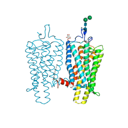
Night blindness causing G90D rhodopsin in the active conformation
Descriptor: ACETATE ION, PALMITIC ACID, RHODOPSIN, ...
Authors:Singhal, A, Ostermaier, M.K, Vishnivetskiy, S.A, Panneels, V, Homan, K.T, Tesmer, J.J.G, Veprintsev, D, Deupi, X, Gurevich, V.V, Schertler, G.F.X, Standfuss, J.
Deposit date:2013-03-12
Release date:2013-04-24
Last modified:2023-12-20
Method:X-RAY DIFFRACTION (3.3 Å)
Cite:Insights Into Congenital Stationary Night Blindness Based on the Structure of G90D Rhodopsin.
Embo Rep., 14, 2013
4BEY
DownloadVisualize
BU of 4bey by Molmil
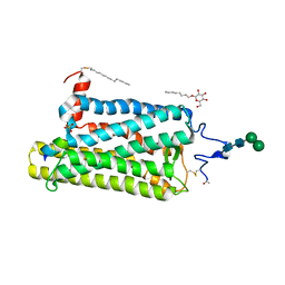
Night blindness causing G90D rhodopsin in complex with GaCT2 peptide
Descriptor: ACETATE ION, Guanine nucleotide-binding protein G(t) subunit alpha-1, PALMITIC ACID, ...
Authors:Singhal, A, Ostermaier, M.K, Vishnivetskiy, S.A, Panneels, V, Homan, K.T, Tesmer, J.J.G, Veprintsev, D, Deupi, X, Gurevich, V.V, Schertler, G.F.X, Standfuss, J.
Deposit date:2013-03-12
Release date:2013-05-08
Last modified:2023-12-20
Method:X-RAY DIFFRACTION (2.9 Å)
Cite:Insights Into Congenital Stationary Night Blindness Based on the Structure of G90D Rhodopsin.
Embo Rep., 14, 2013
2ACX
DownloadVisualize
BU of 2acx by Molmil
Crystal Structure of G protein coupled receptor kinase 6 bound to AMPPNP
Descriptor: G protein-coupled receptor kinase 6, MAGNESIUM ION, PHOSPHATE ION, ...
Authors:Lodowski, D.T, Tesmer, V.M, Benovic, J.L, Tesmer, J.J.
Deposit date:2005-07-19
Release date:2006-04-25
Last modified:2023-08-23
Method:X-RAY DIFFRACTION (2.6 Å)
Cite:The Structure of G Protein-coupled Receptor Kinase (GRK)-6 Defines a Second Lineage of GRKs.
J.Biol.Chem., 281, 2006
3V5W
DownloadVisualize
BU of 3v5w by Molmil
Human G Protein-Coupled Receptor Kinase 2 in Complex with Soluble Gbetagamma Subunits and Paroxetine
Descriptor: G-protein coupled receptor kinase 2, Guanine nucleotide-binding protein G(I)/G(S)/G(O) subunit gamma-2, Guanine nucleotide-binding protein G(I)/G(S)/G(T) subunit beta-1, ...
Authors:Thal, D.M, Tesmer, J.J.G.
Deposit date:2011-12-17
Release date:2012-09-26
Last modified:2023-09-13
Method:X-RAY DIFFRACTION (2.07 Å)
Cite:Paroxetine is a direct inhibitor of g protein-coupled receptor kinase 2 and increases myocardial contractility.
Acs Chem.Biol., 7, 2012

223166

数据于2024-07-31公开中

PDB statisticsPDBj update infoContact PDBjnumon