Loading
PDBj
MenuPDBj@FacebookPDBj@TwitterPDBj@YouTubewwPDB FoundationwwPDB
RCSB PDBPDBeBMRBAdv. SearchSearch help
Search by PDB author
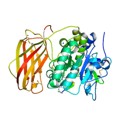
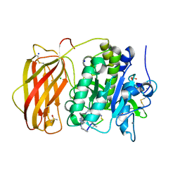
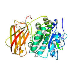
5JXH
DownloadVisualize
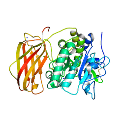
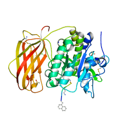
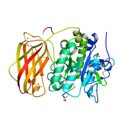
BU of 5jxh by Molmil
Structure the proprotein convertase furin in complex with meta-guanidinomethyl-Phac-RVR-Amba at 2.0 Angstrom resolution.
Descriptor: 2UC-ARG-VAL-ARG-00S, CALCIUM ION, CHLORIDE ION, ...
Authors:Dahms, S.O, Arciniega, M, Steinmetzer, T, Huber, R, Than, M.E.
Deposit date:2016-05-13
Release date:2016-10-05
Last modified:2024-07-10
Method:X-RAY DIFFRACTION (2 Å)
Cite:Structure of the unliganded form of the proprotein convertase furin suggests activation by a substrate-induced mechanism.
Proc.Natl.Acad.Sci.USA, 113, 2016
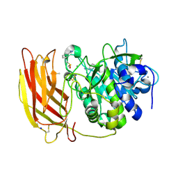
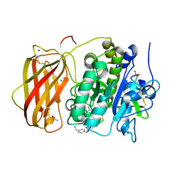
5JXI
DownloadVisualize
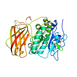
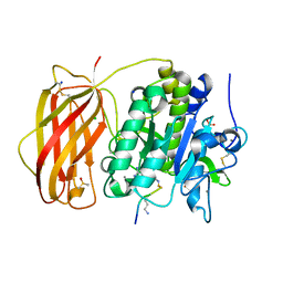
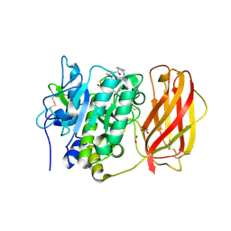
BU of 5jxi by Molmil
Structure of the unliganded form of the proprotein convertase furin in presence of EDTA.
Descriptor: CALCIUM ION, CHLORIDE ION, Furin, ...
Authors:Dahms, S.O, Arciniega, M, Steinmetzer, T, Huber, R, Than, M.E.
Deposit date:2016-05-13
Release date:2016-10-05
Last modified:2024-01-10
Method:X-RAY DIFFRACTION (2 Å)
Cite:Structure of the unliganded form of the proprotein convertase furin suggests activation by a substrate-induced mechanism.
Proc.Natl.Acad.Sci.USA, 113, 2016
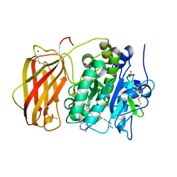
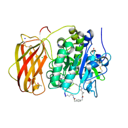
5JXG
DownloadVisualize
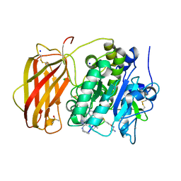
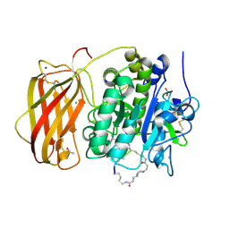
BU of 5jxg by Molmil
Structure of the unliganded form of the proprotein convertase furin.
Descriptor: CALCIUM ION, CHLORIDE ION, Furin, ...
Authors:Dahms, S.O, Arciniega, M, Steinmetzer, T, Huber, R, Than, M.E.
Deposit date:2016-05-13
Release date:2016-10-05
Last modified:2024-01-10
Method:X-RAY DIFFRACTION (1.8 Å)
Cite:Structure of the unliganded form of the proprotein convertase furin suggests activation by a substrate-induced mechanism.
Proc.Natl.Acad.Sci.USA, 113, 2016
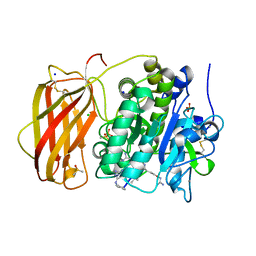
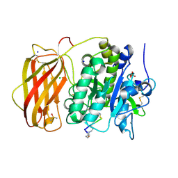
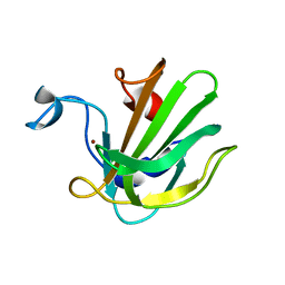
8OYH
DownloadVisualize
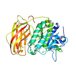
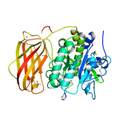
BU of 8oyh by Molmil
X-ray structure of furin (PCSK3) in complex with Guanidinomethyl-Phac-Can-Tle-Can-6-(aminomethyl)-3-amino-isoindol
Descriptor: CALCIUM ION, CHLORIDE ION, DIMETHYL SULFOXIDE, ...
Authors:Dahms, S.O, Brandstetter, H.
Deposit date:2023-05-04
Release date:2024-03-13
Last modified:2024-05-15
Method:X-RAY DIFFRACTION (1.8 Å)
Cite:Fragment-Based Design, Synthesis, and Characterization of Aminoisoindole-Derived Furin Inhibitors.
Chemmedchem, 19, 2024
8B4W
DownloadVisualize
BU of 8b4w by Molmil
X-ray structure of furin (PCSK3) in complex with 1H-isoindol-3-amine
Descriptor: 1H-isoindol-3-amine, 2-acetamido-2-deoxy-beta-D-glucopyranose, CALCIUM ION, ...
Authors:Dahms, S.O, Brandstetter, H.
Deposit date:2022-09-21
Release date:2023-10-04
Last modified:2024-05-15
Method:X-RAY DIFFRACTION (1.6 Å)
Cite:Fragment-Based Design, Synthesis, and Characterization of Aminoisoindole-Derived Furin Inhibitors.
Chemmedchem, 19, 2024
8B4X
DownloadVisualize
BU of 8b4x by Molmil
X-ray structure of furin (PCSK3) in complex with Guanidinomethyl-Phac-R-Tle-K-6-(aminomethyl)-3-amino-isoindol
Descriptor: CALCIUM ION, CHLORIDE ION, DIMETHYL SULFOXIDE, ...
Authors:Dahms, S.O, Brandstetter, H.
Deposit date:2022-09-21
Release date:2023-10-04
Last modified:2024-05-15
Method:X-RAY DIFFRACTION (1.6 Å)
Cite:Fragment-Based Design, Synthesis, and Characterization of Aminoisoindole-Derived Furin Inhibitors.
Chemmedchem, 19, 2024
8B4V
DownloadVisualize
BU of 8b4v by Molmil
X-ray structure of furin (PCSK3) in complex with benzamidine
Descriptor: 2-acetamido-2-deoxy-beta-D-glucopyranose, BENZAMIDINE, CALCIUM ION, ...
Authors:Dahms, S.O, Brandstetter, H.
Deposit date:2022-09-21
Release date:2023-10-04
Last modified:2024-05-15
Method:X-RAY DIFFRACTION (1.6 Å)
Cite:Fragment-Based Design, Synthesis, and Characterization of Aminoisoindole-Derived Furin Inhibitors.
Chemmedchem, 19, 2024
6YD7
DownloadVisualize
BU of 6yd7 by Molmil
X-ray structure of furin in complex with the canavanine-based inhibitor 4-guanidinomethyl-phenylacetyl-Arg-Tle-Canavanine-Amba
Descriptor: 4-guanidinomethyl-phenylacetyl-Arg-Tle-Canavanine-Amba, CALCIUM ION, CHLORIDE ION, ...
Authors:Dahms, S.O.
Deposit date:2020-03-20
Release date:2021-02-17
Last modified:2024-01-31
Method:X-RAY DIFFRACTION (1.8 Å)
Cite:The Basicity Makes the Difference: Improved Canavanine-Derived Inhibitors of the Proprotein Convertase Furin.
Acs Med.Chem.Lett., 12, 2021
6YD4
DownloadVisualize
BU of 6yd4 by Molmil
X-ray structure of furin in complex with the canavanine-based inhibitor 4-guanidinomethyl-phenylacetyl-Canavanine-Tle-Canavanine-Amba
Descriptor: 4-guanidinomethyl-phenylacetyl-Canavanine-Tle-Canavanine-Amba, CALCIUM ION, CHLORIDE ION, ...
Authors:Dahms, S.O.
Deposit date:2020-03-20
Release date:2021-02-17
Last modified:2024-01-31
Method:X-RAY DIFFRACTION (1.7 Å)
Cite:The Basicity Makes the Difference: Improved Canavanine-Derived Inhibitors of the Proprotein Convertase Furin.
Acs Med.Chem.Lett., 12, 2021
6YD3
DownloadVisualize
BU of 6yd3 by Molmil
X-ray structure of furin in complex with the canavanine derived inhibitor 4-guanidinomethyl-phenylacetyl-Canavanine-Tle-Arg-Amba
Descriptor: 4-guanidinomethyl-phenylacetyl-Canavanine-Tle-Arg-Amba, CALCIUM ION, CHLORIDE ION, ...
Authors:Dahms, S.O.
Deposit date:2020-03-20
Release date:2021-02-17
Last modified:2024-01-31
Method:X-RAY DIFFRACTION (2 Å)
Cite:The Basicity Makes the Difference: Improved Canavanine-Derived Inhibitors of the Proprotein Convertase Furin.
Acs Med.Chem.Lett., 12, 2021
6YD2
DownloadVisualize
BU of 6yd2 by Molmil
X-ray structure of furin in complex with the canavanine-based inhibitor 4-aminomethyl-phenylacetyl-canavanine-Tle-Arg-Amba
Descriptor: 4-aminomethyl-phenylacetyl-canavanine-Tle-Arg-Amba, CALCIUM ION, CHLORIDE ION, ...
Authors:Dahms, S.O.
Deposit date:2020-03-20
Release date:2021-02-17
Last modified:2024-01-31
Method:X-RAY DIFFRACTION (1.8 Å)
Cite:The Basicity Makes the Difference: Improved Canavanine-Derived Inhibitors of the Proprotein Convertase Furin.
Acs Med.Chem.Lett., 12, 2021
6HZC
DownloadVisualize
BU of 6hzc by Molmil
X-ray structure of furin in complex with the cyclic inhibitor c[glutaryl-BVK-Lys-Arg-Arg-Tle-Lys]-4-Amba
Descriptor: 2-[4-(aminomethyl)phenyl]ethanoic acid, CALCIUM ION, CHLORIDE ION, ...
Authors:Dahms, S.O.
Deposit date:2018-10-23
Release date:2019-02-06
Last modified:2024-01-24
Method:X-RAY DIFFRACTION (1.9 Å)
Cite:Design, Synthesis, and Characterization of Macrocyclic Inhibitors of the Proprotein Convertase Furin.
Chemmedchem, 14, 2019
6HZB
DownloadVisualize
BU of 6hzb by Molmil
X-ray structure of furin in complex with the cyclic inhibitor c[glutaryl-Arg-Arg-Lys]-Lys-4-Amba
Descriptor: ARG-ARG-LYS-LYS-00S, CALCIUM ION, CHLORIDE ION, ...
Authors:Dahms, S.O.
Deposit date:2018-10-23
Release date:2019-02-06
Last modified:2024-01-24
Method:X-RAY DIFFRACTION (1.9 Å)
Cite:Design, Synthesis, and Characterization of Macrocyclic Inhibitors of the Proprotein Convertase Furin.
Chemmedchem, 14, 2019
6HLD
DownloadVisualize
BU of 6hld by Molmil
X-ray structure of furin in complex with the cyclic peptide c[succinyl-Phe-2-Nal-(Arg)3-Lys]-Lys-4-Amba
Descriptor: ALN-ARG-ARG-ARG-SLL-LYS-00S, CALCIUM ION, CHLORIDE ION, ...
Authors:Dahms, S.O.
Deposit date:2018-09-11
Release date:2019-02-06
Last modified:2024-01-24
Method:X-RAY DIFFRACTION (2.1 Å)
Cite:Design, Synthesis, and Characterization of Macrocyclic Inhibitors of the Proprotein Convertase Furin.
Chemmedchem, 14, 2019
6HLB
DownloadVisualize
BU of 6hlb by Molmil
X-ray structure of furin in complex with the cyclic peptide c[succinyl-Phe-2-Nal-(Arg)4-Lys]-Arg-4-Amba
Descriptor: CALCIUM ION, CHLORIDE ION, DIMETHYL SULFOXIDE, ...
Authors:Dahms, S.O.
Deposit date:2018-09-11
Release date:2019-02-06
Last modified:2024-01-24
Method:X-RAY DIFFRACTION (2 Å)
Cite:Design, Synthesis, and Characterization of Macrocyclic Inhibitors of the Proprotein Convertase Furin.
Chemmedchem, 14, 2019
6HLE
DownloadVisualize
BU of 6hle by Molmil
X-ray structure of furin in complex with the P6-P2-cyclized peptide H-Lys-Arg-Arg-Tle-Lys-4-Amba
Descriptor: 3-[4-(3-oxidanylidenepropyl)piperazin-1-yl]propanal, CALCIUM ION, CHLORIDE ION, ...
Authors:Dahms, S.O.
Deposit date:2018-09-11
Release date:2019-02-06
Last modified:2024-01-24
Method:X-RAY DIFFRACTION (1.994 Å)
Cite:Design, Synthesis, and Characterization of Macrocyclic Inhibitors of the Proprotein Convertase Furin.
Chemmedchem, 14, 2019
6HZD
DownloadVisualize
BU of 6hzd by Molmil
X-ray structure of furin in complex with the cyclic inhibitor c[glutaryl-Arg-Arg-Arg-Lys]-Arg-4-Amba
Descriptor: ARG-ARG-ARG-LYS-ARG-00S, CALCIUM ION, CHLORIDE ION, ...
Authors:Dahms, S.O.
Deposit date:2018-10-23
Release date:2019-02-06
Last modified:2024-01-24
Method:X-RAY DIFFRACTION (1.9 Å)
Cite:Design, Synthesis, and Characterization of Macrocyclic Inhibitors of the Proprotein Convertase Furin.
Chemmedchem, 14, 2019
6HZA
DownloadVisualize
BU of 6hza by Molmil
X-ray structure of furin in complex with the cyclic peptide c[glutaryl-Arg-Arg-Lys]-Arg-4-Amba
Descriptor: ARG-ARG-LYS-ARG-00S, CALCIUM ION, CHLORIDE ION, ...
Authors:Dahms, S.O.
Deposit date:2018-10-23
Release date:2019-02-06
Last modified:2024-01-24
Method:X-RAY DIFFRACTION (1.9 Å)
Cite:Design, Synthesis, and Characterization of Macrocyclic Inhibitors of the Proprotein Convertase Furin.
Chemmedchem, 14, 2019
4RYD
DownloadVisualize
BU of 4ryd by Molmil
X-ray structure of human furin in complex with the competitive inhibitor para-guanidinomethyl-Phac-R-Tle-R-Amba
Descriptor: CALCIUM ION, FORMIC ACID, Furin, ...
Authors:Dahms, S.O, Than, M.E.
Deposit date:2014-12-15
Release date:2015-05-27
Last modified:2024-07-10
Method:X-RAY DIFFRACTION (2.15 Å)
Cite:Novel Furin Inhibitors with Potent Anti-infectious Activity.
Chemmedchem, 10, 2015
4LXC
DownloadVisualize
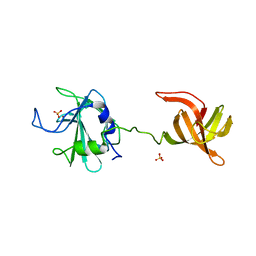
BU of 4lxc by Molmil
The antimicrobial peptidase lysostaphin from Staphylococcus simulans
Descriptor: Lysostaphin, SULFATE ION, ZINC ION
Authors:Sabala, I, Jagielska, E, Bardelang, P.T, Czapinska, H, Dahms, S.O, Sharpe, J.A, James, R, Than, M.E, Thomas, N.R, Bochtler, M.
Deposit date:2013-07-29
Release date:2014-07-09
Last modified:2023-09-20
Method:X-RAY DIFFRACTION (3.5 Å)
Cite:Crystal structure of the antimicrobial peptidase lysostaphin from Staphylococcus simulans.
Febs J., 281, 2014
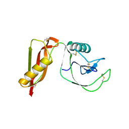
3QO4
DownloadVisualize
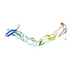
BU of 3qo4 by Molmil
The Crystal Structure of Death Receptor 6
Descriptor: ACETATE ION, SULFATE ION, Tumor necrosis factor receptor superfamily member 21
Authors:Kuester, M, Kemmerzehl, S, Dahms, S.O, Roeser, D, Than, M.E.
Deposit date:2011-02-09
Release date:2011-05-18
Last modified:2012-02-29
Method:X-RAY DIFFRACTION (2.2 Å)
Cite:The crystal structure of death receptor 6 (DR6): a potential receptor of the amyloid precursor protein (APP).
J.Mol.Biol., 409, 2011
4QPB
DownloadVisualize
BU of 4qpb by Molmil
Catalytic domain of the antimicrobial peptidase lysostaphin from Staphylococcus simulans crystallized in the absence of phosphate
Descriptor: 1,2-ETHANEDIOL, Lysostaphin, ZINC ION
Authors:Sabala, I, Jagielska, E, Bardelang, P.T, Czapinska, H, Dahms, S.O, Sharpe, J.A, James, R, Than, M.E, Thomas, N.R, Bochtler, M.
Deposit date:2014-06-22
Release date:2014-07-09
Last modified:2023-09-20
Method:X-RAY DIFFRACTION (1.78 Å)
Cite:Crystal structure of the antimicrobial peptidase lysostaphin from Staphylococcus simulans.
Febs J., 281, 2014
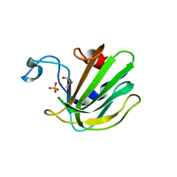
4QP5
DownloadVisualize
BU of 4qp5 by Molmil
Catalytic domain of the antimicrobial peptidase lysostaphin from Staphylococcus simulans crystallized in the presence of phosphate
Descriptor: GLYCEROL, Lysostaphin, PHOSPHATE ION, ...
Authors:Sabala, I, Jagielska, E, Bardelang, P.T, Czapinska, H, Dahms, S.O, Sharpe, J.A, James, R, Than, M.E, Thomas, N.R, Bochtler, M.
Deposit date:2014-06-22
Release date:2014-07-16
Last modified:2023-09-20
Method:X-RAY DIFFRACTION (1.26 Å)
Cite:Crystal structure of the antimicrobial peptidase lysostaphin from Staphylococcus simulans.
Febs J., 281, 2014
4PWQ
DownloadVisualize
BU of 4pwq by Molmil
HIGH-RESOLUTION CRYSTAL STRUCTURE OF THE E1-DOMAIN of THE AMYLOID PRECURSOR PROTEIN
Descriptor: Amyloid beta A4 protein
Authors:Hoefgen, S, Dahms, S.O, Than, M.E.
Deposit date:2014-03-21
Release date:2015-02-04
Method:X-RAY DIFFRACTION (1.4 Å)
Cite:The Amyloid Precursor Protein Shows a pH-Dependent Conformational Switch in Its E1 Domain.
J.Mol.Biol., 427, 2015
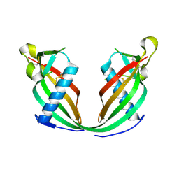
6FK0
DownloadVisualize
BU of 6fk0 by Molmil
Xray structure of domain-swapped cystatin E dimer
Descriptor: Cystatin-M
Authors:Dall, E, Brandstetter, H.
Deposit date:2018-01-23
Release date:2018-07-11
Last modified:2024-01-17
Method:X-RAY DIFFRACTION (2.9 Å)
Cite:Structural and functional analysis of cystatin E reveals enzymologically relevant dimer and amyloid fibril states.
J. Biol. Chem., 293, 2018

224572

数据于2024-09-04公开中

PDB statisticsPDBj update infoContact PDBjnumon