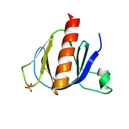
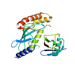
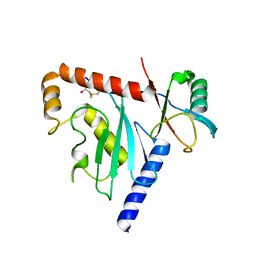
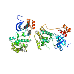
3RZ3
 
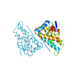
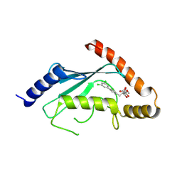 | | Human Cdc34 E2 in complex with CC0651 inhibitor | | 分子名称: | 4,5-dideoxy-5-(3',5'-dichlorobiphenyl-4-yl)-4-[(methoxyacetyl)amino]-L-arabinonic acid, Ubiquitin-conjugating enzyme E2 R1 | | 著者 | Ceccarelli, D.F, Webb, D.R, Sicheri, F. | | 登録日 | 2011-05-11 | | 公開日 | 2011-07-06 | | 最終更新日 | 2023-09-13 | | 実験手法 | X-RAY DIFFRACTION (2.3 Å) | | 主引用文献 | An allosteric inhibitor of the human cdc34 ubiquitin conjugating enzyme
Cell(Cambridge,Mass.), 145, 2011
|
|
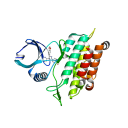
6DRM
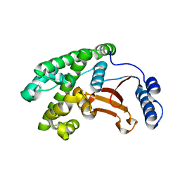 | | OTU domain of Fam105A | | 分子名称: | Inactive ubiquitin thioesterase FAM105A | | 著者 | Ceccarelli, D.F, Sicheri, F, Cordes, S. | | 登録日 | 2018-06-12 | | 公開日 | 2019-05-08 | | 最終更新日 | 2023-10-11 | | 実験手法 | X-RAY DIFFRACTION (2.06 Å) | | 主引用文献 | FAM105A/OTULINL Is a Pseudodeubiquitinase of the OTU-Class that Localizes to the ER Membrane.
Structure, 27, 2019
|
|
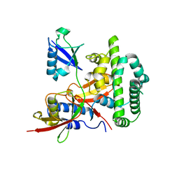
2AEH
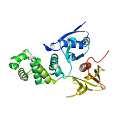 | | Focal adhesion kinase 1 | | 分子名称: | Focal adhesion kinase 1 | | 著者 | Ceccarelli, D.F, Song, H.K, Poy, F, Schaller, M.D, Eck, M.J. | | 登録日 | 2005-07-22 | | 公開日 | 2005-10-18 | | 最終更新日 | 2024-02-14 | | 実験手法 | X-RAY DIFFRACTION (2.53 Å) | | 主引用文献 | Crystal Structure of the FERM Domain of Focal Adhesion Kinase
J.Biol.Chem., 281, 2006
|
|
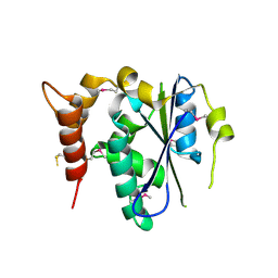
2AL6
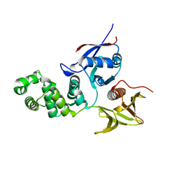 | | FERM domain of Focal Adhesion Kinase | | 分子名称: | Focal adhesion kinase 1 | | 著者 | Ceccarelli, D.F, Song, H.K, Poy, F, Schaller, M.D, Eck, M.J. | | 登録日 | 2005-08-04 | | 公開日 | 2005-10-18 | | 最終更新日 | 2023-08-23 | | 実験手法 | X-RAY DIFFRACTION (2.35 Å) | | 主引用文献 | Crystal Structure of the FERM Domain of Focal Adhesion Kinase
J.Biol.Chem., 281, 2006
|
|
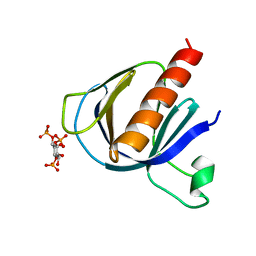
2P0F
 | | ArhGAP9 PH domain in complex with Ins(1,3,5)P3 | | 分子名称: | PHOSPHATE ION, Rho GTPase-activating protein 9 | | 著者 | Ceccarelli, D.F.J, Blasutig, I, Goudreault, M, Ruston, J, Pawson, T, Sicheri, F. | | 登録日 | 2007-02-28 | | 公開日 | 2007-03-27 | | 最終更新日 | 2023-08-30 | | 実験手法 | X-RAY DIFFRACTION (1.91 Å) | | 主引用文献 | Non-canonical Interaction of Phosphoinositides with Pleckstrin Homology Domains of Tiam1 and ArhGAP9.
J.Biol.Chem., 282, 2007
|
|
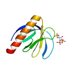
2P0H
 | | ArhGAP9 PH domain in complex with Ins(1,3,4)P3 | | 分子名称: | (1S,3S,4S)-1,3,4-TRIPHOSPHO-MYO-INOSITOL, Rho GTPase-activating protein 9 | | 著者 | Ceccarelli, D.F.J, Blasutig, I, Goudreault, M, Ruston, J, Pawson, T, Sicheri, F. | | 登録日 | 2007-02-28 | | 公開日 | 2007-03-27 | | 最終更新日 | 2023-08-30 | | 実験手法 | X-RAY DIFFRACTION (1.9 Å) | | 主引用文献 | Non-canonical Interaction of Phosphoinositides with Pleckstrin Homology Domains of Tiam1 and ArhGAP9.
J.Biol.Chem., 282, 2007
|
|
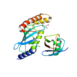
2P0D
 | | ArhGAP9 PH domain in complex with Ins(1,4,5)P3 | | 分子名称: | D-MYO-INOSITOL-1,4,5-TRIPHOSPHATE, Rho GTPase-activating protein 9 | | 著者 | Ceccarelli, D.F.J, Blasutig, I, Goudreault, M, Ruston, J, Pawson, T, Sicheri, F. | | 登録日 | 2007-02-28 | | 公開日 | 2007-03-27 | | 最終更新日 | 2024-02-21 | | 実験手法 | X-RAY DIFFRACTION (1.811 Å) | | 主引用文献 | Non-canonical Interaction of Phosphoinositides with Pleckstrin Homology Domains of Tiam1 and ArhGAP9.
J.Biol.Chem., 282, 2007
|
|
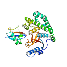
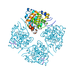
4MDK
 | | Cdc34-ubiquitin-CC0651 complex | | 分子名称: | 4,5-dideoxy-5-(3',5'-dichlorobiphenyl-4-yl)-4-[(methoxyacetyl)amino]-L-arabinonic acid, Ubiquitin, Ubiquitin-conjugating enzyme E2 R1 | | 著者 | Ceccarelli, D.F, Orlicky, S, Tyers, M, Sicheri, F. | | 登録日 | 2013-08-22 | | 公開日 | 2013-12-11 | | 最終更新日 | 2023-09-20 | | 実験手法 | X-RAY DIFFRACTION (2.6095 Å) | | 主引用文献 | E2 enzyme inhibition by stabilization of a low-affinity interface with ubiquitin.
Nat.Chem.Biol., 10, 2014
|
|
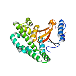
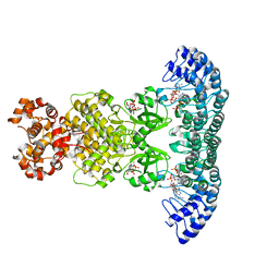
7M2K
 | | CDC34A-Ubiquitin-2ab inhibitor complex | | 分子名称: | 4-[(3',5'-dichloro[1,1'-biphenyl]-4-yl)methyl]-N-ethyl-1-(methoxyacetyl)piperidine-4-carboxamide, Ubiquitin, Ubiquitin-conjugating enzyme E2 R1 | | 著者 | Ceccarelli, D.F, St-Cyr, D, Tyers, M, Sicheri, F. | | 登録日 | 2021-03-16 | | 公開日 | 2021-11-03 | | 最終更新日 | 2023-10-18 | | 実験手法 | X-RAY DIFFRACTION (2.47 Å) | | 主引用文献 | Identification and optimization of molecular glue compounds that inhibit a noncovalent E2 enzyme-ubiquitin complex.
Sci Adv, 7, 2021
|
|
4KSK
 | |
4KSJ
 | |
6D4P
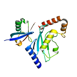 | | Ube2D1 in complex with ubiquitin variant Ubv.D1.1 | | 分子名称: | Ubiquitin Variant Ubv.D1.1, Ubiquitin-conjugating enzyme E2 D1 | | 著者 | Ceccarelli, D.F, Garg, P, Sidhu, S, Sicheri, F. | | 登録日 | 2018-04-18 | | 公開日 | 2019-07-17 | | 最終更新日 | 2023-10-04 | | 実験手法 | X-RAY DIFFRACTION (2.11 Å) | | 主引用文献 | Structural and Functional Analysis of Ubiquitin-based Inhibitors That Target the Backsides of E2 Enzymes.
J.Mol.Biol., 432, 2020
|
|
6D68
 | | Ube2G1 in complex with ubiquitin variant Ubv.G1.1 | | 分子名称: | Ubiquitin-conjugating enzyme E2 G1, Ubv.G1.1 | | 著者 | Ceccarelli, D.F, Garg, P, Sidhu, S, Sicheri, F. | | 登録日 | 2018-04-20 | | 公開日 | 2019-07-17 | | 最終更新日 | 2023-10-04 | | 実験手法 | X-RAY DIFFRACTION (2.36 Å) | | 主引用文献 | Structural and Functional Analysis of Ubiquitin-based Inhibitors That Target the Backsides of E2 Enzymes.
J.Mol.Biol., 432, 2020
|
|
6D6I
 | | Ube2V1 in complex with ubiquitin variant Ubv.V1.1 and Ube2N/Ubc13 | | 分子名称: | Ubiquitin-conjugating enzyme E2 N, Ubiquitin-conjugating enzyme E2 variant 1, Ubv.V1.1 | | 著者 | Ceccarelli, D.F, Garg, P, Keszei, A, Sidhu, S, Sicheri, F. | | 登録日 | 2018-04-21 | | 公開日 | 2019-07-17 | | 最終更新日 | 2023-10-04 | | 実験手法 | X-RAY DIFFRACTION (2.551 Å) | | 主引用文献 | Structural and Functional Analysis of Ubiquitin-based Inhibitors That Target the Backsides of E2 Enzymes.
J.Mol.Biol., 432, 2020
|
|
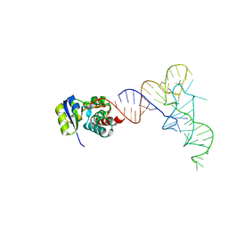
7KJU
 | | Cgi121-tRNA complex | | 分子名称: | MAGNESIUM ION, RNA (75-MER) | | 著者 | Ceccarelli, D.F, Beenstock, J, Wan, L.C.K, Sicheri, F. | | 登録日 | 2020-10-26 | | 公開日 | 2020-12-02 | | 最終更新日 | 2023-10-18 | | 実験手法 | X-RAY DIFFRACTION (3.102 Å) | | 主引用文献 | A substrate binding model for the KEOPS tRNA modifying complex.
Nat Commun, 11, 2020
|
|
7KJT
 | | KEOPS tRNA modifying sub-complex of archaeal Cgi121 and tRNA | | 分子名称: | RNA (70-MER), Regulatory protein Cgi121 | | 著者 | Ceccarelli, D.F, Beenstock, J, Mao, D.Y.L, Sicheri, F. | | 登録日 | 2020-10-26 | | 公開日 | 2020-12-02 | | 最終更新日 | 2023-10-18 | | 実験手法 | X-RAY DIFFRACTION (3.34 Å) | | 主引用文献 | A substrate binding model for the KEOPS tRNA modifying complex.
Nat Commun, 11, 2020
|
|
2QNJ
 | |
3TZM
 | | TGF-beta Receptor type 1 in complex with SB431542 | | 分子名称: | 4-[5-(1,3-benzodioxol-5-yl)-4-(pyridin-2-yl)-1H-imidazol-2-yl]benzamide, TGF-beta receptor type-1 | | 著者 | Ogunjimi, A.A, Zeqiraj, E, Ceccarelli, D.F, Sicheri, F. | | 登録日 | 2011-09-27 | | 公開日 | 2012-05-23 | | 最終更新日 | 2023-09-13 | | 実験手法 | X-RAY DIFFRACTION (1.7 Å) | | 主引用文献 | Structural Basis for Specificity of TGFbeta Family Receptor Small Molecule Inhibitors
Cell Signal, 24, 2012
|
|
4KSL
 | |
3ENP
 | | Crystal structure of human cgi121 | | 分子名称: | TP53RK-binding protein | | 著者 | Haffani, Y.Z, Ceccarelli, D.F, Neculai, D, Mao, D.Y, Sicheri, F. | | 登録日 | 2008-09-25 | | 公開日 | 2008-11-04 | | 最終更新日 | 2011-07-13 | | 実験手法 | X-RAY DIFFRACTION (2.48 Å) | | 主引用文献 | Atomic structure of the KEOPS complex: an ancient protein kinase-containing molecular machine.
Mol.Cell, 32, 2008
|
|
2HJN
 | |
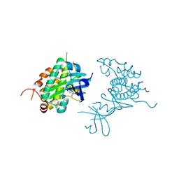
7LUO
 | | N-terminus of Skp2 bound to Cyclin A | | 分子名称: | S-phase kinase-associated protein 2,Cyclin-A2, Skp2 Motif 1 uncharacterized fragment 1, Skp2 Motif 1 uncharacterized fragment 2 | | 著者 | Kelso, S, Ceccarelli, D.F, Sicheri, F. | | 登録日 | 2021-02-22 | | 公開日 | 2021-05-12 | | 最終更新日 | 2023-10-18 | | 実験手法 | X-RAY DIFFRACTION (3.17 Å) | | 主引用文献 | Bipartite binding of the N terminus of Skp2 to cyclin A.
Structure, 29, 2021
|
|
4O1P
 | | Crystal Structure of RNase L in complex with 2-5A and AMP-PNP | | 分子名称: | MAGNESIUM ION, PHOSPHOAMINOPHOSPHONIC ACID-ADENYLATE ESTER, Ribonuclease L, ... | | 著者 | Huang, H, Zeqiraj, E, Ceccarelli, D.F, Sicheri, F. | | 登録日 | 2013-12-16 | | 公開日 | 2014-02-05 | | 最終更新日 | 2023-09-20 | | 実験手法 | X-RAY DIFFRACTION (2.5 Å) | | 主引用文献 | Dimeric structure of pseudokinase RNase L bound to 2-5A reveals a basis for interferon-induced antiviral activity.
Mol.Cell, 53, 2014
|
|
4O1O
 | | Crystal Structure of RNase L in complex with 2-5A | | 分子名称: | Ribonuclease L, [[(2R,3R,4R,5R)-5-(6-aminopurin-9-yl)-4-[[(2R,3R,4R,5R)-5-(6-aminopurin-9-yl)-4-[[(2R,3S,4R,5R)-5-(6-aminopurin-9-yl)-3,4-dihydroxy-oxolan-2-yl]methoxy-hydroxy-phosphoryl]oxy-3-hydroxy-oxolan-2-yl]methoxy-hydroxy-phosphoryl]oxy-3-hydroxy-oxolan-2-yl]methoxy-hydroxy-phosphoryl] phosphono hydrogen phosphate | | 著者 | Huang, H, Zeqiraj, E, Ceccarelli, D.F, Sicheri, F. | | 登録日 | 2013-12-16 | | 公開日 | 2014-02-05 | | 最終更新日 | 2023-09-20 | | 実験手法 | X-RAY DIFFRACTION (3.27 Å) | | 主引用文献 | Dimeric structure of pseudokinase RNase L bound to 2-5A reveals a basis for interferon-induced antiviral activity.
Mol.Cell, 53, 2014
|
|
4PDS
 | | Crystal structure of Rad53 kinase domain and SCD2 in complex with AMPPNP | | 分子名称: | PHOSPHOAMINOPHOSPHONIC ACID-ADENYLATE ESTER, Serine/threonine-protein kinase RAD53 | | 著者 | Ho, C.S, Wybenga-Groot, L.E, Ceccarelli, D.F, Sicheri, F. | | 登録日 | 2014-04-21 | | 公開日 | 2014-05-28 | | 最終更新日 | 2023-09-27 | | 実験手法 | X-RAY DIFFRACTION (2.9 Å) | | 主引用文献 | Structural basis of Rad53 kinase activation by dimerization and activation segment exchange.
Cell Signal., 26, 2014
|
|


