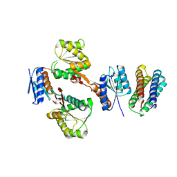
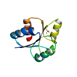
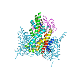
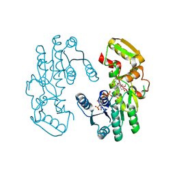
7D3B
 
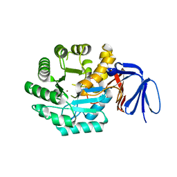
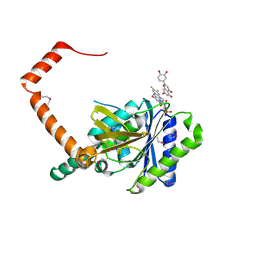 | | flavone reductase | | 分子名称: | 2-(3,4-dihydroxyphenyl)-5,7-dihydroxy-4H-chromen-4-one, Cd1, FLAVIN MONONUCLEOTIDE | | 著者 | Hong, S, Yang, G.H, Zhang, P. | | 登録日 | 2020-09-18 | | 公開日 | 2021-03-03 | | 実験手法 | X-RAY DIFFRACTION (2.25 Å) | | 主引用文献 | Discovery of an ene-reductase for initiating flavone and flavonol catabolism in gut bacteria.
Nat Commun, 12, 2021
|
|
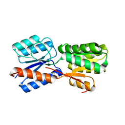
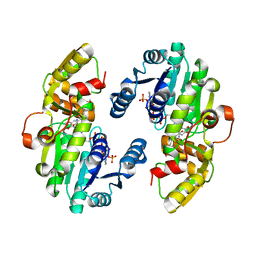
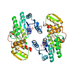
7D3A
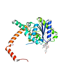 | | flavone reductase | | 分子名称: | 5,7-dihydroxy-2-(4-hydroxyphenyl)-4H-chromen-4-one, Cd1, FLAVIN MONONUCLEOTIDE | | 著者 | Hong, S, Yang, G.H, Zhang, P. | | 登録日 | 2020-09-18 | | 公開日 | 2021-03-03 | | 最終更新日 | 2021-09-29 | | 実験手法 | X-RAY DIFFRACTION (2.552 Å) | | 主引用文献 | Discovery of an ene-reductase for initiating flavone and flavonol catabolism in gut bacteria.
Nat Commun, 12, 2021
|
|
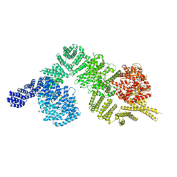
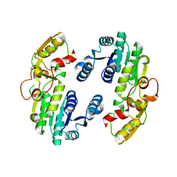
6BYR
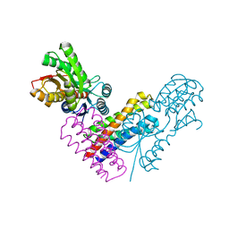
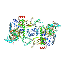 | | Structures of the PKA RI alpha holoenzyme with the FLHCC driver J-PKAc alpha or native PKAc alpha | | 分子名称: | ADENOSINE-5'-TRIPHOSPHATE, DnaJ homolog subfamily B member 1,cAMP-dependent protein kinase catalytic subunit alpha chimera, MAGNESIUM ION, ... | | 著者 | Cao, B, Lu, T.W, Martinez Fiesco, J.A, Tomasini, M, Fan, L, Simon, S.M, Taylor, S.S, Zhang, P. | | 登録日 | 2017-12-21 | | 公開日 | 2019-04-03 | | 最終更新日 | 2023-10-04 | | 実験手法 | X-RAY DIFFRACTION (3.661 Å) | | 主引用文献 | Structures of the PKA RI alpha Holoenzyme with the FLHCC Driver J-PKAc alpha or Wild-Type PKAc alpha.
Structure, 27, 2019
|
|
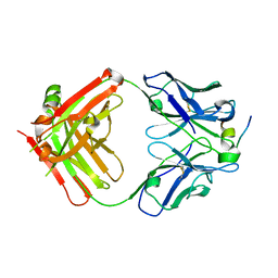
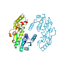
4PBV
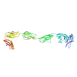
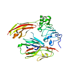 | | Crystal structure of chicken receptor protein tyrosine phosphatase sigma in complex with TrkC | | 分子名称: | 2-acetamido-2-deoxy-beta-D-glucopyranose, NT-3 growth factor receptor, Protein-tyrosine phosphatase CRYPalpha1 isoform, ... | | 著者 | Coles, C.H, Mitakidis, N, Zhang, P, Elegheert, J, Lu, W, Stoker, A.W, Nakagawa, T, Craig, A.M, Jones, E.Y, Aricescu, A.R. | | 登録日 | 2014-04-14 | | 公開日 | 2014-11-12 | | 最終更新日 | 2023-12-20 | | 実験手法 | X-RAY DIFFRACTION (2.5 Å) | | 主引用文献 | Structural basis for extracellular cis and trans RPTP sigma signal competition in synaptogenesis.
Nat Commun, 5, 2014
|
|
4PBX
 | | Crystal structure of the six N-terminal domains of human receptor protein tyrosine phosphatase sigma | | 分子名称: | 2-acetamido-2-deoxy-beta-D-glucopyranose, Receptor-type tyrosine-protein phosphatase S | | 著者 | Coles, C.H, Mitakidis, N, Zhang, P, Elegheert, J, Lu, W, Stoker, A.W, Nakagawa, T, Craig, A.M, Jones, E.Y, Aricescu, A.R. | | 登録日 | 2014-04-14 | | 公開日 | 2014-11-12 | | 最終更新日 | 2023-12-20 | | 実験手法 | X-RAY DIFFRACTION (3.15 Å) | | 主引用文献 | Structural basis for extracellular cis and trans RPTP sigma signal competition in synaptogenesis.
Nat Commun, 5, 2014
|
|
1XRT
 | | The Crystal Structure of a Novel, Latent Dihydroorotase from Aquifex Aeolicus at 1.7 A Resolution | | 分子名称: | Dihydroorotase, ZINC ION | | 著者 | Martin, P.D, Purcarea, C, Zhang, P, Vaishnav, A, Sadecki, S, Guy-Evans, H.I, Evans, D.R, Edwards, B.F. | | 登録日 | 2004-10-15 | | 公開日 | 2005-07-05 | | 最終更新日 | 2023-08-23 | | 実験手法 | X-RAY DIFFRACTION (1.609 Å) | | 主引用文献 | The crystal structure of a novel, latent dihydroorotase from Aquifex aeolicus at 1.7A resolution
J.Mol.Biol., 348, 2005
|
|
3P4F
 | | Structural and biochemical insights into MLL1 core complex assembly and regulation. | | 分子名称: | Histone-lysine N-methyltransferase MLL, Retinoblastoma-binding protein 5, WD repeat-containing protein 5 | | 著者 | Avdic, V, Zhang, P, Lanouette, S, Groulx, A, Tremblay, V, Brunzelle, J.B, Couture, J.-F. | | 登録日 | 2010-10-06 | | 公開日 | 2010-12-08 | | 最終更新日 | 2023-09-06 | | 実験手法 | X-RAY DIFFRACTION (2.35 Å) | | 主引用文献 | Structural and biochemical insights into MLL1 core complex assembly.
Structure, 19, 2011
|
|
5XSJ
 | | XylFII-LytSN complex | | 分子名称: | Periplasmic binding protein/LacI transcriptional regulator, Signal transduction histidine kinase, LytS, ... | | 著者 | Li, J.X, Wang, C.Y, Zhang, P. | | 登録日 | 2017-06-14 | | 公開日 | 2017-08-02 | | 最終更新日 | 2024-05-29 | | 実験手法 | X-RAY DIFFRACTION (2.202 Å) | | 主引用文献 | Molecular mechanism of environmental d-xylose perception by a XylFII-LytS complex in bacteria
Proc. Natl. Acad. Sci. U.S.A., 114, 2017
|
|
5XSD
 | | XylFII-LytSN complex mutant - D103A | | 分子名称: | Periplasmic binding protein/LacI transcriptional regulator, Signal transduction histidine kinase, LytS | | 著者 | Li, J.X, Wang, C.Y, Zhang, P. | | 登録日 | 2017-06-13 | | 公開日 | 2017-08-02 | | 最終更新日 | 2024-03-27 | | 実験手法 | X-RAY DIFFRACTION (2.5 Å) | | 主引用文献 | Molecular mechanism of environmental d-xylose perception by a XylFII-LytS complex in bacteria
Proc. Natl. Acad. Sci. U.S.A., 114, 2017
|
|
5XSS
 | | XylFII molecule | | 分子名称: | Periplasmic binding protein/LacI transcriptional regulator, beta-D-xylopyranose | | 著者 | Li, J.X, Wang, C.Y, Zhang, P. | | 登録日 | 2017-06-15 | | 公開日 | 2017-08-02 | | 最終更新日 | 2024-03-27 | | 実験手法 | X-RAY DIFFRACTION (2.093 Å) | | 主引用文献 | Molecular mechanism of environmental d-xylose perception by a XylFII-LytS complex in bacteria
Proc. Natl. Acad. Sci. U.S.A., 114, 2017
|
|
4HZL
 | |
6K9K
 | | Monomeric human ATM (Ataxia telangiectasia mutated) kinase | | 分子名称: | Serine-protein kinase ATM | | 著者 | Xiao, J, Liu, M, Qi, Y, Yuriy, C, Zhang, P, Xu, Y. | | 登録日 | 2019-06-16 | | 公開日 | 2019-12-25 | | 最終更新日 | 2024-03-27 | | 実験手法 | ELECTRON MICROSCOPY (7.82 Å) | | 主引用文献 | Structural insights into the activation of ATM kinase.
Cell Res., 29, 2019
|
|
7E1H
 | | crystal structure of RD-BEF | | 分子名称: | BERYLLIUM TRIFLUORIDE ION, DNA-binding response regulator, MAGNESIUM ION | | 著者 | Hong, S, Zhang, X, Zhang, P. | | 登録日 | 2021-02-01 | | 公開日 | 2022-02-09 | | 最終更新日 | 2023-11-29 | | 実験手法 | X-RAY DIFFRACTION (2.805 Å) | | 主引用文献 | Structural basis of phosphorylation-induced activation of the response regulator VbrR.
Acta Biochim.Biophys.Sin., 2023
|
|
7E1B
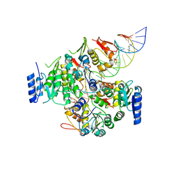 | | Crystal structure of VbrR-DNA complex | | 分子名称: | DNA (26-MER), DNA-binding response regulator | | 著者 | Hong, S, Zhang, X, Zhang, P. | | 登録日 | 2021-02-01 | | 公開日 | 2022-02-09 | | 最終更新日 | 2024-05-29 | | 実験手法 | X-RAY DIFFRACTION (4.587 Å) | | 主引用文献 | Structural basis of phosphorylation-induced activation of the response regulator VbrR.
Acta Biochim.Biophys.Sin., 2023
|
|
8A1S
 | | Structure of murine perforin-2 (Mpeg1) pore in twisted form | | 分子名称: | 2-acetamido-2-deoxy-beta-D-glucopyranose, Macrophage-expressed gene 1 protein | | 著者 | Yu, X, Ni, T, Zhang, P, Gilbert, R. | | 登録日 | 2022-06-02 | | 公開日 | 2022-07-20 | | 最終更新日 | 2023-10-18 | | 実験手法 | ELECTRON MICROSCOPY (4 Å) | | 主引用文献 | Cryo-EM structures of perforin-2 in isolation and assembled on a membrane suggest a mechanism for pore formation.
Embo J., 41, 2022
|
|
8A1D
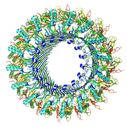 | | Structure of murine perforin-2 (Mpeg1) pore in ring form | | 分子名称: | 2-acetamido-2-deoxy-beta-D-glucopyranose, CYCLOHEXYL-HEXYL-BETA-D-MALTOSIDE, Macrophage-expressed gene 1 protein | | 著者 | Yu, X, Ni, T, Zhang, P, Gilbert, R. | | 登録日 | 2022-06-01 | | 公開日 | 2022-07-20 | | 最終更新日 | 2022-12-14 | | 実験手法 | ELECTRON MICROSCOPY (3 Å) | | 主引用文献 | Cryo-EM structures of perforin-2 in isolation and assembled on a membrane suggest a mechanism for pore formation.
Embo J., 41, 2022
|
|
7EGK
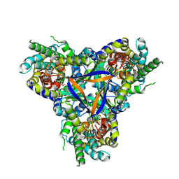 | | Bicarbonate transporter complex SbtA-SbtB bound to AMP | | 分子名称: | ADENOSINE MONOPHOSPHATE, Membrane-associated protein SbtB, SODIUM ION, ... | | 著者 | Fang, S, Huang, X, Zhang, X, Zhang, P. | | 登録日 | 2021-03-24 | | 公開日 | 2021-05-26 | | 最終更新日 | 2021-06-16 | | 実験手法 | ELECTRON MICROSCOPY (2.7 Å) | | 主引用文献 | Molecular mechanism underlying transport and allosteric inhibition of bicarbonate transporter SbtA.
Proc.Natl.Acad.Sci.USA, 118, 2021
|
|
7EGL
 | | Bicarbonate transporter complex SbtA-SbtB bound to HCO3- | | 分子名称: | BICARBONATE ION, Membrane-associated protein SbtB, SODIUM ION, ... | | 著者 | Fang, S, Huang, X, Zhang, X, Zhang, P. | | 登録日 | 2021-03-24 | | 公開日 | 2021-05-26 | | 最終更新日 | 2023-11-29 | | 実験手法 | X-RAY DIFFRACTION (3.2 Å) | | 主引用文献 | Molecular mechanism underlying transport and allosteric inhibition of bicarbonate transporter SbtA.
Proc.Natl.Acad.Sci.USA, 118, 2021
|
|
3S32
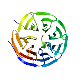
 | | Crystal structure of Ash2L N-terminal domain | | 分子名称: | Set1/Ash2 histone methyltransferase complex subunit ASH2, ZINC ION | | 著者 | Sarvan, S, Avdic, V, Tremblay, V, Chaturvedi, C.-P, Zhang, P, Lanouette, S, Blais, A, Brunzelle, J.S, Brand, M, Couture, J.-F. | | 登録日 | 2011-05-17 | | 公開日 | 2011-06-08 | | 最終更新日 | 2012-01-11 | | 実験手法 | X-RAY DIFFRACTION (2.45 Å) | | 主引用文献 | Crystal structure of the trithorax group protein ASH2L reveals a forkhead-like DNA binding domain.
Nat.Struct.Mol.Biol., 18, 2011
|
|
3PSL
 | | Fine-tuning the stimulation of MLL1 methyltransferase activity by a histone H3 based peptide mimetic | | 分子名称: | N-alpha acetylated form of histone H3, WD repeat-containing protein 5 | | 著者 | Avdic, V, Zhang, P, Lanouette, S, Voronova, A, Skerjanc, I, Couture, J.-F. | | 登録日 | 2010-12-01 | | 公開日 | 2010-12-22 | | 最終更新日 | 2023-09-06 | | 実験手法 | X-RAY DIFFRACTION (1.7 Å) | | 主引用文献 | Fine-tuning the stimulation of MLL1 methyltransferase activity by a histone H3-based peptide mimetic.
Faseb J., 25, 2011
|
|
7CSD
 | | AtPrR1 with NADP+ and (+)lariciresinol | | 分子名称: | 4-[[(3R,4R,5S)-4-(hydroxymethyl)-5-(3-methoxy-4-oxidanyl-phenyl)oxolan-3-yl]methyl]-2-methoxy-phenol, NADPH DIHYDRO-NICOTINAMIDE-ADENINE-DINUCLEOTIDE PHOSPHATE, Pinoresinol reductase 1 | | 著者 | Shao, K, Zhang, P. | | 登録日 | 2020-08-14 | | 公開日 | 2021-06-09 | | 最終更新日 | 2023-11-29 | | 実験手法 | X-RAY DIFFRACTION (1.79709971 Å) | | 主引用文献 | Structure-based engineering of substrate specificity for pinoresinol-lariciresinol reductases.
Nat Commun, 12, 2021
|
|
7CS2
 | | Apo structure of dimeric IiPLR1 | | 分子名称: | Pinoresinol-lariciresinol reductase | | 著者 | Shao, K, Zhang, P. | | 登録日 | 2020-08-14 | | 公開日 | 2021-06-09 | | 最終更新日 | 2023-11-29 | | 実験手法 | X-RAY DIFFRACTION (2.68800473 Å) | | 主引用文献 | Structure-based engineering of substrate specificity for pinoresinol-lariciresinol reductases.
Nat Commun, 12, 2021
|
|
7CS6
 | | IiPLR1 with NADP+ and (-)lariciresinol | | 分子名称: | 4-[[(3S,4S,5R)-4-(hydroxymethyl)-5-(3-methoxy-4-oxidanyl-phenyl)oxolan-3-yl]methyl]-2-methoxy-phenol, NADPH DIHYDRO-NICOTINAMIDE-ADENINE-DINUCLEOTIDE PHOSPHATE, Pinoresinol-lariciresinol reductase | | 著者 | Shao, K, Zhang, P. | | 登録日 | 2020-08-14 | | 公開日 | 2021-06-09 | | 最終更新日 | 2023-11-29 | | 実験手法 | X-RAY DIFFRACTION (2.20139647 Å) | | 主引用文献 | Structure-based engineering of substrate specificity for pinoresinol-lariciresinol reductases.
Nat Commun, 12, 2021
|
|
7CS8
 | | IiPLR1 with NADP+ and (-)secoisolariciresinol | | 分子名称: | (2R,3R)-2,3-bis[(3-methoxy-4-oxidanyl-phenyl)methyl]butane-1,4-diol, NADPH DIHYDRO-NICOTINAMIDE-ADENINE-DINUCLEOTIDE PHOSPHATE, Pinoresinol-lariciresinol reductase | | 著者 | Shao, K, Zhang, P. | | 登録日 | 2020-08-14 | | 公開日 | 2021-06-09 | | 最終更新日 | 2023-11-29 | | 実験手法 | X-RAY DIFFRACTION (2.60003257 Å) | | 主引用文献 | Structure-based engineering of substrate specificity for pinoresinol-lariciresinol reductases.
Nat Commun, 12, 2021
|
|
7CSC
 | | AtPrR1 with NADP+ and (-)pinoresinol | | 分子名称: | 4-[(3R,3aS,6R,6aS)-6-(3-methoxy-4-oxidanyl-phenyl)-1,3,3a,4,6,6a-hexahydrofuro[3,4-c]furan-3-yl]-2-methoxy-phenol, NADPH DIHYDRO-NICOTINAMIDE-ADENINE-DINUCLEOTIDE PHOSPHATE, Pinoresinol reductase 1 | | 著者 | Shao, K, Zhang, P. | | 登録日 | 2020-08-14 | | 公開日 | 2021-06-09 | | 最終更新日 | 2023-11-29 | | 実験手法 | X-RAY DIFFRACTION (2.5151186 Å) | | 主引用文献 | Structure-based engineering of substrate specificity for pinoresinol-lariciresinol reductases.
Nat Commun, 12, 2021
|
|


