Loading
PDBj
メニューPDBj@FacebookPDBj@TwitterPDBj@YouTubewwPDB FoundationwwPDB
RCSB PDBPDBeBMRBAdv. SearchSearch help
Search by PDB author
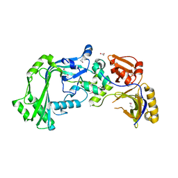
2IAY
DownloadVisualize
BU of 2iay by Molmil
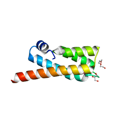
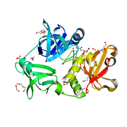
Crystal structure of a duf1831 family protein (lp2179) from lactobacillus plantarum at 1.20 A resolution
分子名称: CHLORIDE ION, GLYCEROL, Hypothetical protein
著者Joint Center for Structural Genomics (JCSG)
登録日2006-09-08
公開日2006-10-10
最終更新日2023-01-25
実験手法X-RAY DIFFRACTION (1.2 Å)
主引用文献Structure of LP2179, the first representative of Pfam family PF08866, suggests a new fold with a role in amino-acid metabolism.
Acta Crystallogr.,Sect.F, 66, 2010
3KK7
DownloadVisualize
BU of 3kk7 by Molmil
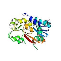
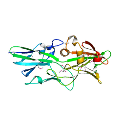
Crystal structure of Putative cell invasion protein with MAC/Perforin domain (NP_812351.1) from BACTERIODES THETAIOTAOMICRON VPI-5482 at 2.46 A resolution
分子名称: (4S)-2-METHYL-2,4-PENTANEDIOL, 1,2-ETHANEDIOL, CHLORIDE ION, ...
著者Joint Center for Structural Genomics (JCSG)
登録日2009-11-04
公開日2009-11-24
最終更新日2023-02-01
実験手法X-RAY DIFFRACTION (2.46 Å)
主引用文献Structure of a membrane-attack complex/perforin (MACPF) family protein from the human gut symbiont Bacteroides thetaiotaomicron.
Acta Crystallogr.,Sect.F, 66, 2010
2OOC
DownloadVisualize
BU of 2ooc by Molmil
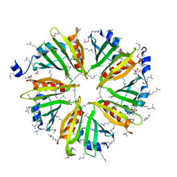
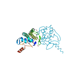
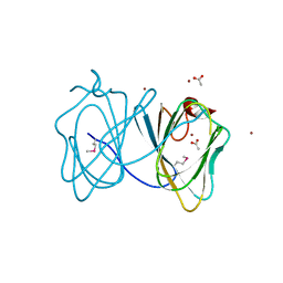
Crystal structure of Histidine Phosphotransferase ShpA (NP_419930.1) from Caulobacter crescentus at 1.52 A resolution
分子名称: GLYCEROL, Histidine phosphotransferase, TETRAETHYLENE GLYCOL
著者Joint Center for Structural Genomics (JCSG)
登録日2007-01-25
公開日2007-02-06
最終更新日2023-01-25
実験手法X-RAY DIFFRACTION (1.52 Å)
主引用文献Crystal structure of histidine phosphotransfer protein ShpA, an essential regulator of stalk biogenesis in Caulobacter crescentus.
J.Mol.Biol., 390, 2009
3L5O
DownloadVisualize
BU of 3l5o by Molmil
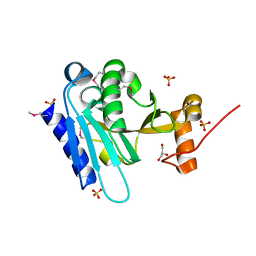
Crystal structure of protein with unknown function from DUF364 family (ZP_00559375.1) from Desulfitobacterium hafniense DCB-2 at 2.01 A resolution
分子名称: 1,2-ETHANEDIOL, CHLORIDE ION, IMIDAZOLE, ...
著者Joint Center for Structural Genomics, Joint Center for Structural Genomics (JCSG)
登録日2009-12-22
公開日2010-02-02
最終更新日2023-02-01
実験手法X-RAY DIFFRACTION (2.01 Å)
主引用文献Structure of the first representative of Pfam family PF04016 (DUF364) reveals enolase and Rossmann-like folds that combine to form a unique active site with a possible role in heavy-metal chelation.
Acta Crystallogr.,Sect.F, 66, 2010
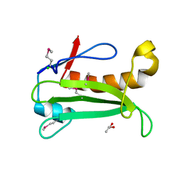
3HSA
DownloadVisualize
BU of 3hsa by Molmil
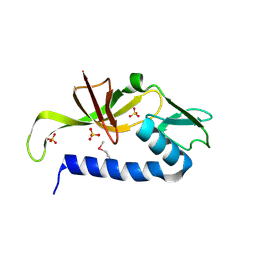
Crystal structure of pleckstrin homology domain (YP_926556.1) from SHEWANELLA AMAZONENSIS SB2B at 1.99 A resolution
分子名称: DI(HYDROXYETHYL)ETHER, GLYCEROL, pleckstrin homology domain
著者Joint Center for Structural Genomics (JCSG)
登録日2009-06-10
公開日2009-06-23
最終更新日2023-02-01
実験手法X-RAY DIFFRACTION (1.99 Å)
主引用文献Bacterial pleckstrin homology domains: a prokaryotic origin for the PH domain.
J.Mol.Biol., 396, 2010
3K5J
DownloadVisualize
BU of 3k5j by Molmil
Crystal structure of Putative SUFU (suppressor of fused protein) homolog (YP_208451.1) from Neisseria gonorrhoeae FA 1090 at 1.40 A resolution
分子名称: GLYCEROL, SULFATE ION, Suppressor of fused family protein
著者Joint Center for Structural Genomics (JCSG)
登録日2009-10-07
公開日2010-01-26
最終更新日2019-07-17
実験手法X-RAY DIFFRACTION (1.4 Å)
主引用文献The crystal structure of a bacterial Sufu-like protein defines a novel group of bacterial proteins that are similar to the N-terminal domain of human Sufu.
Protein Sci., 19, 2010
3GF8
DownloadVisualize
BU of 3gf8 by Molmil
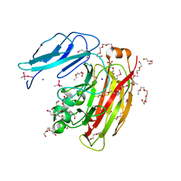
Crystal structure of putative polysaccharide binding proteins (DUF1812) (NP_809975.1) from BACTEROIDES THETAIOTAOMICRON VPI-5482 at 2.20 A resolution
分子名称: 1,2-ETHANEDIOL, putative polysaccharide binding proteins (DUF1812)
著者Joint Center for Structural Genomics (JCSG)
登録日2009-02-26
公開日2009-03-24
最終更新日2023-02-01
実験手法X-RAY DIFFRACTION (2.2 Å)
主引用文献A conserved fold for fimbrial components revealed by the crystal structure of a putative fimbrial assembly protein (BT1062) from Bacteroides thetaiotaomicron at 2.2 A resolution
Acta Crystallogr.,Sect.F, 66, 2010
3H0N
DownloadVisualize
BU of 3h0n by Molmil
Crystal structure of a duf1470 family protein (jann_2411) from jannaschia sp. ccs1 at 1.45 A resolution
分子名称: ACETATE ION, GLYCEROL, NICKEL (II) ION, ...
著者Joint Center for Structural Genomics (JCSG)
登録日2009-04-09
公開日2009-04-21
最終更新日2023-02-01
実験手法X-RAY DIFFRACTION (1.45 Å)
主引用文献The structure of Jann_2411 (DUF1470) from Jannaschia sp. at 1.45  resolution reveals a new fold (the ABATE domain) and suggests its possible role as a transcription regulator.
Acta Crystallogr.,Sect.F, 66, 2010
3HBZ
DownloadVisualize
BU of 3hbz by Molmil
Crystal structure of a putative glycoside hydrolase (bt_2081) from bacteroides thetaiotaomicron vpi-5482 at 2.05 A resolution
分子名称: ACETATE ION, CACODYLATE ION, CALCIUM ION, ...
著者Joint Center for Structural Genomics (JCSG)
登録日2009-05-05
公開日2009-05-26
最終更新日2023-02-01
実験手法X-RAY DIFFRACTION (2.05 Å)
主引用文献Structure of Bacteroides thetaiotaomicron BT2081 at 2.05 A resolution: the first structural representative of a new protein family that may play a role in carbohydrate metabolism.
Acta Crystallogr.,Sect.F, 66, 2010
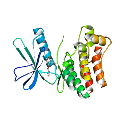
4X7Q
DownloadVisualize
BU of 4x7q by Molmil
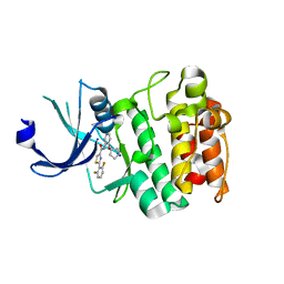
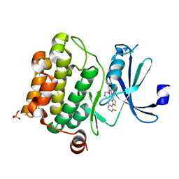
PIM2 kinase in complex with Compound 1s
分子名称: 2-(2,6-difluorophenyl)-N-{4-[(3S)-pyrrolidin-3-yloxy]pyridin-3-yl}-1,3-thiazole-4-carboxamide, PHOSPHATE ION, Serine/threonine-protein kinase pim-2
著者Marcotte, D.J, Silvian, L.F.
登録日2014-12-09
公開日2015-02-11
最終更新日2023-09-27
実験手法X-RAY DIFFRACTION (2.33 Å)
主引用文献Structure-based design of low-nanomolar PIM kinase inhibitors.
Bioorg.Med.Chem.Lett., 25, 2015
3H41
DownloadVisualize
BU of 3h41 by Molmil
CRYSTAL STRUCTURE OF A NLPC/P60 FAMILY PROTEIN (BCE_2878) FROM BACILLUS CEREUS ATCC 10987 AT 1.79 A RESOLUTION
分子名称: ALANINE, D-GLUTAMIC ACID, NLP/P60 family protein, ...
著者Joint Center for Structural Genomics (JCSG)
登録日2009-04-17
公開日2009-05-05
最終更新日2023-02-01
実験手法X-RAY DIFFRACTION (1.79 Å)
主引用文献Structure of the gamma-D-glutamyl-L-diamino acid endopeptidase YkfC from Bacillus cereus in complex with L-Ala-[gamma]-D-Glu: insights into substrate recognition by NlpC/P60 cysteine peptidases
Acta Crystallogr.,Sect.F, 66, 2010
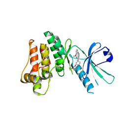
4XHK
DownloadVisualize
BU of 4xhk by Molmil
PIM1 kinase in complex with Compound 1s
分子名称: 2-(2,6-difluorophenyl)-N-{4-[(3S)-pyrrolidin-3-yloxy]pyridin-3-yl}-1,3-thiazole-4-carboxamide, Serine/threonine-protein kinase pim-1
著者Marcotte, D.J, Silvian, L.F.
登録日2015-01-05
公開日2015-02-11
最終更新日2023-09-27
実験手法X-RAY DIFFRACTION (1.9 Å)
主引用文献Structure-based design of low-nanomolar PIM kinase inhibitors.
Bioorg.Med.Chem.Lett., 25, 2015
3H50
DownloadVisualize
BU of 3h50 by Molmil
CRYSTAL STRUCTURE OF A TETRACENOMYCIN POLYKETIDE SYNTHESIS PROTEIN (TCMJ) FROM XANTHOMONAS CAMPESTRIS PV. CAMPESTRIS AT 1.60 A RESOLUTION
分子名称: ACETATE ION, Tetracenomycin polyketide synthesis protein, ZINC ION
著者Joint Center for Structural Genomics (JCSG)
登録日2009-04-21
公開日2009-05-05
最終更新日2023-02-01
実験手法X-RAY DIFFRACTION (1.6 Å)
主引用文献Conformational changes associated with the binding of zinc acetate at the putative active site of XcTcmJ, a cupin from Xanthomonas campestris pv. campestris.
Acta Crystallogr.,Sect.F, 66, 2010
3IRB
DownloadVisualize
BU of 3irb by Molmil
Crystal structure of protein with unknown function from DUF35 family (13815350) from SULFOLOBUS SOLFATARICUS at 1.80 A resolution
分子名称: ACETIC ACID, SULFATE ION, ZINC ION, ...
著者Joint Center for Structural Genomics (JCSG)
登録日2009-08-21
公開日2009-09-01
最終更新日2023-02-01
実験手法X-RAY DIFFRACTION (1.8 Å)
主引用文献The structure of SSO2064, the first representative of Pfam family PF01796, reveals a novel two-domain zinc-ribbon OB-fold architecture with a potential acyl-CoA-binding role.
Acta Crystallogr.,Sect.F, 66, 2010
6TYM
DownloadVisualize
BU of 6tym by Molmil
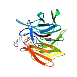
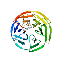
KEAP1 Kelch domain in complex with Compound 9
分子名称: (3S)-3-[2-(benzenecarbonyl)-5-methyl-1,2,3,4-tetrahydroisoquinolin-7-yl]-3-(1-ethyl-4-methyl-1H-benzotriazol-5-yl)propanoic acid, DIMETHYL SULFOXIDE, GLYCEROL, ...
著者Marcotte, D.J.
登録日2019-08-09
公開日2020-01-15
最終更新日2023-10-11
実験手法X-RAY DIFFRACTION (1.422 Å)
主引用文献Design, synthesis and identification of novel, orally bioavailable non-covalent Nrf2 activators.
Bioorg.Med.Chem.Lett., 30, 2020
6TYP
DownloadVisualize
BU of 6typ by Molmil
KEAP1 Kelch domain in complex with Compound 2
分子名称: (3S)-3-[2-(benzenecarbonyl)-1,2,3,4-tetrahydroisoquinolin-7-yl]-3-(1-ethyl-4-methyl-1H-benzotriazol-5-yl)propanoic acid, FORMIC ACID, Kelch-like ECH-associated protein 1
著者Marcotte, D.J.
登録日2019-08-09
公開日2020-01-15
最終更新日2023-10-11
実験手法X-RAY DIFFRACTION (2.5 Å)
主引用文献Design, synthesis and identification of novel, orally bioavailable non-covalent Nrf2 activators.
Bioorg.Med.Chem.Lett., 30, 2020
1NF1
DownloadVisualize
BU of 1nf1 by Molmil
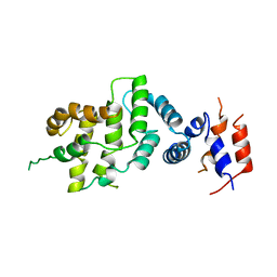
THE GAP RELATED DOMAIN OF NEUROFIBROMIN
分子名称: PROTEIN (NEUROFIBROMIN)
著者Scheffzek, K, Ahmadian, M.R, Wiesmueller, L, Kabsch, W, Stege, P, Schmitz, F, Wittinghofer, A.
登録日1998-07-08
公開日1999-07-20
最終更新日2023-12-27
実験手法X-RAY DIFFRACTION (2.5 Å)
主引用文献Structural analysis of the GAP-related domain from neurofibromin and its implications.
EMBO J., 17, 1998
6VQF
DownloadVisualize
BU of 6vqf by Molmil
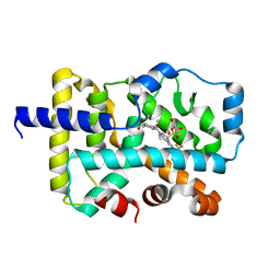
CRYSTAL STRUCTURE OF RAR-RELATED ORPHAN RECEPTOR C (NHIS-RORGT(244- 487)-L6-SRC1(678-692)) IN COMPLEX WITH AN INVERSE AGONIST
分子名称: (1R,3S,4R)-4-[(3aR,9bR)-9b-[(4-fluorophenyl)sulfonyl]-7-(1,1,1,2,3,3,3-heptafluoropropan-2-yl)-1,2,3a,4,5,9b-hexahydro-3H-benzo[e]indole-3-carbonyl]-3-methylcyclohexane-1-carboxylic acid, GLYCEROL, Nuclear receptor ROR-gamma
著者Sack, J.S.
登録日2020-02-05
公開日2020-04-08
最終更新日2023-10-11
実験手法X-RAY DIFFRACTION (2 Å)
主引用文献Discovery of BMS-986251: A Clinically Viable, Potent, and Selective ROR gamma t Inverse Agonist.
Acs Med.Chem.Lett., 11, 2020
6GK8
DownloadVisualize
BU of 6gk8 by Molmil
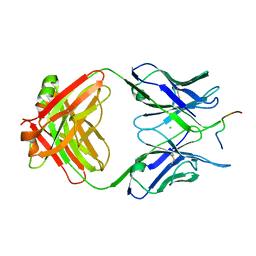
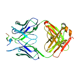
Crystal structure of anti-tau antibody dmCBTAU-28.1, double mutant (S32R, E35K) of CBTAU-28.1, in complex with Tau peptide A7731 (residues 52-71)
分子名称: CHLORIDE ION, HUMAN FAB ANTIBODY FRAGMENT OF CBTAU-28.1(S32R;E35K), TAU PEPTIDE A7731 (RESIDUES 52-71)
著者Steinbacher, S, Mrosek, M, Juraszek, J.
登録日2018-05-18
公開日2018-06-13
実験手法X-RAY DIFFRACTION (2.85 Å)
主引用文献A common antigenic motif recognized by naturally occurring human VH5-51/VL4-1 anti-tau antibodies with distinct functionalities.
Acta Neuropathol Commun, 6, 2018
6GK7
DownloadVisualize
BU of 6gk7 by Molmil
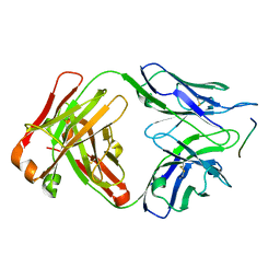
Crystal structure of anti-tau antibody dmCBTAU-27.1, double mutant (S31Y, T100I) of CBTAU-27.1, in complex with Tau peptide A8119B (residues 299-318)
分子名称: CHLORIDE ION, HUMAN FAB ANTIBODY FRAGMENT OF CBTAU-27.1(S31Y,T100I), HUMAN TAU PEPTIDE A8119 RESIDUES 299-318
著者Steinbacher, S, Mrosek, M, Juraszek, J.
登録日2018-05-18
公開日2018-06-13
実験手法X-RAY DIFFRACTION (2.95 Å)
主引用文献A common antigenic motif recognized by naturally occurring human VH5-51/VL4-1 anti-tau antibodies with distinct functionalities.
Acta Neuropathol Commun, 6, 2018
1OCE
DownloadVisualize
BU of 1oce by Molmil
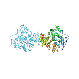
ACETYLCHOLINESTERASE (E.C. 3.1.1.7) COMPLEXED WITH MF268
分子名称: ACETYLCHOLINESTERASE, CIS-2,6-DIMETHYLMORPHOLINOOCTYLCARBAMYLESEROLINE
著者Bartolucci, C, Perola, E, Cellai, L, Brufani, M, Lamba, D.
登録日1998-06-12
公開日1999-05-18
最終更新日2023-08-09
実験手法X-RAY DIFFRACTION (2.7 Å)
主引用文献"Back door" opening implied by the crystal structure of a carbamoylated acetylcholinesterase.
Biochemistry, 38, 1999
3O2M
DownloadVisualize
BU of 3o2m by Molmil
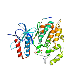
Crystal Structure of JNK1-alpha1 isoform complex with a biaryl tetrazol (A-82118)
分子名称: C-Jun-amino-terminal kinase-interacting protein 1, JIP1, 10MER PEPTIDE, ...
著者Abad-Zapatero, C.
登録日2010-07-22
公開日2011-01-12
最終更新日2024-02-21
実験手法X-RAY DIFFRACTION (2.7 Å)
主引用文献Discovery and characterization of non-ATP site inhibitors of the mitogen activated protein (MAP) kinases.
Acs Chem.Biol., 6, 2011
5ZV3
DownloadVisualize
BU of 5zv3 by Molmil
Crystal structure of human anti-tau antibody CBTAU-28.1 in complex with its tau peptide
分子名称: Heavy chain of antibody CBTAU28.1, Light chain (kappa) of antibody CBTAU28.1, Peptide from Microtubule-associated protein tau
著者Zhang, H, Wilson, I.A.
登録日2018-05-09
公開日2018-06-20
最終更新日2023-11-22
実験手法X-RAY DIFFRACTION (2.093 Å)
主引用文献A common antigenic motif recognized by naturally occurring human VH5-51/VL4-1 anti-tau antibodies with distinct functionalities.
Acta Neuropathol Commun, 6, 2018
5KO1
DownloadVisualize
BU of 5ko1 by Molmil
Pseudokinase Domain of MLKL bound to Compound 4.
分子名称: Mixed lineage kinase domain-like protein, [(1~{R})-2-[(4-fluorophenyl)amino]-2-oxidanylidene-1-phenyl-ethyl] 3-azanylpyrazine-2-carboxylate
著者Marcotte, D.J.
登録日2016-06-29
公開日2016-11-16
最終更新日2023-09-27
実験手法X-RAY DIFFRACTION (2.16 Å)
主引用文献ATP-Competitive MLKL Binders Have No Functional Impact on Necroptosis.
Plos One, 11, 2016
5KNJ
DownloadVisualize
BU of 5knj by Molmil
Pseudokinase Domain of MLKL bound to Compound 1.
分子名称: 1-[4-[methyl-[2-[(3-sulfamoylphenyl)amino]pyrimidin-4-yl]amino]phenyl]-3-[4-(trifluoromethyloxy)phenyl]urea, Mixed lineage kinase domain-like protein
著者Marcotte, D.J.
登録日2016-06-28
公開日2016-11-16
最終更新日2023-09-27
実験手法X-RAY DIFFRACTION (2.88 Å)
主引用文献ATP-Competitive MLKL Binders Have No Functional Impact on Necroptosis.
Plos One, 11, 2016

224931

件を2024-09-11に公開中

PDB statisticsPDBj update infoContact PDBjnumon