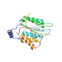
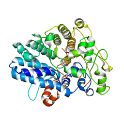
6NS7
 
 | |
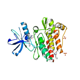
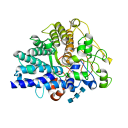
3PYY
 | | Discovery and Characterization of a Cell-Permeable, Small-molecule c-Abl Kinase Activator that Binds to the Myristoyl Binding Site | | 分子名称: | (5R)-5-[3-(4-fluorophenyl)-1-phenyl-1H-pyrazol-4-yl]imidazolidine-2,4-dione, 4-(4-METHYL-PIPERAZIN-1-YLMETHYL)-N-[4-METHYL-3-(4-PYRIDIN-3-YL-PYRIMIDIN-2-YLAMINO)-PHENYL]-BENZAMIDE, GLYCEROL, ... | | 著者 | Yang, J, Campobasso, N, Biju, M.P, Fisher, K, Pan, X.Q, Cottom, J, Galbraith, S, Ho, T, Zhang, H, Hong, X, Ward, P, Hofmann, G, Siegfried, B. | | 登録日 | 2010-12-13 | | 公開日 | 2011-03-09 | | 最終更新日 | 2024-02-21 | | 実験手法 | X-RAY DIFFRACTION (1.85 Å) | | 主引用文献 | Discovery and Characterization of a Cell-Permeable, Small-Molecule c-Abl Kinase Activator that Binds to the Myristoyl Binding Site.
Chem.Biol., 18, 2011
|
|
4NNI
 | | Structural basis for targeting the ribosomal protein S1 of Mycobacterium tuberculosis by pyrazinamide | | 分子名称: | 30S ribosomal protein S1, PYRAZINE-2-CARBOXYLIC ACID | | 著者 | Yang, J, Liu, Y, Cai, Q, Lin, D. | | 登録日 | 2013-11-18 | | 公開日 | 2014-12-24 | | 最終更新日 | 2024-03-20 | | 実験手法 | X-RAY DIFFRACTION (2.64 Å) | | 主引用文献 | Structural basis for targeting the ribosomal protein S1 of Mycobacterium tuberculosis by pyrazinamide.
Mol.Microbiol., 95, 2015
|
|
4NNG
 | |
4NNH
 | |
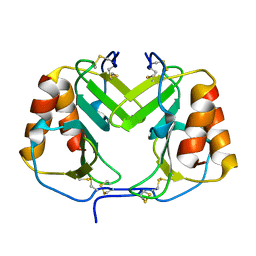
1F9S
 | | CRYSTAL STRUCTURE OF PLATELET FACTOR 4 MUTANT 2 | | 分子名称: | PLATELET FACTOR 4 | | 著者 | Yang, J, Doyle, M, Faulk, T, Visentin, G, Aster, R, Edwards, B. | | 登録日 | 2000-07-11 | | 公開日 | 2003-08-26 | | 最終更新日 | 2021-11-03 | | 実験手法 | X-RAY DIFFRACTION (2.38 Å) | | 主引用文献 | Structure Comparison of Two Platelet Factor 4 Mutants with the Wild-type Reveals the Epitopes for the Heparin-induced Thrombocytopenia Antibodies
To be Published
|
|
4NNK
 | |
1F9P
 | | CRYSTAL STRUCTURE OF CONNECTIVE TISSUE ACTIVATING PEPTIDE-III(CTAP-III) COMPLEXED WITH POLYVINYLSULFONIC ACID | | 分子名称: | CONNECTIVE TISSUE ACTIVATING PEPTIDE-III, ETHANESULFONIC ACID | | 著者 | Yang, J, Faulk, T, Aster, R, Visentin, G, Edwards, B, Castor, C. | | 登録日 | 2000-07-11 | | 公開日 | 2003-08-26 | | 最終更新日 | 2017-10-04 | | 実験手法 | X-RAY DIFFRACTION (1.93 Å) | | 主引用文献 | Structure of the CXC Chemokine, Connective Tissue Activating Peptide-III, Complexed with the Heparin Analogue, Polyvinylsulfonic Acid
To be Published
|
|
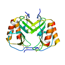
1F9Q
 | | CRYSTAL STRUCTURE OF PLATELET FACTOR 4 | | 分子名称: | PLATELET FACTOR 4 | | 著者 | Yang, J, Doyle, M, Faulk, T, Visentin, G, Aster, R, Edwards, B. | | 登録日 | 2000-07-11 | | 公開日 | 2003-08-26 | | 最終更新日 | 2017-10-04 | | 実験手法 | X-RAY DIFFRACTION (2 Å) | | 主引用文献 | Structure Comparison of Two Platelet Factor 4 Mutants with the Wild-Type Reveals the Epitopes for the Heparin-Induced Thrombocytopenia Antibodies
To be Published
|
|
1F9R
 | | CRYSTAL STRUCTURE OF PLATELET FACTOR 4 MUTANT 1 | | 分子名称: | PLATELET FACTOR 4 | | 著者 | Yang, J, Doyle, M, Faulk, T, Visentin, G, Aster, R, Edwards, B. | | 登録日 | 2000-07-11 | | 公開日 | 2003-08-26 | | 最終更新日 | 2021-11-03 | | 実験手法 | X-RAY DIFFRACTION (2 Å) | | 主引用文献 | Structure Comparison of Two Platelet Factor 4 Mutants with the Wild-type Reveals the Epitopes for the Heparin-induced Thrombocytopenia Antibodies
To be Published
|
|
1FPR
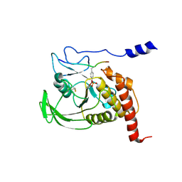 | | CRYSTAL STRUCTURE OF THE COMPLEX FORMED BETWEEN THE CATALYTIC DOMAIN OF SHP-1 AND AN IN VITRO PEPTIDE SUBSTRATE PY469 DERIVED FROM SHPS-1. | | 分子名称: | PEPTIDE PY469, PROTEIN-TYROSINE PHOSPHATASE 1C | | 著者 | Yang, J, Cheng, Z, Niu, Z, Zhao, Z.J, Zhou, G.W. | | 登録日 | 2000-08-31 | | 公開日 | 2001-03-07 | | 最終更新日 | 2021-11-03 | | 実験手法 | X-RAY DIFFRACTION (2.5 Å) | | 主引用文献 | Structural basis for substrate specificity of protein-tyrosine phosphatase SHP-1.
J.Biol.Chem., 275, 2000
|
|
8D9X
 | |
1RE5
 | | Crystal structure of 3-carboxy-cis,cis-muconate lactonizing enzyme from Pseudomonas putida | | 分子名称: | 2,3-DIHYDROXY-1,4-DITHIOBUTANE, 3-carboxy-cis,cis-muconate cycloisomerase, CITRIC ACID | | 著者 | Yang, J, Wang, Y, Woolridge, E.M, Petsko, G.A, Kozarich, J.W, Ringe, D. | | 登録日 | 2003-11-06 | | 公開日 | 2004-06-08 | | 最終更新日 | 2023-08-23 | | 実験手法 | X-RAY DIFFRACTION (2.6 Å) | | 主引用文献 | Crystal Structure of 3-Carboxy-cis,cis-muconate Lactonizing Enzyme from Pseudomonas putida, a Fumarase Class II Type Cycloisomerase: Enzyme Evolution in Parallel Pathways.
Biochemistry, 43, 2004
|
|
1RXT
 | | Crystal structure of human myristoyl-CoA:protein N-myristoyltransferase. | | 分子名称: | COBALT (II) ION, Glycylpeptide N-tetradecanoyltransferase 1, SULFATE ION | | 著者 | Yang, J, Wang, Y, Frey, G, Abeles, R.H, Petsko, G.A, Ringe, D. | | 登録日 | 2003-12-18 | | 公開日 | 2005-04-12 | | 最終更新日 | 2024-02-14 | | 実験手法 | X-RAY DIFFRACTION (3 Å) | | 主引用文献 | Crystal structure of human myristoyl-CoA:protein N-myristoyltransferase
To be Published
|
|
2MWN
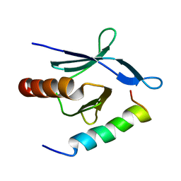 | | Talin-F3 / RIAM N-terminal Peptide complex | | 分子名称: | Amyloid beta A4 precursor protein-binding family B member 1-interacting protein, Talin-1 | | 著者 | Yang, J, Zhu, L, Zhang, H, Hirbawi, J, Fukuda, K, Dwivedi, P, Liu, J, Byzova, T, Plow, E.F, Wu, J, Qin, J. | | 登録日 | 2014-11-13 | | 公開日 | 2014-12-17 | | 最終更新日 | 2024-05-15 | | 実験手法 | SOLUTION NMR | | 主引用文献 | Conformational activation of talin by RIAM triggers integrin-mediated cell adhesion.
Nat Commun, 5, 2014
|
|
5HM2
 | |
1O6K
 | | Structure of activated form of PKB kinase domain S474D with GSK3 peptide and AMP-PNP | | 分子名称: | GLYCOGEN SYNTHASE KINASE-3 BETA, MANGANESE (II) ION, PHOSPHOAMINOPHOSPHONIC ACID-ADENYLATE ESTER, ... | | 著者 | Yang, J, Cron, P, Good, V.M, Thompson, V, Hemmings, B.A, Barford, D. | | 登録日 | 2002-10-08 | | 公開日 | 2002-11-19 | | 最終更新日 | 2024-05-01 | | 実験手法 | X-RAY DIFFRACTION (1.7 Å) | | 主引用文献 | Crystal Structure of an Activated Akt/Protein Kinase B Ternary Complex with Gsk-3 Peptide and AMP-Pnp
Nat.Struct.Biol., 9, 2002
|
|
1O6L
 | | Crystal structure of an activated Akt/protein kinase B (PKB-PIF chimera) ternary complex with AMP-PNP and GSK3 peptide | | 分子名称: | GLYCOGEN SYNTHASE KINASE-3 BETA, MANGANESE (II) ION, PHOSPHOAMINOPHOSPHONIC ACID-ADENYLATE ESTER, ... | | 著者 | Yang, J, Cron, P, Good, V.M, Thompson, V, Hemmings, B.A, Barford, D. | | 登録日 | 2002-10-08 | | 公開日 | 2002-11-19 | | 最終更新日 | 2024-05-01 | | 実験手法 | X-RAY DIFFRACTION (1.6 Å) | | 主引用文献 | Crystal Structure of an Activated Akt/Protein Kinase B Ternary Complex with Gsk-3 Peptide and AMP-Pnp
Nat.Struct.Biol., 9, 2002
|
|
7VQM
 | | GH2 beta-galacturonate AqGalA in complex with galacturonide | | 分子名称: | CHLORIDE ION, GH2 beta-galacturonate AqGalA, beta-D-galactopyranuronic acid | | 著者 | Yang, J. | | 登録日 | 2021-10-20 | | 公開日 | 2021-11-24 | | 最終更新日 | 2023-11-29 | | 実験手法 | X-RAY DIFFRACTION (2.4 Å) | | 主引用文献 | Structural and Biochemical Basis of a Marine Bacterial Glycoside Hydrolase Family 2 beta-Glycosidase with Broad Substrate Specificity
Appl.Environ.Microbiol., 88, 2022
|
|
7VW2
 | |
7VW1
 | |
7VW0
 | | Structure of a dimeric periplasmic protein | | 分子名称: | DUF305 domain-containing protein | | 著者 | Yang, J, Liu, L. | | 登録日 | 2021-11-09 | | 公開日 | 2022-01-26 | | 最終更新日 | 2023-11-29 | | 実験手法 | X-RAY DIFFRACTION (1.447 Å) | | 主引用文献 | Structural basis of copper binding by a dimeric periplasmic protein forming a six-helical bundle.
J.Inorg.Biochem., 229, 2022
|
|
8J0P
 | |
8JXZ
 | | Chitin binding SusD-like protein AqSusD in complex with (GlcNAc)3 | | 分子名称: | 2-acetamido-2-deoxy-beta-D-glucopyranose-(1-4)-2-acetamido-2-deoxy-beta-D-glucopyranose-(1-4)-2-acetamido-2-deoxy-beta-D-glucopyranose, SusD-like protein AqSusD | | 著者 | Yang, J. | | 登録日 | 2023-07-01 | | 公開日 | 2023-11-01 | | 最終更新日 | 2024-02-28 | | 実験手法 | X-RAY DIFFRACTION (2.2 Å) | | 主引用文献 | Structural insights of a SusD-like protein in marine Bacteroidetes bacteria reveal the molecular basis for chitin recognition and acquisition.
Febs J., 291, 2024
|
|
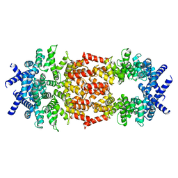
8W78
 | | Structure of Drosophila melanogaster L-2-hydroxyglutarate dehydrogenase in complex with FAD and 2-oxoglutarate | | 分子名称: | 2-OXOGLUTARIC ACID, DODECYL-BETA-D-MALTOSIDE, FI05204p, ... | | 著者 | Yang, J, Chen, X, Jin, S, Ding, J. | | 登録日 | 2023-08-30 | | 公開日 | 2023-11-29 | | 最終更新日 | 2023-12-20 | | 実験手法 | X-RAY DIFFRACTION (2.81 Å) | | 主引用文献 | Structure and biochemical characterization of l-2-hydroxyglutarate dehydrogenase and its role in the pathogenesis of l-2-hydroxyglutaric aciduria.
J.Biol.Chem., 300, 2023
|
|


