 Movie
Movie Controller

[English] 日本語
 Yorodumi
- PDB-2x3g: Crystal Structure of the hypothetical protein ORF119 from Sulfolo... -
+ Open data

- Basic information
| Entry | Database: PDB / ID: 2x3g | ||||||
|---|---|---|---|---|---|---|---|
| Title | Crystal Structure of the hypothetical protein ORF119 from Sulfolobus islandicus rod-shaped virus 1 | ||||||
 Components Components | SIRV1 HYPOTHETICAL PROTEIN ORF119 | ||||||
 Keywords Keywords |  VIRAL PROTEIN / HOST MEMBRANE VIRAL PROTEIN / HOST MEMBRANE | ||||||
| Function / homology | Alpha-Beta Plaits - #1910 / Alpha-Beta Plaits / 2-Layer Sandwich / Alpha Beta / Uncharacterized protein Function and homology information Function and homology information | ||||||
| Biological species |   SULFOLOBUS ISLANDICUS RUDIVIRUS 1 VARIANT XX SULFOLOBUS ISLANDICUS RUDIVIRUS 1 VARIANT XX | ||||||
| Method | X-RAY DIFFRACTION / SYNCHROTRON / SAD / Resolution: 1.8 Å | ||||||
 Authors Authors | Oke, M. / Carter, L.G. / Johnson, K.A. / Liu, H. / Mcmahon, S.A. / White, M.F. / Naismith, J.H. | ||||||
 Citation Citation | Journal: J.Struct.Funct.Genom. / Year: 2010 Title: The Scottish Structural Proteomics Facility: Targets, Methods and Outputs. Authors: Oke, M. / Carter, L.G. / Johnson, K.A. / Liu, H. / Mcmahon, S.A. / Yan, X. / Kerou, M. / Weikart, N.D. / Kadi, N. / Sheikh, M.A. / Schmelz, S. / Dorward, M. / Zawadzki, M. / Cozens, C. / ...Authors: Oke, M. / Carter, L.G. / Johnson, K.A. / Liu, H. / Mcmahon, S.A. / Yan, X. / Kerou, M. / Weikart, N.D. / Kadi, N. / Sheikh, M.A. / Schmelz, S. / Dorward, M. / Zawadzki, M. / Cozens, C. / Falconer, H. / Powers, H. / Overton, I.M. / Van Niekerk, C.A.J. / Peng, X. / Patel, P. / Garrett, R.A. / Prangishvili, D. / Botting, C.H. / Coote, P.J. / Dryden, D.T.F. / Barton, G.J. / Schwarz-Linek, U. / Challis, G.L. / Taylor, G.L. / White, M.F. / Naismith, J.H. | ||||||
| History |
|
- Structure visualization
| Structure viewer | Molecule: MolmilJmol/JSmol |
|---|
- Downloads & links
-Download
| PDBx/mmCIF format | 2x3g.cif.gz | 58.2 KB | Display | PDBx/mmCIF format |
|---|---|---|---|---|
| PDB format | pdb2x3g.ent.gz | 47.2 KB | Display | PDB format |
| PDBx/mmJSON format | 2x3g.json.gz | Tree view | PDBx/mmJSON format | |
| Others |  Other downloads Other downloads |
-Validation report
| Arichive directory | https://data.pdbj.org/pub/pdb/validation_reports/x3/2x3gftp://data.pdbj.org/pub/pdb/validation_reports/x3/2x3g | HTTPS FTP |
|---|
-Related structure data
| Related structure data | 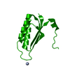 2ivyC  2jg5C 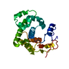 2jg6C 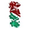 2vw8C 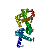 2vxzC 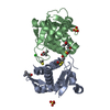 2wj9C 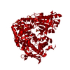 2x0oC 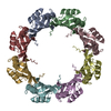 2x3dC  2x3eC  2x3fC  2x3lC  2x3mC 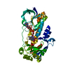 2x3nC 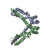 2x3oC  2x48C 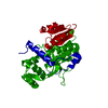 2x4gC 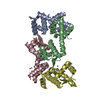 2x4hC 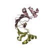 2x4iC 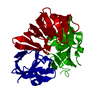 2x4jC 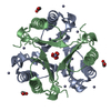 2x4kC 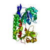 2x4lC 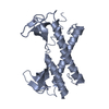 2x5cC 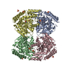 2x5dC 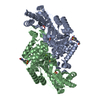 2x5fC 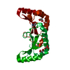 2x5gC 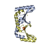 2x5hC 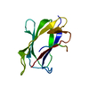 2x5pC 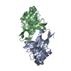 2x5qC 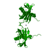 2x5rC  2x5tC 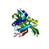 2x7bC 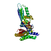 2x7iC 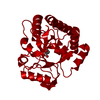 2xu2C C: citing same article ( |
|---|---|
| Similar structure data |


-Links
 PDBj
PDBj- Assembly
| Deposited unit | 
| ||||||||
|---|---|---|---|---|---|---|---|---|---|
| 1 | 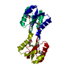
| ||||||||
| Unit cell |
|
-Components
| #1: Protein | Mass: 14520.341 Da / Num. of mol.: 1 / Fragment: RESIDUES 2-119 Source method: isolated from a genetically manipulated source Source: (gene. exp.) SULFOLOBUS ISLANDICUS RUDIVIRUS 1 VARIANT XXPlasmid: PDEST14 / Production host:  ESCHERICHIA COLI BL21(DE3) (bacteria) / Variant (production host): C43 / References: UniProt: Q5TJA9 ESCHERICHIA COLI BL21(DE3) (bacteria) / Variant (production host): C43 / References: UniProt: Q5TJA9 |
|---|---|
| #2: Water | ChemComp-HOH / Water Mass: 18.015 Da / Num. of mol.: 88 / Source method: isolated from a natural source / Formula: H2O Mass: 18.015 Da / Num. of mol.: 88 / Source method: isolated from a natural source / Formula: H2O |
-Experimental details
-Experiment
| Experiment | Method: X-RAY DIFFRACTION / Number of used crystals: 1 |
|---|
- Sample preparation
| Crystal | Density Matthews: 2.31 Å3/Da / Density % sol: 47 % / Description: NONE |
|---|---|
| Crystal grow | pH: 6 Details: 20% PEG 4000, 0.1 M SODIUM CITRATE PH 6.0. CRYSTALS WERE CRYOPROTECTED WITH 25% PEG400 IN A SOLUTION CONTAINING 22% PEG 4000, 0.1 M SODIUM CITRATE PH 6.0. |
-Data collection
| Diffraction | Mean temperature: 100 K |
|---|---|
| Diffraction source | Source: SYNCHROTRON / Site: ESRF  / Beamline: BM14 / Wavelength: 0.9537 / Beamline: BM14 / Wavelength: 0.9537 |
| Detector | Type: MARRESEARCH / Detector: CCD / Date: Feb 21, 2008 / Details: MIRRORS |
| Radiation | Monochromator: SI(III) / Protocol: SINGLE WAVELENGTH / Monochromatic (M) / Laue (L): M / Scattering type: x-ray |
| Radiation wavelength | Wavelength: 0.9537 Å / Relative weight: 1 |
| Reflection | Resolution: 1.8→38.84 Å / Num. obs: 11671 / % possible obs: 100 % / Observed criterion σ(I): 0 / Redundancy: 6.5 % / Biso Wilson estimate: 0 Å2 / Rmerge(I) obs: 0.08 / Net I/σ(I): 15.8 |
| Reflection shell | Resolution: 1.8→1.85 Å / Redundancy: 6.5 % / Rmerge(I) obs: 0.54 / Mean I/σ(I) obs: 3.8 / % possible all: 100 |
- Processing
| Software |
| ||||||||||||||||||||||||||||||||||||||||||||||||||||||||||||||||||||||||||||||||||||||||||||||||||||||||||||||||||||||||||||||||||||||||||||||||||||||||||||||||||||||||||||||||||||||
|---|---|---|---|---|---|---|---|---|---|---|---|---|---|---|---|---|---|---|---|---|---|---|---|---|---|---|---|---|---|---|---|---|---|---|---|---|---|---|---|---|---|---|---|---|---|---|---|---|---|---|---|---|---|---|---|---|---|---|---|---|---|---|---|---|---|---|---|---|---|---|---|---|---|---|---|---|---|---|---|---|---|---|---|---|---|---|---|---|---|---|---|---|---|---|---|---|---|---|---|---|---|---|---|---|---|---|---|---|---|---|---|---|---|---|---|---|---|---|---|---|---|---|---|---|---|---|---|---|---|---|---|---|---|---|---|---|---|---|---|---|---|---|---|---|---|---|---|---|---|---|---|---|---|---|---|---|---|---|---|---|---|---|---|---|---|---|---|---|---|---|---|---|---|---|---|---|---|---|---|---|---|---|---|
| Refinement | Method to determine structure: SAD Starting model: NONE Resolution: 1.8→38.84 Å / Cor.coef. Fo:Fc: 0.954 / Cor.coef. Fo:Fc free: 0.926 / SU B: 6.192 / SU ML: 0.104 / Cross valid method: THROUGHOUT / ESU R: 0.137 / ESU R Free: 0.133 / Stereochemistry target values: MAXIMUM LIKELIHOOD Details: HYDROGENS HAVE BEEN ADDED IN THE RIDING POSITIONS. ATOM RECORD CONTAINS SUM OF TLS AND RESIDUAL B FACTORS ANISOU RECORD CONTAINS SUM OF TLS AND RESIDUAL U FACTORS RESIDUES 57-59 ARE DISORDERED.
| ||||||||||||||||||||||||||||||||||||||||||||||||||||||||||||||||||||||||||||||||||||||||||||||||||||||||||||||||||||||||||||||||||||||||||||||||||||||||||||||||||||||||||||||||||||||
| Solvent computation | Ion probe radii: 0.8 Å / Shrinkage radii: 0.8 Å / VDW probe radii: 1.4 Å / Solvent model: BABINET MODEL WITH MASK | ||||||||||||||||||||||||||||||||||||||||||||||||||||||||||||||||||||||||||||||||||||||||||||||||||||||||||||||||||||||||||||||||||||||||||||||||||||||||||||||||||||||||||||||||||||||
| Displacement parameters | Biso mean: 21.839 Å2
| ||||||||||||||||||||||||||||||||||||||||||||||||||||||||||||||||||||||||||||||||||||||||||||||||||||||||||||||||||||||||||||||||||||||||||||||||||||||||||||||||||||||||||||||||||||||
| Refinement step | Cycle: LAST / Resolution: 1.8→38.84 Å
| ||||||||||||||||||||||||||||||||||||||||||||||||||||||||||||||||||||||||||||||||||||||||||||||||||||||||||||||||||||||||||||||||||||||||||||||||||||||||||||||||||||||||||||||||||||||
| Refine LS restraints |
|