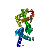
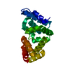
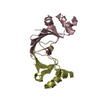
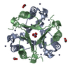
PDB-2ivy: Crystal structure of hypothetical protein sso1404 from Sulfolobus solfataricus P2 Method: X-RAY DIFFRACTION / Resolution: 1.4 Å
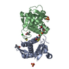
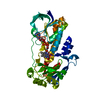
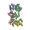
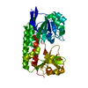
PDB-2jg5: CRYSTAL STRUCTURE OF A PUTATIVE PHOSPHOFRUCTOKINASE FROM STAPHYLOCOCCUS AUREUS Method: X-RAY DIFFRACTION / Resolution: 2.3 Å
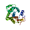
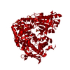
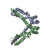
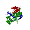
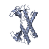
PDB-2jg6: CRYSTAL STRUCTURE OF A 3-METHYLADENINE DNA GLYCOSYLASE I FROM STAPHYLOCOCCUS AUREUS Method: X-RAY DIFFRACTION / Resolution: 1.7 Å
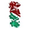
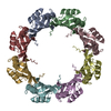
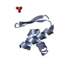
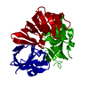
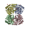
PDB-2vw8: Crystal Structure of Quinolone signal response protein pqsE from Pseudomonas aeruginosa Method: X-RAY DIFFRACTION / Resolution: 1.45 Å
PDB-2vxz: Crystal Structure of hypothetical protein PyrSV_gp04 from Pyrobaculum spherical virus Method: X-RAY DIFFRACTION / Resolution: 1.7 Å
PDB-2wj9: ArdB Method: X-RAY DIFFRACTION / Resolution: 1.62 Å
PDB-2x0o: Apo structure of the Alcaligin biosynthesis protein C (AlcC) from Bordetella bronchiseptica Method: X-RAY DIFFRACTION / Resolution: 2.4 Å
PDB-2x3d: Crystal Structure of SSo6206 from Sulfolobus solfataricus P2 Method: X-RAY DIFFRACTION / Resolution: 2.7 Å
PDB-2x3e: Crystal structure of 3-oxoacyl-(acyl carrier protein) synthase III, FabH from Pseudomonas aeruginosa PAO1 Method: X-RAY DIFFRACTION / Resolution: 1.81 Å
PDB-2x3f: Crystal Structure of the Methicillin-Resistant Staphylococcus aureus Sar2676, a Pantothenate Synthetase. Method: X-RAY DIFFRACTION / Resolution: 1.95 Å
PDB-2x3g: Crystal Structure of the hypothetical protein ORF119 from Sulfolobus islandicus rod-shaped virus 1 Method: X-RAY DIFFRACTION / Resolution: 1.8 Å
PDB-2x3l: Crystal Structure of the Orn_Lys_Arg decarboxylase family protein SAR0482 from Methicillin-resistant Staphylococcus aureus Method: X-RAY DIFFRACTION / Resolution: 2 Å
PDB-2x3m: Crystal Structure of Hypothetical Protein ORF239 from Pyrobaculum Spherical Virus Method: X-RAY DIFFRACTION / Resolution: 1.45 Å
PDB-2x3n: Crystal structure of pqsL, a probable FAD-dependent monooxygenase from Pseudomonas aeruginosa Method: X-RAY DIFFRACTION / Resolution: 1.75 Å
PDB-2x3o: Crystal Structure of the Hypothetical Protein PA0856 from Pseudomonas aeruginosa Method: X-RAY DIFFRACTION / Resolution: 2.9 Å
PDB-2x48: ORF 55 from Sulfolobus islandicus rudivirus 1 Method: X-RAY DIFFRACTION / Resolution: 2.6 Å
PDB-2x4g: Crystal structure of PA4631, a nucleoside-diphosphate-sugar epimerase from Pseudomonas aeruginosa Method: X-RAY DIFFRACTION / Resolution: 2.65 Å
PDB-2x4h: Crystal Structure of the hypothetical protein SSo2273 from Sulfolobus solfataricus Method: X-RAY DIFFRACTION / Resolution: 2.3 Å
PDB-2x4i: ORF 114a from Sulfolobus islandicus rudivirus 1 Method: X-RAY DIFFRACTION / Resolution: 2.2 Å
PDB-2x4j: Crystal structure of ORF137 from Pyrobaculum spherical virus Method: X-RAY DIFFRACTION / Resolution: 1.62 Å
PDB-2x4k: Crystal structure of SAR1376, a putative 4-oxalocrotonate tautomerase from the methicillin-resistant Staphylococcus aureus (MRSA) Method: X-RAY DIFFRACTION / Resolution: 1.1 Å
PDB-2x4l: Crystal structure of DesE, a ferric-siderophore receptor protein from Streptomyces coelicolor Method: X-RAY DIFFRACTION / Resolution: 1.5 Å
PDB-2x5c: Crystal structure of hypothetical protein ORF131 from Pyrobaculum Spherical Virus Method: X-RAY DIFFRACTION / Resolution: 1.8 Å
PDB-2x5d: Crystal Structure of a probable aminotransferase from Pseudomonas aeruginosa Method: X-RAY DIFFRACTION / Resolution: 2.25 Å
PDB-2x5f: Crystal structure of the methicillin-resistant Staphylococcus aureus Sar2028, an aspartate_tyrosine_phenylalanine pyridoxal-5'-phosphate dependent aminotransferase Method: X-RAY DIFFRACTION / Resolution: 1.8 Å
PDB-2x5g: Crystal structure of the ORF131L51M mutant from Sulfolobus islandicus rudivirus 1 Method: X-RAY DIFFRACTION / Resolution: 2 Å
PDB-2x5h: Crystal structure of the ORF131 L26M L51M double mutant from Sulfolobus islandicus rudivirus 1 Method: X-RAY DIFFRACTION / Resolution: 1.8 Å
PDB-2x5p: Crystal structure of the Streptococcus pyogenes fibronectin binding protein Fbab-B Method: X-RAY DIFFRACTION / Resolution: 1.6 Å
PDB-2x5q: Crystal Structure of Hypothetical protein sso1986 from Sulfolobus solfataricus P2 Method: X-RAY DIFFRACTION / Resolution: 2.05 Å
PDB-2x5r: Crystal Structure of the hypothetical protein ORF126 from Pyrobaculum spherical virus Method: X-RAY DIFFRACTION / Resolution: 2 Å
PDB-2x5t: Crystal structure of ORF131 from Sulfolobus islandicus rudivirus 1 Method: X-RAY DIFFRACTION / Resolution: 2.2 Å
PDB-2x7b: Crystal structure of the N-terminal acetylase Ard1 from Sulfolobus solfataricus P2 Method: X-RAY DIFFRACTION / Resolution: 1.95 Å
PDB-2x7i: Crystal structure of mevalonate kinase from methicillin-resistant Staphylococcus aureus MRSA252 Method: X-RAY DIFFRACTION / Resolution: 2.2 Å
PDB-2xu2: Crystal Structure of the hypothetical protein PA4511 from Pseudomonas aeruginosa Method: X-RAY DIFFRACTION / Resolution: 2.3 Å
In the structure databanks used in Yorodumi, some data are registered as the other names, "COVID-19 virus" and "2019-nCoV". Here are the details of the virus and the list of structure data.
Jan 31, 2019. EMDB accession codes are about to change! (news from PDBe EMDB page)
EMDB accession codes are about to change! (news from PDBe EMDB page)
The allocation of 4 digits for EMDB accession codes will soon come to an end. Whilst these codes will remain in use, new EMDB accession codes will include an additional digit and will expand incrementally as the available range of codes is exhausted. The current 4-digit format prefixed with “EMD-” (i.e. EMD-XXXX) will advance to a 5-digit format (i.e. EMD-XXXXX), and so on. It is currently estimated that the 4-digit codes will be depleted around Spring 2019, at which point the 5-digit format will come into force.
The EM Navigator/Yorodumi systems omit the EMD- prefix.
Related info.:Q: What is EMD? / ID/Accession-code notation in Yorodumi/EM Navigator
 Movie
Movie Controller
 Structure viewers
 About Yorodumi Papers


 Authors
 External links
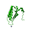


 Keywords
 sulfolobus solfataricus (archaea)
 staphylococcus aureus (bacteria)
 pseudomonas aeruginosa (bacteria)