 Movie
Movie Controller

[English] 日本語
 Yorodumi
- PDB-1ybi: Crystal structure of HA33A, a neurotoxin-associated protein from ... -
+ Open data

- Basic information
| Entry | Database: PDB / ID: 1ybi | ||||||
|---|---|---|---|---|---|---|---|
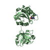
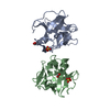
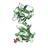
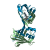
| Title | Crystal structure of HA33A, a neurotoxin-associated protein from Clostridium botulinum type A | ||||||
 Components Components | non-toxin haemagglutinin HA34 | ||||||
 Keywords Keywords | TOXIN / BETA-TREFOIL | ||||||
| Function / homology |  Function and homology information Function and homology informationRicin-type beta-trefoil lectin domain-like / Ricin-type beta-trefoil / Lectin domain of ricin B chain profile. / Ricin B, lectin domain / Ricin B-like lectins / Trefoil (Acidic Fibroblast Growth Factor, subunit A) - #50 / Trefoil (Acidic Fibroblast Growth Factor, subunit A) / Trefoil / Mainly Beta Similarity search - Domain/homology | ||||||
| Biological species |   Clostridium botulinum (bacteria) Clostridium botulinum (bacteria) | ||||||
| Method |  X-RAY DIFFRACTION / SYNCHROTRON / MOLECULAR REPLACEMENT / Resolution: 1.5 Å X-RAY DIFFRACTION / SYNCHROTRON / MOLECULAR REPLACEMENT / Resolution: 1.5 Å | ||||||
 Authors Authors | Arndt, J.W. / Gu, J. / Jaroszewski, L. / Schwarzenbacher, R. / Hanson, M. / Lebeda, F.J. / Stevens, R.C. | ||||||
 Citation Citation | Journal: J.Mol.Biol. / Year: 2005 Title: The Structure of the Neurotoxin-associated Protein HA33/A from Clostridium botulinum Suggests a Reoccurring beta-Trefoil Fold in the Progenitor Toxin Complex. Authors: Arndt, J.W. / Gu, J. / Jaroszewski, L. / Schwarzenbacher, R. / Hanson, M.A. / Lebeda, F.J. / Stevens, R.C. | ||||||
| History |
|
- Structure visualization
| Structure viewer | Molecule: MolmilJmol/JSmol |
|---|
- Downloads & links
-Download
| PDBx/mmCIF format | 1ybi.cif.gz | 137.5 KB | Display | PDBx/mmCIF format |
|---|---|---|---|---|
| PDB format | pdb1ybi.ent.gz | 107.4 KB | Display | PDB format |
| PDBx/mmJSON format | 1ybi.json.gz | Tree view | PDBx/mmJSON format | |
| Others |  Other downloads Other downloads |
-Validation report
| Arichive directory | https://data.pdbj.org/pub/pdb/validation_reports/yb/1ybiftp://data.pdbj.org/pub/pdb/validation_reports/yb/1ybi | HTTPS FTP |
|---|
-Related structure data
| Related structure data |  1qxmS S: Starting model for refinement |
|---|---|
| Similar structure data |


-Links
 PDBj
PDBj

- Assembly
| Deposited unit | 
| ||||||||
|---|---|---|---|---|---|---|---|---|---|
| 1 | 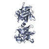
| ||||||||
| 2 | 
| ||||||||
| Unit cell |
|
-Components
| #1: Protein | Mass: 33251.828 Da / Num. of mol.: 2 / Source method: isolated from a natural source / Source: (natural) Clostridium botulinum (bacteria) / Strain: SEROTYPE A, HALL STRAIN / References: UniProt: Q45871#2: Water | ChemComp-HOH / |  Mass: 18.015 Da / Num. of mol.: 564 / Source method: isolated from a natural source / Formula: H2O Mass: 18.015 Da / Num. of mol.: 564 / Source method: isolated from a natural source / Formula: H2O |
|---|
-Experimental details
-Experiment
| Experiment | Method: X-RAY DIFFRACTION / Number of used crystals: 1 |
|---|
- Sample preparation
| Crystal | Density Matthews: 2.1 Å3/Da / Density % sol: 40 % |
|---|---|
| Crystal grow | Temperature: 298 K / Method: vapor diffusion, hanging drop / pH: 7.5 Details: 20% PEG 4000, 15% isopropanol, 200 mM Li2SO4, 0.1M HEPES, pH 7.5, VAPOR DIFFUSION, HANGING DROP, temperature 298K |
-Data collection
| Diffraction | Mean temperature: 100 K |
|---|---|
| Diffraction source | Source: SYNCHROTRON / Site: SSRL  / Beamline: BL9-1 / Wavelength: 0.9179 / Wavelength: 0.9179 Å / Beamline: BL9-1 / Wavelength: 0.9179 / Wavelength: 0.9179 Å |
| Detector | Type: ADSC / Detector: CCD |
| Radiation | Protocol: SINGLE WAVELENGTH / Monochromatic (M) / Laue (L): M / Scattering type: x-ray |
| Radiation wavelength | Wavelength: 0.9179 Å / Relative weight: 1 |
| Reflection | Resolution: 1.5→25 Å / Num. obs: 88654 / % possible obs: 87 % / Observed criterion σ(F): 2 / Observed criterion σ(I): 2 / Rsym value: 0.066 / Net I/σ(I): 13.6 |
| Reflection shell | Resolution: 1.5→1.539 Å / Mean I/σ(I) obs: 3.7 / Num. unique all: 88654 / Rsym value: 0.299 / % possible all: 78.6 |
- Processing
| Software |
| ||||||||||||||||||||||||||||||||||||||||||||||||||||||||||||||||||||||||||||||||||||||||||||||||||||||||||||||||||||||||||||||||||
|---|---|---|---|---|---|---|---|---|---|---|---|---|---|---|---|---|---|---|---|---|---|---|---|---|---|---|---|---|---|---|---|---|---|---|---|---|---|---|---|---|---|---|---|---|---|---|---|---|---|---|---|---|---|---|---|---|---|---|---|---|---|---|---|---|---|---|---|---|---|---|---|---|---|---|---|---|---|---|---|---|---|---|---|---|---|---|---|---|---|---|---|---|---|---|---|---|---|---|---|---|---|---|---|---|---|---|---|---|---|---|---|---|---|---|---|---|---|---|---|---|---|---|---|---|---|---|---|---|---|---|---|
| Refinement | Method to determine structure: MOLECULAR REPLACEMENT Starting model: pdb entry 1QXM Resolution: 1.5→21.22 Å / Cor.coef. Fo:Fc: 0.962 / Cor.coef. Fo:Fc free: 0.953 / SU B: 2.297 / SU ML: 0.047 / Cross valid method: THROUGHOUT / σ(F): 1 / ESU R: 0.085 / ESU R Free: 0.083 / Stereochemistry target values: MAXIMUM LIKELIHOOD / Details: HYDROGENS HAVE BEEN ADDED IN THE RIDING POSITIONS
| ||||||||||||||||||||||||||||||||||||||||||||||||||||||||||||||||||||||||||||||||||||||||||||||||||||||||||||||||||||||||||||||||||
| Solvent computation | Ion probe radii: 0.8 Å / Shrinkage radii: 0.8 Å / VDW probe radii: 1.2 Å / Solvent model: MASK | ||||||||||||||||||||||||||||||||||||||||||||||||||||||||||||||||||||||||||||||||||||||||||||||||||||||||||||||||||||||||||||||||||
| Displacement parameters | Biso mean: 19.526 Å2
| ||||||||||||||||||||||||||||||||||||||||||||||||||||||||||||||||||||||||||||||||||||||||||||||||||||||||||||||||||||||||||||||||||
| Refinement step | Cycle: LAST / Resolution: 1.5→21.22 Å
| ||||||||||||||||||||||||||||||||||||||||||||||||||||||||||||||||||||||||||||||||||||||||||||||||||||||||||||||||||||||||||||||||||
| Refine LS restraints |
| ||||||||||||||||||||||||||||||||||||||||||||||||||||||||||||||||||||||||||||||||||||||||||||||||||||||||||||||||||||||||||||||||||
| LS refinement shell | Resolution: 1.5→1.539 Å / Total num. of bins used: 20
|
