 Movie
Movie Controller

[English] 日本語
 Yorodumi
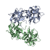
- PDB-1qxm: Crystal structure of a hemagglutinin component (HA1) from type C ... -
+ Open data

- Basic information
| Entry | Database: PDB / ID: 1qxm | ||||||
|---|---|---|---|---|---|---|---|
| Title | Crystal structure of a hemagglutinin component (HA1) from type C Clostridium botulinum | ||||||
 Components Components | HA1 | ||||||
 Keywords Keywords | TOXIN / Clostridium botulinum / hemagglutinin / HA1 / trefoil | ||||||
| Function / homology |  Function and homology information Function and homology information | ||||||
| Biological species |  Clostridium botulinum D phage (virus) Clostridium botulinum D phage (virus) | ||||||
| Method |  X-RAY DIFFRACTION / SIRAS / Resolution: 1.7 Å X-RAY DIFFRACTION / SIRAS / Resolution: 1.7 Å | ||||||
 Authors Authors | Inoue, K. / Sobhany, M. / Transue, T.R. / Oguma, K. / Pedersen, L.C. / Negishi, M. | ||||||
 Citation Citation | Journal: Microbiology / Year: 2003 Title: Structural analysis by X-ray crystallography and calorimetry of a haemagglutinin component (HA1) of the progenitor toxin from Clostridium botulinum. Authors: Inoue, K. / Sobhany, M. / Transue, T.R. / Oguma, K. / Pedersen, L.C. / Negishi, M. | ||||||
| History |
|
- Structure visualization
| Structure viewer | Molecule: MolmilJmol/JSmol |
|---|
- Downloads & links
-Download
| PDBx/mmCIF format | 1qxm.cif.gz | 142.6 KB | Display | PDBx/mmCIF format |
|---|---|---|---|---|
| PDB format | pdb1qxm.ent.gz | 111.7 KB | Display | PDB format |
| PDBx/mmJSON format | 1qxm.json.gz | Tree view | PDBx/mmJSON format | |
| Others |  Other downloads Other downloads |
-Validation report
| Arichive directory | https://data.pdbj.org/pub/pdb/validation_reports/qx/1qxmftp://data.pdbj.org/pub/pdb/validation_reports/qx/1qxm | HTTPS FTP |
|---|
-Related structure data
| Similar structure data |
|---|


-Links
 PDBj
PDBj


- Assembly
| Deposited unit | 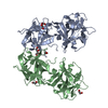
| ||||||||
|---|---|---|---|---|---|---|---|---|---|
| 1 |
| ||||||||
| Unit cell |
| ||||||||
| Details | not known |
-Components
| #1: Protein | Mass: 35549.348 Da / Num. of mol.: 2 / Fragment: hemagglutinin component HA1 / Mutation: type C Source method: isolated from a genetically manipulated source Source: (gene. exp.) Clostridium botulinum D phage (virus) / Species: Clostridium phage c-st / Plasmid: pGEX-4T-3 / Species (production host): Escherichia coli / Production host:  #2: Chemical | ChemComp-EDO /   Mass: 62.068 Da / Num. of mol.: 7 / Source method: obtained synthetically / Formula: C2H6O2 Mass: 62.068 Da / Num. of mol.: 7 / Source method: obtained synthetically / Formula: C2H6O2#3: Water | ChemComp-HOH / |  Mass: 18.015 Da / Num. of mol.: 651 / Source method: isolated from a natural source / Formula: H2O Mass: 18.015 Da / Num. of mol.: 651 / Source method: isolated from a natural source / Formula: H2O |
|---|
-Experimental details
-Experiment
| Experiment | Method: X-RAY DIFFRACTION / Number of used crystals: 1 |
|---|
- Sample preparation
| Crystal | Density Matthews: 2.41 Å3/Da / Density % sol: 49.03 % | ||||||||||||||||||||||||||||||||||||||||||
|---|---|---|---|---|---|---|---|---|---|---|---|---|---|---|---|---|---|---|---|---|---|---|---|---|---|---|---|---|---|---|---|---|---|---|---|---|---|---|---|---|---|---|---|
| Crystal grow | Temperature: 296 K / Method: sirsas / pH: 5.5 Details: MES, PEG8000, MgCl2, pH 5.5, SIRSAS, temperature 296K | ||||||||||||||||||||||||||||||||||||||||||
| Crystal grow | *PLUS Method: vapor diffusion, hanging drop | ||||||||||||||||||||||||||||||||||||||||||
| Components of the solutions | *PLUS
|
-Data collection
| Diffraction | Mean temperature: 100 K |
|---|---|
| Diffraction source | Source: ROTATING ANODE / Type: RIGAKU RU300 / Wavelength: 1.5418 Å |
| Radiation | Monochromator: Mirrors / Protocol: SINGLE WAVELENGTH / Monochromatic (M) / Laue (L): M / Scattering type: x-ray |
| Radiation wavelength | Wavelength: 1.5418 Å / Relative weight: 1 |
| Reflection | Resolution: 1.7→25 Å / Num. all: 71843 / Num. obs: 71843 / % possible obs: 96.4 % / Observed criterion σ(F): 0 / Observed criterion σ(I): -3 / Redundancy: 2.8 % / Biso Wilson estimate: 15.7 Å2 / Rsym value: 0.042 / Net I/σ(I): 23.2 |
| Reflection shell | Resolution: 1.7→1.76 Å / Redundancy: 2 % / Mean I/σ(I) obs: 8 / Num. unique all: 6546 / Rsym value: 0.15 / % possible all: 87.6 |
| Reflection | *PLUS Num. measured all: 203352 / Rmerge(I) obs: 0.042 |
| Reflection shell | *PLUS % possible obs: 87.6 % / Rmerge(I) obs: 0.15 |
- Processing
| Software |
| ||||||||||||||||||||||||||||||||||||
|---|---|---|---|---|---|---|---|---|---|---|---|---|---|---|---|---|---|---|---|---|---|---|---|---|---|---|---|---|---|---|---|---|---|---|---|---|---|
| Refinement | Method to determine structure: SIRAS / Resolution: 1.7→24.58 Å / Rfactor Rfree error: 0.003 / Isotropic thermal model: RESTRAINED / Cross valid method: THROUGHOUT / σ(F): 0 / Stereochemistry target values: Engh & Huber
| ||||||||||||||||||||||||||||||||||||
| Solvent computation | Solvent model: FLAT MODEL / Bsol: 42.5967 Å2 / ksol: 0.361759 e/Å3 | ||||||||||||||||||||||||||||||||||||
| Displacement parameters | Biso mean: 17.2 Å2
| ||||||||||||||||||||||||||||||||||||
| Refine analyze | Luzzati coordinate error free: 0.2 Å / Luzzati sigma a free: 0.11 Å | ||||||||||||||||||||||||||||||||||||
| Refinement step | Cycle: LAST / Resolution: 1.7→24.58 Å
| ||||||||||||||||||||||||||||||||||||
| Refine LS restraints |
| ||||||||||||||||||||||||||||||||||||
| LS refinement shell | Resolution: 1.7→1.81 Å / Rfactor Rfree error: 0.011 / Total num. of bins used: 6
| ||||||||||||||||||||||||||||||||||||
| Xplor file |
| ||||||||||||||||||||||||||||||||||||
| Refinement | *PLUS Highest resolution: 1.7 Å / Lowest resolution: 25 Å / % reflection Rfree: 5 % | ||||||||||||||||||||||||||||||||||||
| Solvent computation | *PLUS | ||||||||||||||||||||||||||||||||||||
| Displacement parameters | *PLUS | ||||||||||||||||||||||||||||||||||||
| Refine LS restraints | *PLUS
|
