 Movie
Movie Controller

[English] 日本語
 Yorodumi
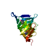
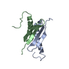
- PDB-3tlp: Crystal structure of the fourth bromodomain of human poly-bromodo... -
+ Open data

- Basic information
| Entry | Database: PDB / ID: 3tlp | ||||||
|---|---|---|---|---|---|---|---|
| Title | Crystal structure of the fourth bromodomain of human poly-bromodomain containing protein 1 (PB1) | ||||||
 Components Components | Protein polybromo-1 | ||||||
 Keywords Keywords | TRANSCRIPTION / PB1 / polybromo 1 isoform 1 / BAF180 / Polybromo-1D / PBRM1 / BRG1-associated factor 180 / Structural Genomics Consortium / SGC / Bromodomain | ||||||
| Function / homology |  Function and homology information Function and homology informationFormation of the polybromo-BAF (pBAF) complex / regulation of G0 to G1 transition / RSC-type complex / regulation of nucleotide-excision repair / regulation of mitotic metaphase/anaphase transition / SWI/SNF complex / positive regulation of T cell differentiation / positive regulation of double-strand break repair / nuclear chromosome / RUNX1 interacts with co-factors whose precise effect on RUNX1 targets is not known ...Formation of the polybromo-BAF (pBAF) complex / regulation of G0 to G1 transition / RSC-type complex / regulation of nucleotide-excision repair / regulation of mitotic metaphase/anaphase transition / SWI/SNF complex / positive regulation of T cell differentiation / positive regulation of double-strand break repair / nuclear chromosome / RUNX1 interacts with co-factors whose precise effect on RUNX1 targets is not known / regulation of G1/S transition of mitotic cell cycle / positive regulation of myoblast differentiation / positive regulation of cell differentiation / transcription elongation by RNA polymerase II / kinetochore / nuclear matrix / RMTs methylate histone arginines / mitotic cell cycle / chromatin remodeling / negative regulation of cell population proliferation / chromatin binding / regulation of transcription by RNA polymerase II / chromatin / DNA binding / nucleoplasm / nucleus Similarity search - Function | ||||||
| Biological species |  Homo sapiens (human) Homo sapiens (human) | ||||||
| Method |  X-RAY DIFFRACTION / SYNCHROTRON / MOLECULAR REPLACEMENT / molecular replacement / Resolution: 2.13 Å X-RAY DIFFRACTION / SYNCHROTRON / MOLECULAR REPLACEMENT / molecular replacement / Resolution: 2.13 Å | ||||||
 Authors Authors | Filippakopoulos, P. / Felletar, I. / Picaud, S. / Keates, T. / Muniz, J. / Krojer, T. / Allerston, C.K. / Latwiel, S. / von Delft, F. / Arrowsmith, C.H. ...Filippakopoulos, P. / Felletar, I. / Picaud, S. / Keates, T. / Muniz, J. / Krojer, T. / Allerston, C.K. / Latwiel, S. / von Delft, F. / Arrowsmith, C.H. / Edwards, A.M. / Weigelt, J. / Bountra, C. / Knapp, S. / Structural Genomics Consortium (SGC) | ||||||
 Citation Citation | Journal: Cell(Cambridge,Mass.) / Year: 2012 Title: Histone recognition and large-scale structural analysis of the human bromodomain family. Authors: Filippakopoulos, P. / Picaud, S. / Mangos, M. / Keates, T. / Lambert, J.P. / Barsyte-Lovejoy, D. / Felletar, I. / Volkmer, R. / Muller, S. / Pawson, T. / Gingras, A.C. / Arrowsmith, C.H. / Knapp, S. | ||||||
| History |
|
- Structure visualization
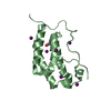
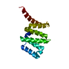
| Structure viewer | Molecule: MolmilJmol/JSmol |
|---|
- Downloads & links
-Download
| PDBx/mmCIF format | 3tlp.cif.gz | 112.5 KB | Display | PDBx/mmCIF format |
|---|---|---|---|---|
| PDB format | pdb3tlp.ent.gz | 84.5 KB | Display | PDB format |
| PDBx/mmJSON format | 3tlp.json.gz | Tree view | PDBx/mmJSON format | |
| Others |  Other downloads Other downloads |
-Validation report
| Arichive directory | https://data.pdbj.org/pub/pdb/validation_reports/tl/3tlpftp://data.pdbj.org/pub/pdb/validation_reports/tl/3tlp | HTTPS FTP |
|---|
-Related structure data
| Related structure data | 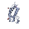 2nxbC 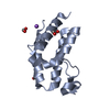 2oo1SC 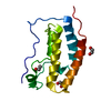 2ossSC 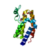 2ouoSC 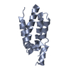 2rfjC 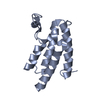 3d7cSC 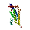 3daiSC 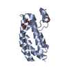 3dwySC 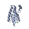 3gg3C 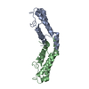 3hmeC 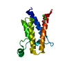 3hmfSC 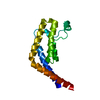 3hmhSC 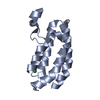 3i3jC 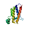 3iu5C 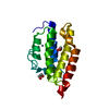 3iu6C 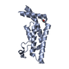 3lxjC 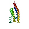 3mb3C 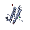 3mb4C 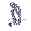 3mqmC 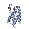 3nxbC 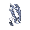 3p1cC 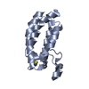 3p1dC 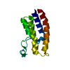 3q2eC 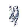 3rcwC 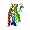 3uv2C  3uv4C 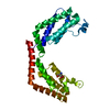 3uv5C 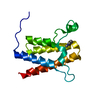 3uvdC 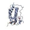 3uvwC 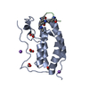 3uvxC 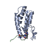 3uvyC  3uw9C S: Starting model for refinement C: citing same article ( |
|---|---|
| Similar structure data |


-Links
 PDBj
PDBj


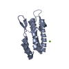
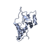
- Assembly
| Deposited unit | 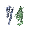
| ||||||||
|---|---|---|---|---|---|---|---|---|---|
| 1 | 
| ||||||||
| 2 | 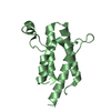
| ||||||||
| Unit cell |
|
-Components
| #1: Protein | Mass: 17464.084 Da / Num. of mol.: 2 / Fragment: UNP residues 496-637 Source method: isolated from a genetically manipulated source Source: (gene. exp.) Homo sapiens (human) / Gene: BAF180, PB1, PBRM1 / Plasmid: pNIC-CTHF / Production host:  #2: Chemical | ChemComp-NI /   Mass: 58.693 Da / Num. of mol.: 4 / Source method: obtained synthetically / Formula: Ni Mass: 58.693 Da / Num. of mol.: 4 / Source method: obtained synthetically / Formula: Ni#3: Water | ChemComp-HOH / |  Mass: 18.015 Da / Num. of mol.: 78 / Source method: isolated from a natural source / Formula: H2O Mass: 18.015 Da / Num. of mol.: 78 / Source method: isolated from a natural source / Formula: H2O |
|---|
-Experimental details
-Experiment
| Experiment | Method: X-RAY DIFFRACTION / Number of used crystals: 1 |
|---|
- Sample preparation
| Crystal | Density Matthews: 2.34 Å3/Da / Density % sol: 47.47 % |
|---|---|
| Crystal grow | Temperature: 277 K / Method: vapor diffusion, sitting drop / pH: 8.5 Details: 25% PEG_MME_2000, 0.015M NiCl, 0.1M Tris pH 8.5, VAPOR DIFFUSION, SITTING DROP, temperature 277K |
-Data collection
| Diffraction | Mean temperature: 100 K | ||||||||||||||||||||||||||||||||||||||||||||||||||||||||||||||||||||||||||||||||||||||||
|---|---|---|---|---|---|---|---|---|---|---|---|---|---|---|---|---|---|---|---|---|---|---|---|---|---|---|---|---|---|---|---|---|---|---|---|---|---|---|---|---|---|---|---|---|---|---|---|---|---|---|---|---|---|---|---|---|---|---|---|---|---|---|---|---|---|---|---|---|---|---|---|---|---|---|---|---|---|---|---|---|---|---|---|---|---|---|---|---|---|
| Diffraction source | Source: SYNCHROTRON / Site: Diamond  / Beamline: I03 / Wavelength: 1 Å / Beamline: I03 / Wavelength: 1 Å | ||||||||||||||||||||||||||||||||||||||||||||||||||||||||||||||||||||||||||||||||||||||||
| Detector | Type: PSI PILATUS 6M / Detector: PIXEL / Date: Jul 8, 2011 | ||||||||||||||||||||||||||||||||||||||||||||||||||||||||||||||||||||||||||||||||||||||||
| Radiation | Protocol: SINGLE WAVELENGTH / Monochromatic (M) / Laue (L): M / Scattering type: x-ray | ||||||||||||||||||||||||||||||||||||||||||||||||||||||||||||||||||||||||||||||||||||||||
| Radiation wavelength | Wavelength: 1 Å / Relative weight: 1 | ||||||||||||||||||||||||||||||||||||||||||||||||||||||||||||||||||||||||||||||||||||||||
| Reflection | Redundancy: 4.4 % / Av σ(I) over netI: 9.9 / Number: 82363 / Rsym value: 0.062 / D res high: 2.13 Å / D res low: 19.747 Å / Num. obs: 18871 / % possible obs: 99.4 | ||||||||||||||||||||||||||||||||||||||||||||||||||||||||||||||||||||||||||||||||||||||||
| Diffraction reflection shell |
| ||||||||||||||||||||||||||||||||||||||||||||||||||||||||||||||||||||||||||||||||||||||||
| Reflection | Resolution: 2.13→19.75 Å / Num. all: 18984 / Num. obs: 18871 / % possible obs: 99.4 % / Redundancy: 4.4 % / Biso Wilson estimate: 44.7 Å2 / Rmerge(I) obs: 0.062 / Rsym value: 0.062 / Net I/σ(I): 14.3 | ||||||||||||||||||||||||||||||||||||||||||||||||||||||||||||||||||||||||||||||||||||||||
| Reflection shell | Diffraction-ID: 1
|
-Phasing
| Phasing | Method: molecular replacement | |||||||||
|---|---|---|---|---|---|---|---|---|---|---|
| Phasing MR | Rfactor: 52.22 / Model details: Phaser MODE: MR_AUTO
|
- Processing
| Software |
| |||||||||||||||||||||||||||||||||||||||||||||||||||||||||||||||||||||||||||
|---|---|---|---|---|---|---|---|---|---|---|---|---|---|---|---|---|---|---|---|---|---|---|---|---|---|---|---|---|---|---|---|---|---|---|---|---|---|---|---|---|---|---|---|---|---|---|---|---|---|---|---|---|---|---|---|---|---|---|---|---|---|---|---|---|---|---|---|---|---|---|---|---|---|---|---|---|
| Refinement | Method to determine structure: MOLECULAR REPLACEMENT Starting model: PDB entries 3HMF, 2OO1, 3DAI, 3HMH, 2OSS, 2OUO, 3D7C, 3DWY Resolution: 2.13→19.75 Å / Cor.coef. Fo:Fc: 0.958 / Cor.coef. Fo:Fc free: 0.938 / WRfactor Rfree: 0.2298 / WRfactor Rwork: 0.188 / Occupancy max: 1 / Occupancy min: 1 / FOM work R set: 0.8454 / SU B: 9.063 / SU ML: 0.124 / SU R Cruickshank DPI: 0.1948 / SU Rfree: 0.1796 / Cross valid method: THROUGHOUT / σ(F): 0 / ESU R Free: 0.18 / Stereochemistry target values: MAXIMUM LIKELIHOOD Details: HYDROGENS HAVE BEEN ADDED IN THE RIDING POSITIONS U VALUES: WITH TLS ADDED
| |||||||||||||||||||||||||||||||||||||||||||||||||||||||||||||||||||||||||||
| Solvent computation | Ion probe radii: 0.8 Å / Shrinkage radii: 0.8 Å / VDW probe radii: 1.2 Å / Solvent model: MASK | |||||||||||||||||||||||||||||||||||||||||||||||||||||||||||||||||||||||||||
| Displacement parameters | Biso max: 194.25 Å2 / Biso mean: 50.8461 Å2 / Biso min: 21.35 Å2
| |||||||||||||||||||||||||||||||||||||||||||||||||||||||||||||||||||||||||||
| Refinement step | Cycle: LAST / Resolution: 2.13→19.75 Å
| |||||||||||||||||||||||||||||||||||||||||||||||||||||||||||||||||||||||||||
| Refine LS restraints |
| |||||||||||||||||||||||||||||||||||||||||||||||||||||||||||||||||||||||||||
| LS refinement shell | Resolution: 2.13→2.185 Å / Total num. of bins used: 20
| |||||||||||||||||||||||||||||||||||||||||||||||||||||||||||||||||||||||||||
| Refinement TLS params. | Method: refined / Refine-ID: X-RAY DIFFRACTION
| |||||||||||||||||||||||||||||||||||||||||||||||||||||||||||||||||||||||||||
| Refinement TLS group |
|
