 Movie
Movie Controller

[English] 日本語
 Yorodumi
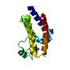
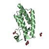
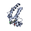
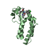
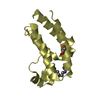
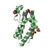
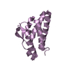
- PDB-3q2e: Crystal structure of the second bromodomain of human bromodomain ... -
+ Open data

- Basic information
| Entry | Database: PDB / ID: 3q2e | ||||||
|---|---|---|---|---|---|---|---|
| Title | Crystal structure of the second bromodomain of human bromodomain and WD repeat-containing protein 1 isoform A (WDR9) | ||||||
 Components Components | Bromodomain and WD repeat-containing protein 1 | ||||||
 Keywords Keywords | SIGNALING PROTEIN / Structural Genomics Consortium / SGC / Bromodomain / cell cycle progression / signal transduction / apoptosis / gene regulation / Down syndrome region-2 on chromosome 21 | ||||||
| Function / homology |  Function and homology information Function and homology informationmotile cilium / ciliary membrane / axoneme / Chromatin modifying enzymes / cytoskeleton organization / Interleukin-7 signaling / regulation of cell shape / regulation of transcription by RNA polymerase II / nucleolus / nucleoplasm ...motile cilium / ciliary membrane / axoneme / Chromatin modifying enzymes / cytoskeleton organization / Interleukin-7 signaling / regulation of cell shape / regulation of transcription by RNA polymerase II / nucleolus / nucleoplasm / nucleus / cytosol Similarity search - Function | ||||||
| Biological species |  Homo sapiens (human) Homo sapiens (human) | ||||||
| Method |  X-RAY DIFFRACTION / SYNCHROTRON / MOLECULAR REPLACEMENT / molecular replacement / Resolution: 1.74 Å X-RAY DIFFRACTION / SYNCHROTRON / MOLECULAR REPLACEMENT / molecular replacement / Resolution: 1.74 Å | ||||||
 Authors Authors | Filippakopoulos, P. / Felletar, I. / Picaud, S. / Keates, T. / Krojer, T. / Muniz, J. / Gileadi, O. / Von Delft, F. / Arrowsmith, C.H. / Edwards, A. ...Filippakopoulos, P. / Felletar, I. / Picaud, S. / Keates, T. / Krojer, T. / Muniz, J. / Gileadi, O. / Von Delft, F. / Arrowsmith, C.H. / Edwards, A. / Weigelt, J. / Bountra, C. / Knapp, S. / Structural Genomics Consortium (SGC) | ||||||
 Citation Citation | Journal: Cell(Cambridge,Mass.) / Year: 2012 Title: Histone recognition and large-scale structural analysis of the human bromodomain family. Authors: Filippakopoulos, P. / Picaud, S. / Mangos, M. / Keates, T. / Lambert, J.P. / Barsyte-Lovejoy, D. / Felletar, I. / Volkmer, R. / Muller, S. / Pawson, T. / Gingras, A.C. / Arrowsmith, C.H. / Knapp, S. | ||||||
| History |
|
- Structure visualization
| Structure viewer | Molecule: MolmilJmol/JSmol |
|---|
- Downloads & links
-Download
| PDBx/mmCIF format | 3q2e.cif.gz | 61.6 KB | Display | PDBx/mmCIF format |
|---|---|---|---|---|
| PDB format | pdb3q2e.ent.gz | 44 KB | Display | PDB format |
| PDBx/mmJSON format | 3q2e.json.gz | Tree view | PDBx/mmJSON format | |
| Others |  Other downloads Other downloads |
-Validation report
| Arichive directory | https://data.pdbj.org/pub/pdb/validation_reports/q2/3q2eftp://data.pdbj.org/pub/pdb/validation_reports/q2/3q2e | HTTPS FTP |
|---|
-Related structure data
| Related structure data | 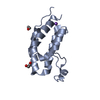 2nxbC 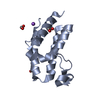 2oo1SC 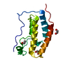 2ossSC 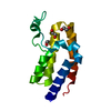 2ouoSC 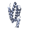 2rfjC 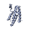 3d7cSC 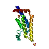 3daiSC 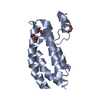 3dwySC  3gg3C  3hmeC 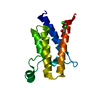 3hmfC  3hmhC 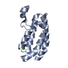 3i3jC 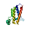 3iu5C 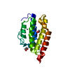 3iu6C 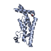 3lxjC 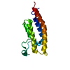 3mb3SC 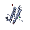 3mb4C 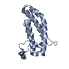 3mqmC 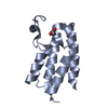 3nxbC  3p1cC  3p1dC  3rcwC 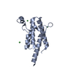 3tlpC 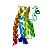 3uv2C  3uv4C  3uv5C 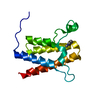 3uvdC 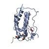 3uvwC 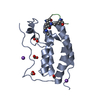 3uvxC 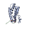 3uvyC 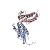 3uw9C 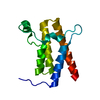 2grcS S: Starting model for refinement C: citing same article ( |
|---|---|
| Similar structure data |


-Links
 PDBj
PDBj- Assembly
| Deposited unit | 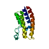
| ||||||||
|---|---|---|---|---|---|---|---|---|---|
| 1 |
| ||||||||
| Unit cell |
| ||||||||
| Components on special symmetry positions |
|
-Components
| #1: Protein | Mass: 14447.411 Da / Num. of mol.: 1 / Fragment: UNP RESIDUES 1310-1430 Source method: isolated from a genetically manipulated source Source: (gene. exp.) Homo sapiens (human) / Gene: BRWD1, C21orf107, WDR9 / Plasmid: pNIC28-Bsa4 / Production host:  |
|---|---|
| #2: Chemical | ChemComp-ACT /   Mass: 59.044 Da / Num. of mol.: 1 / Source method: obtained synthetically / Formula: C2H3O2 Mass: 59.044 Da / Num. of mol.: 1 / Source method: obtained synthetically / Formula: C2H3O2 |
| #3: Water | ChemComp-HOH /  Mass: 18.015 Da / Num. of mol.: 105 / Source method: isolated from a natural source / Formula: H2O Mass: 18.015 Da / Num. of mol.: 105 / Source method: isolated from a natural source / Formula: H2O |
-Experimental details
-Experiment
| Experiment | Method: X-RAY DIFFRACTION / Number of used crystals: 1 |
|---|
- Sample preparation
| Crystal | Density Matthews: 2.02 Å3/Da / Density % sol: 39.04 % / Mosaicity: 0.28 ° |
|---|---|
| Crystal grow | Temperature: 277 K / Method: vapor diffusion, sitting drop / pH: 5.5 Details: 0.2M (NH4)COOCH3, 0.1M BisTris, 25% PEG 3350 , pH 5.5, VAPOR DIFFUSION, SITTING DROP, temperature 277K |
-Data collection
| Diffraction | Mean temperature: 100 K | ||||||||||||||||||||||||||||||||||||||||||||||||||||||||||||||||||||||||||||||||||||||||||||||||||||||||||||||||||||||||||||||||||||
|---|---|---|---|---|---|---|---|---|---|---|---|---|---|---|---|---|---|---|---|---|---|---|---|---|---|---|---|---|---|---|---|---|---|---|---|---|---|---|---|---|---|---|---|---|---|---|---|---|---|---|---|---|---|---|---|---|---|---|---|---|---|---|---|---|---|---|---|---|---|---|---|---|---|---|---|---|---|---|---|---|---|---|---|---|---|---|---|---|---|---|---|---|---|---|---|---|---|---|---|---|---|---|---|---|---|---|---|---|---|---|---|---|---|---|---|---|---|---|---|---|---|---|---|---|---|---|---|---|---|---|---|---|---|
| Diffraction source | Source: SYNCHROTRON / Site: Diamond  / Beamline: I02 / Wavelength: 0.97 Å / Beamline: I02 / Wavelength: 0.97 Å | ||||||||||||||||||||||||||||||||||||||||||||||||||||||||||||||||||||||||||||||||||||||||||||||||||||||||||||||||||||||||||||||||||||
| Detector | Type: ADSC QUANTUM 315 / Detector: CCD / Date: Nov 27, 2010 | ||||||||||||||||||||||||||||||||||||||||||||||||||||||||||||||||||||||||||||||||||||||||||||||||||||||||||||||||||||||||||||||||||||
| Radiation | Protocol: SINGLE WAVELENGTH / Monochromatic (M) / Laue (L): M / Scattering type: x-ray | ||||||||||||||||||||||||||||||||||||||||||||||||||||||||||||||||||||||||||||||||||||||||||||||||||||||||||||||||||||||||||||||||||||
| Radiation wavelength | Wavelength: 0.97 Å / Relative weight: 1 | ||||||||||||||||||||||||||||||||||||||||||||||||||||||||||||||||||||||||||||||||||||||||||||||||||||||||||||||||||||||||||||||||||||
| Reflection | Redundancy: 6.7 % / Av σ(I) over netI: 5.6 / Number: 87081 / Rsym value: 0.088 / D res high: 1.74 Å / D res low: 28.892 Å / Num. obs: 12968 / % possible obs: 99.5 | ||||||||||||||||||||||||||||||||||||||||||||||||||||||||||||||||||||||||||||||||||||||||||||||||||||||||||||||||||||||||||||||||||||
| Diffraction reflection shell |
| ||||||||||||||||||||||||||||||||||||||||||||||||||||||||||||||||||||||||||||||||||||||||||||||||||||||||||||||||||||||||||||||||||||
| Reflection | Resolution: 1.74→28.892 Å / Num. all: 13033 / Num. obs: 12968 / % possible obs: 99.5 % / Redundancy: 6.7 % / Biso Wilson estimate: 25.9 Å2 / Rmerge(I) obs: 0.088 / Rsym value: 0.088 / Net I/σ(I): 12.9 | ||||||||||||||||||||||||||||||||||||||||||||||||||||||||||||||||||||||||||||||||||||||||||||||||||||||||||||||||||||||||||||||||||||
| Reflection shell | Diffraction-ID: 1
|
-Phasing
| Phasing | Method: molecular replacement | |||||||||
|---|---|---|---|---|---|---|---|---|---|---|
| Phasing MR | Rfactor: 49.93 / Model details: Phaser MODE: MR_AUTO
|
- Processing
| Software |
| |||||||||||||||||||||||||||||||||||||||||||||||||||||||||||||||||||||||||||||||||||||||||||||||||||||||||||||||||||||||||||||
|---|---|---|---|---|---|---|---|---|---|---|---|---|---|---|---|---|---|---|---|---|---|---|---|---|---|---|---|---|---|---|---|---|---|---|---|---|---|---|---|---|---|---|---|---|---|---|---|---|---|---|---|---|---|---|---|---|---|---|---|---|---|---|---|---|---|---|---|---|---|---|---|---|---|---|---|---|---|---|---|---|---|---|---|---|---|---|---|---|---|---|---|---|---|---|---|---|---|---|---|---|---|---|---|---|---|---|---|---|---|---|---|---|---|---|---|---|---|---|---|---|---|---|---|---|---|---|
| Refinement | Method to determine structure: MOLECULAR REPLACEMENT Starting model: 3MB3, 2OSS, 2OUO, 2GRC, 2OO1, 3DAI, 3D7C, 3DWY Resolution: 1.74→28.89 Å / Cor.coef. Fo:Fc: 0.96 / Cor.coef. Fo:Fc free: 0.954 / WRfactor Rfree: 0.2165 / WRfactor Rwork: 0.1822 / Occupancy max: 1 / Occupancy min: 0.5 / FOM work R set: 0.8744 / SU B: 4.247 / SU ML: 0.071 / SU R Cruickshank DPI: 0.1125 / SU Rfree: 0.1098 / Cross valid method: THROUGHOUT / σ(F): 0 / ESU R Free: 0.11 / Stereochemistry target values: MAXIMUM LIKELIHOOD Details: HYDROGENS HAVE BEEN ADDED IN THE RIDING POSITIONS U VALUES: WITH TLS ADDED
| |||||||||||||||||||||||||||||||||||||||||||||||||||||||||||||||||||||||||||||||||||||||||||||||||||||||||||||||||||||||||||||
| Solvent computation | Ion probe radii: 0.8 Å / Shrinkage radii: 0.8 Å / VDW probe radii: 1.4 Å / Solvent model: MASK | |||||||||||||||||||||||||||||||||||||||||||||||||||||||||||||||||||||||||||||||||||||||||||||||||||||||||||||||||||||||||||||
| Displacement parameters | Biso max: 89.96 Å2 / Biso mean: 29.4449 Å2 / Biso min: 11.64 Å2
| |||||||||||||||||||||||||||||||||||||||||||||||||||||||||||||||||||||||||||||||||||||||||||||||||||||||||||||||||||||||||||||
| Refinement step | Cycle: LAST / Resolution: 1.74→28.89 Å
| |||||||||||||||||||||||||||||||||||||||||||||||||||||||||||||||||||||||||||||||||||||||||||||||||||||||||||||||||||||||||||||
| Refine LS restraints |
| |||||||||||||||||||||||||||||||||||||||||||||||||||||||||||||||||||||||||||||||||||||||||||||||||||||||||||||||||||||||||||||
| LS refinement shell | Resolution: 1.74→1.785 Å / Total num. of bins used: 20
| |||||||||||||||||||||||||||||||||||||||||||||||||||||||||||||||||||||||||||||||||||||||||||||||||||||||||||||||||||||||||||||
| Refinement TLS params. | Method: refined / Refine-ID: X-RAY DIFFRACTION
| |||||||||||||||||||||||||||||||||||||||||||||||||||||||||||||||||||||||||||||||||||||||||||||||||||||||||||||||||||||||||||||
| Refinement TLS group |
|
