[English] 日本語
Yorodumi
- PDB-5a7c: Crystal structure of the second bromodomain of human BRD3 in comp... -

+
Open data


ID or keywords:

Loading...

-
Basic information

Entry
Database: PDB / ID: 5a7c
TitleCrystal structure of the second bromodomain of human BRD3 in complex with compound
ComponentsBROMODOMAIN-CONTAINING PROTEIN 3
KeywordsDNA BINDING PROTEIN / BRD3 / BROMODOMAIN CONTAINING PROTEIN 3 / RING3-LIKE PROTEIN / BRD3 DOMAIN 2
Function / homology
Function and homology information


lncRNA binding / endodermal cell differentiation / protein localization to chromatin / molecular condensate scaffold activity / lysine-acetylated histone binding / chromatin remodeling / chromatin binding / chromatin / regulation of transcription by RNA polymerase II / positive regulation of transcription by RNA polymerase II / nucleus
Similarity search - Function
NET domain superfamily / NET domain profile. / Brdt, bromodomain, repeat II / Brdt, bromodomain, repeat I / NET domain / Bromodomain extra-terminal - transcription regulation / Bromodomain-like / Histone Acetyltransferase; Chain A / Bromodomain, conserved site / Bromodomain signature. ...NET domain superfamily / NET domain profile. / Brdt, bromodomain, repeat II / Brdt, bromodomain, repeat I / NET domain / Bromodomain extra-terminal - transcription regulation / Bromodomain-like / Histone Acetyltransferase; Chain A / Bromodomain, conserved site / Bromodomain signature. / Bromodomain / Bromodomain profile. / bromo domain / Bromodomain / Bromodomain-like superfamily / Up-down Bundle / Mainly Alpha
Similarity search - Domain/homology
N-(6-ACETAMIDOHEXYL)ACETAMIDE / Bromodomain-containing protein 3
Similarity search - Component
Biological speciesHOMO SAPIENS (human)
MethodX-RAY DIFFRACTION / SYNCHROTRON / MOLECULAR REPLACEMENT / Resolution: 1.9 Å
AuthorsWelin, M. / Kimbung, R. / Diehl, C. / Hakansson, M. / Logan, D.T. / Walse, B.
CitationJournal: Cancer Res. / Year: 2016
Title: Cancer Differentiating Agent Hexamethylene Bisacetamide Inhibits Bet Bromodomain Proteins.
Authors: Nilsson, L.M. / Green, L.C. / Muralidharan, S.V. / Demir, D. / Welin, M. / Bhadury, J. / Logan, D.T. / Walse, B. / Nilsson, J.A.
History
DepositionJul 3, 2015Deposition site: PDBE / Processing site: PDBE
Revision 1.0Mar 16, 2016Provider: repository / Type: Initial release
Revision 1.1Sep 28, 2016Group: Database references
Revision 1.2Jan 17, 2018Group: Data collection / Category: diffrn_source / Item: _diffrn_source.pdbx_synchrotron_site
Revision 1.3Jan 10, 2024Group: Data collection / Database references ...Data collection / Database references / Derived calculations / Other / Refinement description
Category: chem_comp_atom / chem_comp_bond ...chem_comp_atom / chem_comp_bond / database_2 / pdbx_database_status / pdbx_initial_refinement_model / struct_ncs_dom_lim / struct_site
Item: _database_2.pdbx_DOI / _database_2.pdbx_database_accession ..._database_2.pdbx_DOI / _database_2.pdbx_database_accession / _pdbx_database_status.status_code_sf / _struct_ncs_dom_lim.beg_auth_comp_id / _struct_ncs_dom_lim.beg_label_asym_id / _struct_ncs_dom_lim.beg_label_comp_id / _struct_ncs_dom_lim.beg_label_seq_id / _struct_ncs_dom_lim.end_auth_comp_id / _struct_ncs_dom_lim.end_label_asym_id / _struct_ncs_dom_lim.end_label_comp_id / _struct_ncs_dom_lim.end_label_seq_id / _struct_site.pdbx_auth_asym_id / _struct_site.pdbx_auth_comp_id / _struct_site.pdbx_auth_seq_id

-
Structure visualization

Structure viewerMolecule:
MolmilJmol/JSmol

Downloads & links

-
Assembly

Deposited unit
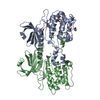
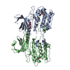
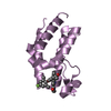
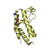
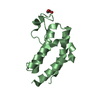
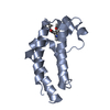
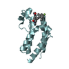
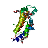
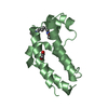
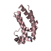
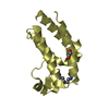
A: BROMODOMAIN-CONTAINING PROTEIN 3
B: BROMODOMAIN-CONTAINING PROTEIN 3
C: BROMODOMAIN-CONTAINING PROTEIN 3
D: BROMODOMAIN-CONTAINING PROTEIN 3
hetero molecules


Theoretical massNumber of molelcules
Total (without water)54,34113
Polymers53,2304
Non-polymers1,1119
Water6,593366
1
A: BROMODOMAIN-CONTAINING PROTEIN 3
hetero molecules


Theoretical massNumber of molelcules
Total (without water)13,6324
Polymers13,3071
Non-polymers3243
Water181
TypeNameSymmetry operationNumber
identity operation1_555x,y,z1
MethodPISA
2
B: BROMODOMAIN-CONTAINING PROTEIN 3
hetero molecules


Theoretical massNumber of molelcules
Total (without water)13,5703
Polymers13,3071
Non-polymers2622
Water181
TypeNameSymmetry operationNumber
identity operation1_555x,y,z1
MethodPISA
3
C: BROMODOMAIN-CONTAINING PROTEIN 3
hetero molecules


Theoretical massNumber of molelcules
Total (without water)13,5703
Polymers13,3071
Non-polymers2622
Water181
TypeNameSymmetry operationNumber
identity operation1_555x,y,z1
MethodPISA
4
D: BROMODOMAIN-CONTAINING PROTEIN 3
hetero molecules


Theoretical massNumber of molelcules
Total (without water)13,5703
Polymers13,3071
Non-polymers2622
Water181
TypeNameSymmetry operationNumber
identity operation1_555x,y,z1
MethodPISA
Unit cell
Length a, b, c (Å)103.365, 92.013, 64.898
Angle α, β, γ (deg.)90.00, 95.35, 90.00
Int Tables number5
Space group name H-MC121
Noncrystallographic symmetry (NCS)NCS domain:
IDEns-IDDetails
11A
21B
12A
22C
13A
23D
14B
24C
15B
25D
16C
26D

NCS domain segments:

Component-ID: _ / Refine code: _

Dom-IDEns-IDBeg auth comp-IDBeg label comp-IDEnd auth comp-IDEnd label comp-IDAuth asym-IDLabel asym-IDAuth seq-IDLabel seq-ID
11GLYGLYMETMETAA306 - 4153 - 112
21GLYGLYMETMETBB306 - 4153 - 112
12LEULEUMETMETAA308 - 4155 - 112
22LEULEUMETMETCC308 - 4155 - 112
13LEULEUMETMETAA308 - 4155 - 112
23LEULEUMETMETDD308 - 4155 - 112
14LEULEUMETMETBB308 - 4155 - 112
24LEULEUMETMETCC308 - 4155 - 112
15LEULEUMETMETBB308 - 4155 - 112
25LEULEUMETMETDD308 - 4155 - 112
16LEULEUPROPROCC308 - 4165 - 113
26LEULEUPROPRODD308 - 4165 - 113

NCS ensembles :
ID
1
2
3
4
5
6

NCS oper:
IDCodeMatrixVector
1given(0.582, -0.679, 0.448), (-0.668, -0.713, -0.214), (0.464, -0.174, -0.868)-24.627, -18.204, 58.751
2given(0.582, -0.679, 0.448), (-0.668, -0.713, -0.214), (0.464, -0.174, -0.868)-24.627, -18.204, 58.751
3given(-0.668, 0.669, 0.327), (0.679, 0.727, -0.101), (-0.306, 0.155, -0.94)11.39, -30.85, 112.24

-
Components

#1: Protein
BROMODOMAIN-CONTAINING PROTEIN 3 / RING3-LIKE PROTEIN


Mass: 13307.399 Da / Num. of mol.: 4 / Fragment: BROMO 2 DOMAIN, RESIDUES 306-416
Source method: isolated from a genetically manipulated source
Source: (gene. exp.) HOMO SAPIENS (human) / Plasmid: PNIC28-BSA4 / Production host: ESCHERICHIA COLI (E. coli) / Strain (production host): BL21(DE3) / Variant (production host): T1R PRARE2 / References: UniProt: Q15059
#2: Chemical
ChemComp-5D4 / N-(6-ACETAMIDOHEXYL)ACETAMIDE


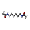
Mass: 200.278 Da / Num. of mol.: 4 / Source method: obtained synthetically / Formula: C10H20N2O2
#3: Chemical
ChemComp-EDO / 1,2-ETHANEDIOL / ETHYLENE GLYCOL


Mass: 62.068 Da / Num. of mol.: 5 / Source method: obtained synthetically / Formula: C2H6O2
#4: Water ChemComp-HOH / water


Mass: 18.015 Da / Num. of mol.: 366 / Source method: isolated from a natural source / Formula: H2O

-
Experimental details

-
Experiment

ExperimentMethod: X-RAY DIFFRACTION / Number of used crystals: 1

-
Sample preparation

CrystalDensity Matthews: 2.89 Å3/Da / Density % sol: 57.5 % / Description: NONE
Crystal growpH: 7 / Details: 19% PEG 6000, 0.1M HEPES PH 7.0

-
Data collection

DiffractionMean temperature: 100 K
Diffraction sourceSource: SYNCHROTRON / Site: MAX II / Beamline: I911-3 / Wavelength: 1
DetectorType: MARMOSAIC 225 mm CCD / Detector: CCD / Date: Jun 25, 2015 / Details: MIRRORS
RadiationMonochromator: SI (111) DOUBLE CRYSTAL MONOCHROMATOR / Protocol: SINGLE WAVELENGTH / Monochromatic (M) / Laue (L): M / Scattering type: x-ray
Radiation wavelengthWavelength: 1 Å / Relative weight: 1
ReflectionResolution: 1.9→48.56 Å / Num. obs: 47219 / % possible obs: 99.5 % / Redundancy: 3.4 % / Rmerge(I) obs: 0.16 / Net I/σ(I): 7
Reflection shellResolution: 1.9→1.95 Å / Redundancy: 2.5 % / Rmerge(I) obs: 0.77 / Mean I/σ(I) obs: 1.4 / % possible all: 94.6

-
Processing

Software
NameVersionClassification
REFMAC5.8.0135refinement
XDSdata reduction
XDSdata scaling
PHASERphasing
RefinementMethod to determine structure: MOLECULAR REPLACEMENT
Starting model: PDB ENTRY 3S92
Resolution: 1.9→50.01 Å / Cor.coef. Fo:Fc: 0.915 / Cor.coef. Fo:Fc free: 0.895 / SU B: 4.846 / SU ML: 0.131 / Cross valid method: THROUGHOUT / ESU R: 0.168 / ESU R Free: 0.15 / Stereochemistry target values: MAXIMUM LIKELIHOOD
Details: HYDROGENS HAVE BEEN ADDED IN THE RIDING POSITIONS. U VALUES REFINED INDIVIDUALLY
RfactorNum. reflection% reflectionSelection details
Rfree0.25558 2311 4.9 %RANDOM
Rwork0.22813 ---
obs0.22947 44884 99.36 %-
Solvent computationIon probe radii: 0.8 Å / Shrinkage radii: 0.8 Å / VDW probe radii: 1.2 Å / Solvent model: MASK
Displacement parametersBiso mean: 20.218 Å2
Baniso -1Baniso -2Baniso -3
1-1.75 Å20 Å20.24 Å2
2---0.69 Å20 Å2
3----1.08 Å2
Refinement stepCycle: LAST / Resolution: 1.9→50.01 Å
ProteinNucleic acidLigandSolventTotal
Num. atoms3650 0 76 366 4092
Refine LS restraints
Refine-IDTypeDev idealDev ideal targetNumber
X-RAY DIFFRACTIONr_bond_refined_d0.0150.0193837
X-RAY DIFFRACTIONr_bond_other_d0.010.023646
X-RAY DIFFRACTIONr_angle_refined_deg1.651.9745148
X-RAY DIFFRACTIONr_angle_other_deg1.50138423
X-RAY DIFFRACTIONr_dihedral_angle_1_deg4.9455439
X-RAY DIFFRACTIONr_dihedral_angle_2_deg35.64122.963189
X-RAY DIFFRACTIONr_dihedral_angle_3_deg12.73615674
X-RAY DIFFRACTIONr_dihedral_angle_4_deg21.8011528
X-RAY DIFFRACTIONr_chiral_restr0.090.2510
X-RAY DIFFRACTIONr_gen_planes_refined0.0110.0214235
X-RAY DIFFRACTIONr_gen_planes_other0.0090.02907
X-RAY DIFFRACTIONr_nbd_refined
X-RAY DIFFRACTIONr_nbd_other
X-RAY DIFFRACTIONr_nbtor_refined
X-RAY DIFFRACTIONr_nbtor_other
X-RAY DIFFRACTIONr_xyhbond_nbd_refined
X-RAY DIFFRACTIONr_xyhbond_nbd_other
X-RAY DIFFRACTIONr_metal_ion_refined
X-RAY DIFFRACTIONr_metal_ion_other
X-RAY DIFFRACTIONr_symmetry_vdw_refined
X-RAY DIFFRACTIONr_symmetry_vdw_other
X-RAY DIFFRACTIONr_symmetry_hbond_refined
X-RAY DIFFRACTIONr_symmetry_hbond_other
X-RAY DIFFRACTIONr_symmetry_metal_ion_refined
X-RAY DIFFRACTIONr_symmetry_metal_ion_other
X-RAY DIFFRACTIONr_mcbond_it1.4941.7511765
X-RAY DIFFRACTIONr_mcbond_other1.4941.7521766
X-RAY DIFFRACTIONr_mcangle_it2.2912.6252213
X-RAY DIFFRACTIONr_mcangle_other2.2772.6252211
X-RAY DIFFRACTIONr_scbond_it2.532.0932071
X-RAY DIFFRACTIONr_scbond_other2.532.0932071
X-RAY DIFFRACTIONr_scangle_it
X-RAY DIFFRACTIONr_scangle_other4.03132936
X-RAY DIFFRACTIONr_long_range_B_refined5.53114.744851
X-RAY DIFFRACTIONr_long_range_B_other5.5314.7434852
X-RAY DIFFRACTIONr_rigid_bond_restr
X-RAY DIFFRACTIONr_sphericity_free
X-RAY DIFFRACTIONr_sphericity_bonded
Refine LS restraints NCS

Refine-ID: X-RAY DIFFRACTION / Type: interatomic distance / Weight position: 0.05

Ens-IDDom-IDAuth asym-IDNumberRms dev position (Å)
11A134080.06
12B134080.06
21A134060.04
22C134060.04
31A132900.06
32D132900.06
41B131300.06
42C131300.06
51B133160.04
52D133160.04
61C133620.06
62D133620.06
LS refinement shellHighest resolution: 1.902 Å / Total num. of bins used: 20
RfactorNum. reflection% reflection
Rfree0.401 158 -
Rwork0.402 3163 -
obs--94.72 %

+
About Yorodumi

-
News

-
Feb 9, 2022. New format data for meta-information of EMDB entries

New format data for meta-information of EMDB entries

  • Version 3 of the EMDB header file is now the official format.
  • The previous official version 1.9 will be removed from the archive.

Related info.:EMDB header

External links:wwPDB to switch to version 3 of the EMDB data model

-
Aug 12, 2020. Covid-19 info

Covid-19 info

URL: https://pdbjlvh1.pdbj.org/emnavi/covid19.php

New page: Covid-19 featured information page in EM Navigator.

Related info.:Covid-19 info / Mar 5, 2020. Novel coronavirus structure data

+
Mar 5, 2020. Novel coronavirus structure data

Novel coronavirus structure data

Related info.:Yorodumi Speices / Aug 12, 2020. Covid-19 info

External links:COVID-19 featured content - PDBj / Molecule of the Month (242):Coronavirus Proteases

+
Jan 31, 2019. EMDB accession codes are about to change! (news from PDBe EMDB page)

EMDB accession codes are about to change! (news from PDBe EMDB page)

  • The allocation of 4 digits for EMDB accession codes will soon come to an end. Whilst these codes will remain in use, new EMDB accession codes will include an additional digit and will expand incrementally as the available range of codes is exhausted. The current 4-digit format prefixed with “EMD-” (i.e. EMD-XXXX) will advance to a 5-digit format (i.e. EMD-XXXXX), and so on. It is currently estimated that the 4-digit codes will be depleted around Spring 2019, at which point the 5-digit format will come into force.
  • The EM Navigator/Yorodumi systems omit the EMD- prefix.

Related info.:Q: What is EMD? / ID/Accession-code notation in Yorodumi/EM Navigator

External links:EMDB Accession Codes are Changing Soon! / Contact to PDBj

+
Jul 12, 2017. Major update of PDB

Major update of PDB

  • wwPDB released updated PDB data conforming to the new PDBx/mmCIF dictionary.
  • This is a major update changing the version number from 4 to 5, and with Remediation, in which all the entries are updated.
  • In this update, many items about electron microscopy experimental information are reorganized (e.g. em_software).
  • Now, EM Navigator and Yorodumi are based on the updated data.

External links:wwPDB Remediation / Enriched Model Files Conforming to OneDep Data Standards Now Available in the PDB FTP Archive

-
Yorodumi

Thousand views of thousand structures

  • Yorodumi is a browser for structure data from EMDB, PDB, SASBDB, etc.
  • This page is also the successor to EM Navigator detail page, and also detail information page/front-end page for Omokage search.
  • The word "yorodu" (or yorozu) is an old Japanese word meaning "ten thousand". "mi" (miru) is to see.

Related info.:EMDB / PDB / SASBDB / Comparison of 3 databanks / Yorodumi Search / Aug 31, 2016. New EM Navigator & Yorodumi / Yorodumi Papers / Jmol/JSmol / Function and homology information / Changes in new EM Navigator and Yorodumi

Read more