 Movie
Movie Controller

[English] 日本語
 Yorodumi
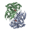
- PDB-5a6n: Crystal structure of human death associated protein kinase 3 (DAP... -
+ Open data

- Basic information
| Entry | Database: PDB / ID: 5a6n | ||||||
|---|---|---|---|---|---|---|---|
| Title | Crystal structure of human death associated protein kinase 3 (DAPK3) in complex with compound 2 | ||||||
 Components Components | DEATH-ASSOCIATED PROTEIN KINASE 3 | ||||||
 Keywords Keywords | TRANSFERASE / DAPK3 / HUMAN DEATH ASSOCIATED PROTEIN KINASE 3 / DAP-LIKE KINASE / DLK / ZIPPER-INTERACTING PROTEIN KINASE / ZIP-KINASE | ||||||
| Function / homology |  Function and homology information Function and homology informationregulation of myosin II filament organization / leucine zipper domain binding / cAMP response element binding protein binding / regulation of smooth muscle contraction / regulation of cell motility / Caspase activation via Dependence Receptors in the absence of ligand / regulation of focal adhesion assembly / regulation of mitotic nuclear division / chromosome, centromeric region / regulation of mitotic cell cycle ...regulation of myosin II filament organization / leucine zipper domain binding / cAMP response element binding protein binding / regulation of smooth muscle contraction / regulation of cell motility / Caspase activation via Dependence Receptors in the absence of ligand / regulation of focal adhesion assembly / regulation of mitotic nuclear division / chromosome, centromeric region / regulation of mitotic cell cycle / regulation of autophagy / regulation of actin cytoskeleton organization / apoptotic signaling pathway / PML body / cellular response to type II interferon / spindle / small GTPase binding / positive regulation of canonical Wnt signaling pathway / protein autophosphorylation / regulation of cell shape / chromatin organization / midbody / regulation of apoptotic process / protein phosphorylation / protein kinase activity / non-specific serine/threonine protein kinase / cilium / negative regulation of translation / intracellular signal transduction / positive regulation of cell migration / positive regulation of apoptotic process / protein serine kinase activity / protein serine/threonine kinase activity / apoptotic process / centrosome / regulation of DNA-templated transcription / protein homodimerization activity / nucleoplasm / ATP binding / identical protein binding / nucleus / cytosol / cytoplasm Similarity search - Function | ||||||
| Biological species |  HOMO SAPIENS (human) HOMO SAPIENS (human) | ||||||
| Method |  X-RAY DIFFRACTION / SYNCHROTRON / MOLECULAR REPLACEMENT / Resolution: 1.7 Å X-RAY DIFFRACTION / SYNCHROTRON / MOLECULAR REPLACEMENT / Resolution: 1.7 Å | ||||||
 Authors Authors | Rodrigues, T. / Reker, D. / Welin, M. / Caldera, M. / Brunner, C. / Gabernet, G. / Schneider, P. / Walse, B. / Schneider, G. | ||||||
 Citation Citation | Journal: Angew.Chem.Int.Ed.Engl. / Year: 2015 Title: De Novo Fragment Design for Drug Discovery and Chemical Biology. Authors: Rodrigues, T. / Reker, D. / Welin, M. / Caldera, M. / Brunner, C. / Gabernet, G. / Schneider, P. / Walse, B. / Schneider, G. | ||||||
| History |
|
- Structure visualization
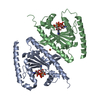
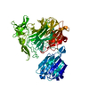
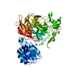
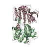
| Structure viewer | Molecule: MolmilJmol/JSmol |
|---|
- Downloads & links
-Download
| PDBx/mmCIF format | 5a6n.cif.gz | 127.9 KB | Display | PDBx/mmCIF format |
|---|---|---|---|---|
| PDB format | pdb5a6n.ent.gz | 99.6 KB | Display | PDB format |
| PDBx/mmJSON format | 5a6n.json.gz | Tree view | PDBx/mmJSON format | |
| Others |  Other downloads Other downloads |
-Validation report
| Arichive directory | https://data.pdbj.org/pub/pdb/validation_reports/a6/5a6nftp://data.pdbj.org/pub/pdb/validation_reports/a6/5a6n | HTTPS FTP |
|---|
-Related structure data
| Related structure data |  5a6oC 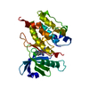 3bhyS C: citing same article ( S: Starting model for refinement |
|---|---|
| Similar structure data |


-Links
 PDBj
PDBj
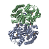
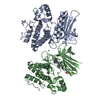
- Assembly
| Deposited unit | 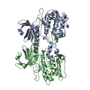
| ||||||||
|---|---|---|---|---|---|---|---|---|---|
| 1 |
| ||||||||
| Unit cell |
| ||||||||
| Noncrystallographic symmetry (NCS) | NCS oper: (Code: given Matrix: (-0.998, 0.049, -0.043), Vector: |
-Components
| #1: Protein | Mass: 32545.123 Da / Num. of mol.: 2 / Fragment: PROTEIN KINASE DOMAIN, UNP RESIDUES 9-289 Source method: isolated from a genetically manipulated source Source: (gene. exp.) HOMO SAPIENS (human) / Plasmid: PNIC28-BSA4 / Production host:  References: UniProt: O43293, non-specific serine/threonine protein kinase #2: Chemical | ChemComp-U7E / | 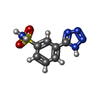  Mass: 225.228 Da / Num. of mol.: 1 / Source method: obtained synthetically / Formula: C7H7N5O2S Mass: 225.228 Da / Num. of mol.: 1 / Source method: obtained synthetically / Formula: C7H7N5O2S#3: Chemical |   Mass: 92.094 Da / Num. of mol.: 2 / Source method: obtained synthetically / Formula: C3H8O3 Mass: 92.094 Da / Num. of mol.: 2 / Source method: obtained synthetically / Formula: C3H8O3#4: Chemical |   Mass: 76.094 Da / Num. of mol.: 2 / Source method: obtained synthetically / Formula: C3H8O2 Mass: 76.094 Da / Num. of mol.: 2 / Source method: obtained synthetically / Formula: C3H8O2#5: Water | ChemComp-HOH / |  Mass: 18.015 Da / Num. of mol.: 298 / Source method: isolated from a natural source / Formula: H2O Mass: 18.015 Da / Num. of mol.: 298 / Source method: isolated from a natural source / Formula: H2O |
|---|
-Experimental details
-Experiment
| Experiment | Method: X-RAY DIFFRACTION / Number of used crystals: 1 |
|---|
- Sample preparation
| Crystal | Density Matthews: 2.49 Å3/Da / Density % sol: 50.6 % / Description: NONE |
|---|---|
| Crystal grow | pH: 6.2 Details: 23% PROPANEDIOL, 0.1M NA/K PHOSPHATE PH 6.2, 10% GLYCEROL |
-Data collection
| Diffraction | Mean temperature: 100 K |
|---|---|
| Diffraction source | Source: SYNCHROTRON / Site: Diamond  / Beamline: I03 / Wavelength: 0.97625 / Beamline: I03 / Wavelength: 0.97625 |
| Detector | Type: DECTRIS PILATUS 6M / Detector: PIXEL / Date: Apr 24, 2015 |
| Radiation | Monochromator: SI(111) DOUBLE CRYSTAL / Protocol: SINGLE WAVELENGTH / Monochromatic (M) / Laue (L): M / Scattering type: x-ray |
| Radiation wavelength | Wavelength: 0.97625 Å / Relative weight: 1 |
| Reflection | Resolution: 1.7→29.71 Å / Num. obs: 71490 / % possible obs: 99.5 % / Redundancy: 4.3 % / Biso Wilson estimate: 19.5 Å2 / Rmerge(I) obs: 0.06 / Net I/σ(I): 15.3 |
| Reflection shell | Resolution: 1.7→1.73 Å / Redundancy: 3.3 % / Rmerge(I) obs: 0.35 / Mean I/σ(I) obs: 3.6 / % possible all: 98.4 |
- Processing
| Software |
| ||||||||||||||||||||||||||||||||||||||||||||||||||||||||||||||||||||||||||||||||||||||||||||||||||||||||||||||||||||||||||||||||||||||||||||||||||||||||||||||||||||||||||||||||||||||
|---|---|---|---|---|---|---|---|---|---|---|---|---|---|---|---|---|---|---|---|---|---|---|---|---|---|---|---|---|---|---|---|---|---|---|---|---|---|---|---|---|---|---|---|---|---|---|---|---|---|---|---|---|---|---|---|---|---|---|---|---|---|---|---|---|---|---|---|---|---|---|---|---|---|---|---|---|---|---|---|---|---|---|---|---|---|---|---|---|---|---|---|---|---|---|---|---|---|---|---|---|---|---|---|---|---|---|---|---|---|---|---|---|---|---|---|---|---|---|---|---|---|---|---|---|---|---|---|---|---|---|---|---|---|---|---|---|---|---|---|---|---|---|---|---|---|---|---|---|---|---|---|---|---|---|---|---|---|---|---|---|---|---|---|---|---|---|---|---|---|---|---|---|---|---|---|---|---|---|---|---|---|---|---|
| Refinement | Method to determine structure: MOLECULAR REPLACEMENT Starting model: PDB ENTRY 3BHY Resolution: 1.7→30 Å / Cor.coef. Fo:Fc: 0.959 / Cor.coef. Fo:Fc free: 0.947 / SU B: 1.875 / SU ML: 0.063 / Cross valid method: THROUGHOUT / ESU R: 0.098 / ESU R Free: 0.097 / Stereochemistry target values: MAXIMUM LIKELIHOOD Details: HYDROGENS HAVE BEEN ADDED IN THE RIDING POSITIONS. U VALUE REFINED INDIVIDUALLY. RESIDUES 170-174 IN CHAIN B ARE DISORDERED.
| ||||||||||||||||||||||||||||||||||||||||||||||||||||||||||||||||||||||||||||||||||||||||||||||||||||||||||||||||||||||||||||||||||||||||||||||||||||||||||||||||||||||||||||||||||||||
| Solvent computation | Ion probe radii: 0.8 Å / Shrinkage radii: 0.8 Å / VDW probe radii: 1.2 Å / Solvent model: MASK | ||||||||||||||||||||||||||||||||||||||||||||||||||||||||||||||||||||||||||||||||||||||||||||||||||||||||||||||||||||||||||||||||||||||||||||||||||||||||||||||||||||||||||||||||||||||
| Displacement parameters | Biso mean: 26.386 Å2
| ||||||||||||||||||||||||||||||||||||||||||||||||||||||||||||||||||||||||||||||||||||||||||||||||||||||||||||||||||||||||||||||||||||||||||||||||||||||||||||||||||||||||||||||||||||||
| Refinement step | Cycle: LAST / Resolution: 1.7→30 Å
| ||||||||||||||||||||||||||||||||||||||||||||||||||||||||||||||||||||||||||||||||||||||||||||||||||||||||||||||||||||||||||||||||||||||||||||||||||||||||||||||||||||||||||||||||||||||
| Refine LS restraints |
|