 Movie
Movie Controller

[English] 日本語
 Yorodumi
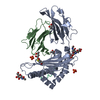
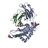
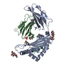
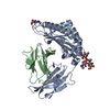
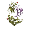
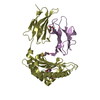
- PDB-2clz: Mhc Class I Natural Mutant H-2Kbm8 Heavy Chain Complexed With bet... -
+ Open data

- Basic information
| Entry | Database: PDB / ID: 2clz | ||||||
|---|---|---|---|---|---|---|---|
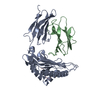
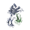
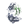
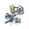
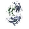
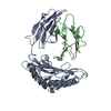
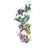
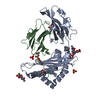
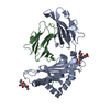
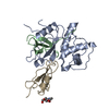
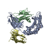
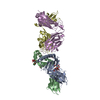
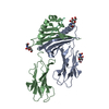
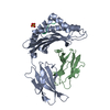
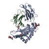
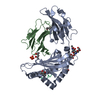
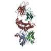
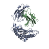
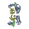
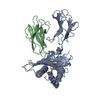
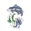
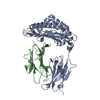
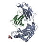
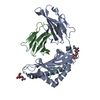
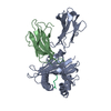
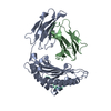
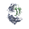
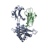
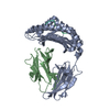
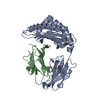
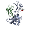
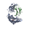
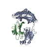
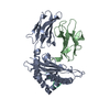
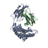
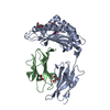
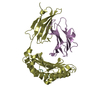
| Title | Mhc Class I Natural Mutant H-2Kbm8 Heavy Chain Complexed With beta-2 Microglobulin and pBM1 peptide | ||||||
 Components Components |
| ||||||
 Keywords Keywords | IMMUNE SYSTEM / IMMUNE RESPONSE / IMMUNOGLOBULIN DOMAIN / GLYCOPROTEIN / TRANSMEMBRANE / ALLOREACTIVITY / MHC I / H-2KBM8 / MEMBRANE / CLASS I MHC / POLYMORPHISM | ||||||
| Function / homology |  Function and homology information Function and homology informationmRNA Polyadenylation / mRNA Splicing - Major Pathway / MHC class Ib protein complex / natural killer cell lectin-like receptor binding / TAP2 binding / TAP1 binding / cis-Golgi network membrane / Endosomal/Vacuolar pathway / DAP12 interactions / Antigen Presentation: Folding, assembly and peptide loading of class I MHC ...mRNA Polyadenylation / mRNA Splicing - Major Pathway / MHC class Ib protein complex / natural killer cell lectin-like receptor binding / TAP2 binding / TAP1 binding / cis-Golgi network membrane / Endosomal/Vacuolar pathway / DAP12 interactions / Antigen Presentation: Folding, assembly and peptide loading of class I MHC / ER-Phagosome pathway / DAP12 signaling / Immunoregulatory interactions between a Lymphoid and a non-Lymphoid cell / perinuclear theca / antigen processing and presentation of endogenous peptide antigen via MHC class I via ER pathway, TAP-dependent / TAP complex binding / antigen processing and presentation of exogenous peptide antigen via MHC class I / Golgi medial cisterna / regulation of alternative mRNA splicing, via spliceosome / regulation of membrane depolarization / inner ear development / CD8 receptor binding / spliceosomal complex assembly / beta-2-microglobulin binding / endoplasmic reticulum exit site / TAP binding / positive regulation of phosphorylation / MHC class I protein binding / antigen processing and presentation of endogenous peptide antigen via MHC class Ib / antigen processing and presentation of endogenous peptide antigen via MHC class I via ER pathway, TAP-independent / cellular defense response / T cell receptor binding / Neutrophil degranulation / 14-3-3 protein binding / lumenal side of endoplasmic reticulum membrane / regulation of iron ion transport / spliceosomal complex / cellular response to iron(III) ion / negative regulation of iron ion transport / negative regulation of forebrain neuron differentiation / antigen processing and presentation of exogenous protein antigen via MHC class Ib, TAP-dependent / centriole / iron ion transport / peptide antigen assembly with MHC class I protein complex / regulation of erythrocyte differentiation / response to molecule of bacterial origin / HFE-transferrin receptor complex / MHC class I peptide loading complex / sperm end piece / transferrin transport / cellular response to iron ion / negative regulation of receptor-mediated endocytosis / mRNA splicing, via spliceosome / positive regulation of T cell cytokine production / antigen processing and presentation of endogenous peptide antigen via MHC class I / MHC class I protein complex / peptide antigen assembly with MHC class II protein complex / negative regulation of neurogenesis / cellular response to nicotine / MHC class II protein complex / positive regulation of receptor-mediated endocytosis / multicellular organismal-level iron ion homeostasis / positive regulation of T cell mediated cytotoxicity / antigen processing and presentation of exogenous peptide antigen via MHC class II / positive regulation of immune response / peptide antigen binding / phagocytic vesicle membrane / negative regulation of epithelial cell proliferation / positive regulation of T cell activation / sensory perception of smell / positive regulation of cellular senescence / MHC class II protein complex binding / T cell differentiation in thymus / late endosome membrane / antimicrobial humoral immune response mediated by antimicrobial peptide / negative regulation of neuron projection development / antibacterial humoral response / protein refolding / protein-folding chaperone binding / sperm principal piece / cellular response to lipopolysaccharide / sperm midpiece / early endosome membrane / amyloid fibril formation / protein homotetramerization / defense response to Gram-negative bacterium / intracellular iron ion homeostasis / learning or memory / early endosome / defense response to bacterium / defense response to Gram-positive bacterium / immune response / positive regulation of apoptotic process / receptor ligand activity / signaling receptor binding / Golgi membrane / external side of plasma membrane / innate immune response / lysosomal membrane / mRNA binding Similarity search - Function | ||||||
| Biological species |  | ||||||
| Method |  X-RAY DIFFRACTION / SYNCHROTRON / MOLECULAR REPLACEMENT / Resolution: 1.9 Å X-RAY DIFFRACTION / SYNCHROTRON / MOLECULAR REPLACEMENT / Resolution: 1.9 Å | ||||||
 Authors Authors | Mazza, C. / Auphan-Anezin, N. / Guimezanes, A. / Barrett-Wilt, G.A. / Montero-Julian, F. / Roussel, A. / Hunt, D.F. / Schmitt-Verhulst, A.M. / Malissen, B. | ||||||
 Citation Citation | Journal: Eur.J.Immunol. / Year: 2006 Title: Distinct Orientation of the Alloreactive Monoclonal Cd8 T Cell Activation Program by Three Different Peptide/Mhc Complexes. Authors: Auphan-Anezin, N. / Mazza, C. / Guimezanes, A. / Barrett-Wilt, G.A. / Montero-Julian, F. / Roussel, A. / Hunt, D.F. / Malissen, B. / Schmitt-Verhulst, A.M. #1: Journal: Nat.Immunol. / Year: 2003Title: Cdr3 Loop Flexibility Contributes to the Degeneracy of Tcr Recognition Authors: Reiser, J.B. / Darnault, C. / Gregoire, C. / Mosser, T. / Mazza, G. / Kearnay, A. / Van Der Merwe, P.A. / Fontecilla-Camps, J.C. / Housset, D. / Malissen, B. | ||||||
| History |
| ||||||
| Remark 700 | SHEET THE SHEET STRUCTURE OF THIS MOLECULE IS BIFURCATED. IN ORDER TO REPRESENT THIS FEATURE IN ... SHEET THE SHEET STRUCTURE OF THIS MOLECULE IS BIFURCATED. IN ORDER TO REPRESENT THIS FEATURE IN THE SHEET RECORDS BELOW, TWO SHEETS ARE DEFINED. |
- Structure visualization
| Structure viewer | Molecule: MolmilJmol/JSmol |
|---|
- Downloads & links
-Download
| PDBx/mmCIF format | 2clz.cif.gz | 179.8 KB | Display | PDBx/mmCIF format |
|---|---|---|---|---|
| PDB format | pdb2clz.ent.gz | 144.2 KB | Display | PDB format |
| PDBx/mmJSON format | 2clz.json.gz | Tree view | PDBx/mmJSON format | |
| Others |  Other downloads Other downloads |
-Validation report
| Arichive directory | https://data.pdbj.org/pub/pdb/validation_reports/cl/2clzftp://data.pdbj.org/pub/pdb/validation_reports/cl/2clz | HTTPS FTP |
|---|
-Related structure data
| Related structure data |  2clvC 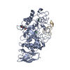 1clvS C: citing same article ( S: Starting model for refinement |
|---|---|
| Similar structure data |


-Links
 PDBj
PDBj


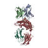
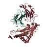
- Assembly
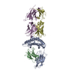
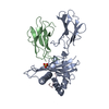
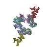
| Deposited unit | 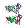
| ||||||||
|---|---|---|---|---|---|---|---|---|---|
| 1 | 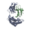
| ||||||||
| 2 | 
| ||||||||
| Unit cell |
|
-Components
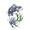
| #1: Protein | Mass: 32078.799 Da / Num. of mol.: 2 Fragment: EXTRACELLULAR DOMAINS (ALPHA1, ALPHA2, ALPHA3), RESIDUES 22-300 Mutation: YES Source method: isolated from a genetically manipulated source Source: (gene. exp.)  #2: Protein | Mass: 11704.359 Da / Num. of mol.: 2 Source method: isolated from a genetically manipulated source Source: (gene. exp.) #3: Protein/peptide | Mass: 983.076 Da / Num. of mol.: 2 / Fragment: RESIDUES 136-143 / Source method: obtained synthetically Details: THE PEPTIDE WAS CHEMICALLY SYNTHESIZED. THE SEQUENCE IS NATURALLY FOUND IN MUS MUCULUS (MOUSE) Source: (synth.) #4: Water | ChemComp-HOH / |  Mass: 18.015 Da / Num. of mol.: 641 / Source method: isolated from a natural source / Formula: H2O Mass: 18.015 Da / Num. of mol.: 641 / Source method: isolated from a natural source / Formula: H2OCompound details | ENGINEERED RESIDUE IN CHAIN A, TYR 43 TO PHE ENGINEERED RESIDUE IN CHAIN A, MET 44 TO ILE ...ENGINEERED | Has protein modification | Y | Sequence details | THE Y22F, M23I, E24S, D30N QUADRUPLE MUTANT IS CALLED BM8, A NATURALLY OCCURING MUTANT IN MICE | |
|---|
-Experimental details
-Experiment
| Experiment | Method: X-RAY DIFFRACTION / Number of used crystals: 1 |
|---|
- Sample preparation
| Crystal | Density Matthews: 2.7 Å3/Da / Density % sol: 54.6 % / Description: STARTING FROM PDB 1CLV |
|---|---|
| Crystal grow | pH: 6.3 / Details: 14% PEG 6000 ; 100 MM MES PH 6.3 |
-Data collection
| Diffraction | Mean temperature: 100 K |
|---|---|
| Diffraction source | Source: SYNCHROTRON / Site: ESRF  / Beamline: ID14-2 / Wavelength: 0.934 / Beamline: ID14-2 / Wavelength: 0.934 |
| Detector | Type: ADSC CCD / Detector: CCD / Date: Apr 15, 2005 |
| Radiation | Protocol: SINGLE WAVELENGTH / Monochromatic (M) / Laue (L): M / Scattering type: x-ray |
| Radiation wavelength | Wavelength: 0.934 Å / Relative weight: 1 |
| Reflection | Resolution: 1.9→51.23 Å / Num. obs: 75377 / % possible obs: 96.8 % / Observed criterion σ(I): 0 / Redundancy: 3.7 % / Rmerge(I) obs: 0.06 / Net I/σ(I): 14.9 |
| Reflection shell | Resolution: 1.9→2 Å / Redundancy: 3.5 % / Rmerge(I) obs: 0.38 / Mean I/σ(I) obs: 3.3 / % possible all: 86.8 |
- Processing
| Software |
| ||||||||||||||||||||||||||||||||||||||||||||||||||||||||||||||||||||||||||||||||||||||||||||||||||||||||||||||||||||||||||||||||||||||||||||||||||||||||||||||||||||||||||||||||||||||
|---|---|---|---|---|---|---|---|---|---|---|---|---|---|---|---|---|---|---|---|---|---|---|---|---|---|---|---|---|---|---|---|---|---|---|---|---|---|---|---|---|---|---|---|---|---|---|---|---|---|---|---|---|---|---|---|---|---|---|---|---|---|---|---|---|---|---|---|---|---|---|---|---|---|---|---|---|---|---|---|---|---|---|---|---|---|---|---|---|---|---|---|---|---|---|---|---|---|---|---|---|---|---|---|---|---|---|---|---|---|---|---|---|---|---|---|---|---|---|---|---|---|---|---|---|---|---|---|---|---|---|---|---|---|---|---|---|---|---|---|---|---|---|---|---|---|---|---|---|---|---|---|---|---|---|---|---|---|---|---|---|---|---|---|---|---|---|---|---|---|---|---|---|---|---|---|---|---|---|---|---|---|---|---|
| Refinement | Method to determine structure: MOLECULAR REPLACEMENT Starting model: PDB ENTRY 1CLV Resolution: 1.9→15 Å / Cor.coef. Fo:Fc: 0.951 / Cor.coef. Fo:Fc free: 0.929 / SU B: 6.905 / SU ML: 0.107 / Cross valid method: THROUGHOUT / ESU R: 0.16 / ESU R Free: 0.15 / Stereochemistry target values: MAXIMUM LIKELIHOOD / Details: HYDROGENS HAVE BEEN ADDED IN THE RIDING POSITIONS.
| ||||||||||||||||||||||||||||||||||||||||||||||||||||||||||||||||||||||||||||||||||||||||||||||||||||||||||||||||||||||||||||||||||||||||||||||||||||||||||||||||||||||||||||||||||||||
| Solvent computation | Ion probe radii: 0.8 Å / Shrinkage radii: 0.8 Å / VDW probe radii: 1.2 Å / Solvent model: MASK | ||||||||||||||||||||||||||||||||||||||||||||||||||||||||||||||||||||||||||||||||||||||||||||||||||||||||||||||||||||||||||||||||||||||||||||||||||||||||||||||||||||||||||||||||||||||
| Displacement parameters | Biso mean: 25.62 Å2
| ||||||||||||||||||||||||||||||||||||||||||||||||||||||||||||||||||||||||||||||||||||||||||||||||||||||||||||||||||||||||||||||||||||||||||||||||||||||||||||||||||||||||||||||||||||||
| Refinement step | Cycle: LAST / Resolution: 1.9→15 Å
| ||||||||||||||||||||||||||||||||||||||||||||||||||||||||||||||||||||||||||||||||||||||||||||||||||||||||||||||||||||||||||||||||||||||||||||||||||||||||||||||||||||||||||||||||||||||
| Refine LS restraints |
|