 Movie
Movie Controller

[English] 日本語
 Yorodumi
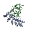
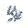
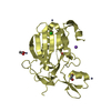
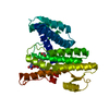
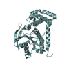
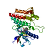
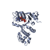
- PDB-1icf: CRYSTAL STRUCTURE OF MHC CLASS II ASSOCIATED P41 II FRAGMENT IN C... -
+ Open data

- Basic information
| Entry | Database: PDB / ID: 1icf | ||||||
|---|---|---|---|---|---|---|---|
| Title | CRYSTAL STRUCTURE OF MHC CLASS II ASSOCIATED P41 II FRAGMENT IN COMPLEX WITH CATHEPSIN L | ||||||
 Components Components |
| ||||||
 Keywords Keywords | HYDROLASE / CYSTEINE PROTEINASE / CATHEPSIN / MHC CLASS II / INVARIANT CHAIN / THYROGLOBULIN TYPE-1 DOMAIN | ||||||
| Function / homology |  Function and homology information Function and homology informationnegative regulation of peptide secretion / macrophage migration inhibitory factor signaling pathway / NOS2-CD74 complex / MHC class II protein binding, via antigen binding groove / antigen processing and presentation of endogenous antigen / positive regulation of dendritic cell antigen processing and presentation / negative regulation of T cell differentiation / enkephalin processing / cathepsin L / CD4-positive, alpha-beta T cell lineage commitment ...negative regulation of peptide secretion / macrophage migration inhibitory factor signaling pathway / NOS2-CD74 complex / MHC class II protein binding, via antigen binding groove / antigen processing and presentation of endogenous antigen / positive regulation of dendritic cell antigen processing and presentation / negative regulation of T cell differentiation / enkephalin processing / cathepsin L / CD4-positive, alpha-beta T cell lineage commitment / positive regulation of macrophage migration inhibitory factor signaling pathway / macrophage apoptotic process / macrophage migration inhibitory factor receptor complex / protein trimerization / macrophage migration inhibitory factor binding / chromaffin granule / positive regulation of cytokine-mediated signaling pathway / antigen processing and presentation of peptide antigen / elastin catabolic process / T cell activation involved in immune response / T cell selection / positive regulation of type 2 immune response / HS-GAG degradation / positive regulation of prostaglandin biosynthetic process / host-mediated suppression of symbiont invasion / RUNX1 regulates transcription of genes involved in differentiation of keratinocytes / MHC class II protein binding / negative thymic T cell selection / negative regulation of mature B cell apoptotic process / endolysosome lumen / positive regulation of monocyte differentiation / positive thymic T cell selection / CD4 receptor binding / prostaglandin biosynthetic process / cellular response to thyroid hormone stimulus / Trafficking and processing of endosomal TLR / vacuole / positive regulation of chemokine (C-X-C motif) ligand 2 production / cytokine receptor activity / proteoglycan binding / positive regulation of neutrophil chemotaxis / Assembly of collagen fibrils and other multimeric structures / positive regulation of macrophage cytokine production / zymogen activation / positive regulation of T cell differentiation / antigen processing and presentation / negative regulation of intrinsic apoptotic signaling pathway in response to DNA damage by p53 class mediator / transport vesicle membrane / regulation of macrophage activation / negative regulation of DNA damage response, signal transduction by p53 class mediator / cytokine binding / immunoglobulin mediated immune response / Collagen degradation / protein autoprocessing / collagen catabolic process / fibronectin binding / nitric-oxide synthase binding / serpin family protein binding / response to type II interferon / Degradation of the extracellular matrix / collagen binding / receptor-mediated endocytosis of virus by host cell / Attachment and Entry / positive regulation of chemokine production / multivesicular body / positive regulation of B cell proliferation / endocytic vesicle lumen / cysteine-type peptidase activity / MHC class II antigen presentation / protein folding chaperone / lysosomal lumen / : / negative regulation of cell migration / Cell surface interactions at the vascular wall / trans-Golgi network membrane / positive regulation of interleukin-8 production / Developmental Lineage of Pancreatic Ductal Cells / Endosomal/Vacuolar pathway / lumenal side of endoplasmic reticulum membrane / ER to Golgi transport vesicle membrane / clathrin-coated endocytic vesicle membrane / intracellular protein transport / MHC class II protein complex / positive regulation of interleukin-6 production / Degradation of CDH1 / antigen processing and presentation of exogenous peptide antigen via MHC class II / positive regulation of fibroblast proliferation / endocytic vesicle membrane / MHC class II protein complex binding / late endosome / amyloid-beta binding / extracellular matrix / protein-containing complex assembly / histone binding / adaptive immune response / Attachment and Entry / positive regulation of ERK1 and ERK2 cascade / positive regulation of canonical NF-kappaB signal transduction / lysosome / apical plasma membrane Similarity search - Function | ||||||
| Biological species |  Homo sapiens (human) Homo sapiens (human) | ||||||
| Method |  X-RAY DIFFRACTION / MOLECULAR REPLACEMENT / Resolution: 2 Å X-RAY DIFFRACTION / MOLECULAR REPLACEMENT / Resolution: 2 Å | ||||||
 Authors Authors | Guncar, G. / Pungercic, G. / Klemencic, I. / Turk, V. / Turk, D. | ||||||
 Citation Citation | Journal: EMBO J. / Year: 1999 Title: Crystal structure of MHC class II-associated p41 Ii fragment bound to cathepsin L reveals the structural basis for differentiation between cathepsins L and S. Authors: Guncar, G. / Pungercic, G. / Klemencic, I. / Turk, V. / Turk, D. | ||||||
| History |
|
- Structure visualization
| Structure viewer | Molecule: MolmilJmol/JSmol |
|---|
- Downloads & links
-Download
| PDBx/mmCIF format | 1icf.cif.gz | 137.1 KB | Display | PDBx/mmCIF format |
|---|---|---|---|---|
| PDB format | pdb1icf.ent.gz | 106.2 KB | Display | PDB format |
| PDBx/mmJSON format | 1icf.json.gz | Tree view | PDBx/mmJSON format | |
| Others |  Other downloads Other downloads |
-Validation report
| Arichive directory | https://data.pdbj.org/pub/pdb/validation_reports/ic/1icfftp://data.pdbj.org/pub/pdb/validation_reports/ic/1icf | HTTPS FTP |
|---|
-Related structure data
| Related structure data | 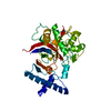 1cjlS S: Starting model for refinement |
|---|---|
| Similar structure data |


-Links
 PDBj
PDBj


- Assembly
| Deposited unit | 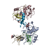
| ||||||||
|---|---|---|---|---|---|---|---|---|---|
| 1 | 
| ||||||||
| 2 | 
| ||||||||
| 3 | 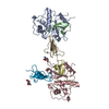
| ||||||||
| Unit cell |
| ||||||||
| Noncrystallographic symmetry (NCS) | NCS oper: (Code: given Matrix: (0.967984, -0.116869, 0.222146), Vector: |
-Components
| #1: Protein | Mass: 19095.020 Da / Num. of mol.: 2 / Source method: isolated from a natural source / Source: (natural) Homo sapiens (human) / Organ: KIDNEY / References: UniProt: P07711, cathepsin L#2: Protein/peptide | Mass: 4783.409 Da / Num. of mol.: 2 / Source method: isolated from a natural source / Source: (natural) Homo sapiens (human) / Organ: KIDNEY / References: UniProt: P07711, cathepsin L#3: Protein | Mass: 7261.075 Da / Num. of mol.: 2 / Fragment: THYROGLOBULIN TYPE-1 DOMAIN / Source method: isolated from a natural source / Source: (natural) Homo sapiens (human) / Organ: KIDNEY / References: UniProt: P04233#4: Sugar |   Type: D-saccharide, beta linking / Mass: 221.208 Da / Num. of mol.: 2 Type: D-saccharide, beta linking / Mass: 221.208 Da / Num. of mol.: 2Source method: isolated from a genetically manipulated source Formula: C8H15NO6 #5: Water | ChemComp-HOH / |  Mass: 18.015 Da / Num. of mol.: 668 / Source method: isolated from a natural source / Formula: H2O Mass: 18.015 Da / Num. of mol.: 668 / Source method: isolated from a natural source / Formula: H2OHas protein modification | Y | |
|---|
-Experimental details
-Experiment
| Experiment | Method: X-RAY DIFFRACTION / Number of used crystals: 1 |
|---|
- Sample preparation
| Crystal | Density Matthews: 2.58 Å3/Da / Density % sol: 52.37 % | |||||||||||||||||||||||||||||||||||
|---|---|---|---|---|---|---|---|---|---|---|---|---|---|---|---|---|---|---|---|---|---|---|---|---|---|---|---|---|---|---|---|---|---|---|---|---|
| Crystal grow | pH: 6.1 Details: SITTING DROP VAPOR DIFFUSION METHOD RESERVOIR CONTAINED 1ML OF 0.2 M NA- ACETATE TRIHYDRATE, 30% W/V PEG 8K AND 0.1M MES, PH 6.1. DROP WAS COMPOSED OF 2 MICRO L OF RESERVOIR SOLUTION AND 2 ...Details: SITTING DROP VAPOR DIFFUSION METHOD RESERVOIR CONTAINED 1ML OF 0.2 M NA- ACETATE TRIHYDRATE, 30% W/V PEG 8K AND 0.1M MES, PH 6.1. DROP WAS COMPOSED OF 2 MICRO L OF RESERVOIR SOLUTION AND 2 MICRO L OF THE COMPLEX (10 MG/ML) IN 20MM NA-ACETATE AND 1MM EDTA, PH 5.0. | |||||||||||||||||||||||||||||||||||
| Crystal grow | *PLUS pH: 5 / Method: vapor diffusion, hanging drop | |||||||||||||||||||||||||||||||||||
| Components of the solutions | *PLUS
|
-Data collection
| Diffraction | Mean temperature: 289 K |
|---|---|
| Diffraction source | Source: ROTATING ANODE / Type: RIGAKU RU200 / Wavelength: 1.5418 |
| Detector | Type: MARRESEARCH / Detector: IMAGE PLATE / Date: Mar 15, 1997 / Details: MIRRORS |
| Radiation | Protocol: SINGLE WAVELENGTH / Monochromatic (M) / Laue (L): M / Scattering type: x-ray |
| Radiation wavelength | Wavelength: 1.5418 Å / Relative weight: 1 |
| Reflection | Resolution: 2→99 Å / Num. obs: 42072 / % possible obs: 97 % / Observed criterion σ(I): 1 / Redundancy: 3.16 % / Rsym value: 0.11 |
| Reflection | *PLUS Num. measured all: 132945 / Rmerge(I) obs: 0.11 |
- Processing
| Software |
| ||||||||||||||||||||||||||||||||||||||||||||||||||||||||||||
|---|---|---|---|---|---|---|---|---|---|---|---|---|---|---|---|---|---|---|---|---|---|---|---|---|---|---|---|---|---|---|---|---|---|---|---|---|---|---|---|---|---|---|---|---|---|---|---|---|---|---|---|---|---|---|---|---|---|---|---|---|---|
| Refinement | Method to determine structure: MOLECULAR REPLACEMENT Starting model: 1CJL Resolution: 2→10 Å / Cross valid method: THROUGHOUT / σ(F): 1
| ||||||||||||||||||||||||||||||||||||||||||||||||||||||||||||
| Refinement step | Cycle: LAST / Resolution: 2→10 Å
| ||||||||||||||||||||||||||||||||||||||||||||||||||||||||||||
| Refine LS restraints |
| ||||||||||||||||||||||||||||||||||||||||||||||||||||||||||||
| Software | *PLUS Name: MAIN / Classification: refinement | ||||||||||||||||||||||||||||||||||||||||||||||||||||||||||||
| Refinement | *PLUS | ||||||||||||||||||||||||||||||||||||||||||||||||||||||||||||
| Solvent computation | *PLUS | ||||||||||||||||||||||||||||||||||||||||||||||||||||||||||||
| Displacement parameters | *PLUS Biso mean: 32.9 Å2 |
