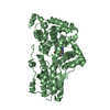
 Movie
Movie Controller


+ Open data

- Basic information
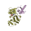
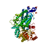
| Entry | Database: PDB / ID: 3p0k | ||||||
|---|---|---|---|---|---|---|---|
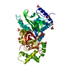
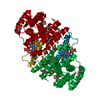
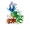
| Title | Structure of Baculovirus Sulfhydryl Oxidase Ac92 | ||||||
 Components Components | sulfhydryl oxidase | ||||||
 Keywords Keywords | Oxidoreductase / Viral protein / 4-helix bundle / 5-helix bundle / flavin adenine dinucleotide / sulfhydryl oxidase | ||||||
| Function / homology |  Function and homology information Function and homology informationthiol oxidase activity / thiol oxidase / host cell cytoplasm / host cell nucleus Similarity search - Function | ||||||
| Biological species |  Autographa californica nucleopolyhedrovirus Autographa californica nucleopolyhedrovirus | ||||||
| Method |  X-RAY DIFFRACTION / SYNCHROTRON / MAD / Resolution: 1.47 Å X-RAY DIFFRACTION / SYNCHROTRON / MAD / Resolution: 1.47 Å | ||||||
 Authors Authors | Hakim, M. / Fass, D. | ||||||
 Citation Citation | Journal: J.Virol. / Year: 2011 Title: Structure of a baculovirus sulfhydryl oxidase, a highly divergent member of the erv flavoenzyme family. Authors: Hakim, M. / Mandelbaum, A. / Fass, D. | ||||||
| History |
|
- Structure visualization
| Structure viewer | Molecule: MolmilJmol/JSmol |
|---|
- Downloads & links
-Download
| PDBx/mmCIF format | 3p0k.cif.gz | 77.8 KB | Display | PDBx/mmCIF format |
|---|---|---|---|---|
| PDB format | pdb3p0k.ent.gz | 56.7 KB | Display | PDB format |
| PDBx/mmJSON format | 3p0k.json.gz | Tree view | PDBx/mmJSON format | |
| Others |  Other downloads Other downloads |
-Validation report
| Arichive directory | https://data.pdbj.org/pub/pdb/validation_reports/p0/3p0kftp://data.pdbj.org/pub/pdb/validation_reports/p0/3p0k | HTTPS FTP |
|---|
-Related structure data


-Links
 PDBj
PDBj


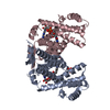
- Assembly
| Deposited unit | 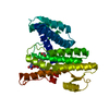
| ||||||||||||
|---|---|---|---|---|---|---|---|---|---|---|---|---|---|
| 1 | 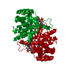
| ||||||||||||
| Unit cell |
| ||||||||||||
| Components on special symmetry positions |
|
-Components
| #1: Protein | Mass: 31682.828 Da / Num. of mol.: 1 Source method: isolated from a genetically manipulated source Source: (gene. exp.) Autographa californica nucleopolyhedrovirusGene: Ac92, P33 / Plasmid: pET-28a / Production host:  |
|---|---|
| #2: Chemical | ChemComp-IMD /   Mass: 69.085 Da / Num. of mol.: 1 / Source method: obtained synthetically / Formula: C3H5N2 Mass: 69.085 Da / Num. of mol.: 1 / Source method: obtained synthetically / Formula: C3H5N2 |
| #3: Chemical | ChemComp-FAD /   Mass: 785.550 Da / Num. of mol.: 1 / Source method: obtained synthetically / Formula: C27H33N9O15P2 / Comment: FAD*YM Mass: 785.550 Da / Num. of mol.: 1 / Source method: obtained synthetically / Formula: C27H33N9O15P2 / Comment: FAD*YM |
| #4: Chemical | ChemComp-ACT /   Mass: 59.044 Da / Num. of mol.: 1 / Source method: obtained synthetically / Formula: C2H3O2 Mass: 59.044 Da / Num. of mol.: 1 / Source method: obtained synthetically / Formula: C2H3O2 |
| #5: Water | ChemComp-HOH /  Mass: 18.015 Da / Num. of mol.: 195 / Source method: isolated from a natural source / Formula: H2O Mass: 18.015 Da / Num. of mol.: 195 / Source method: isolated from a natural source / Formula: H2O |
| Has protein modification | Y |
-Experimental details
-Experiment
| Experiment | Method: X-RAY DIFFRACTION |
|---|
- Sample preparation
| Crystal | Density Matthews: 2.22 Å3/Da / Density % sol: 44.72 % |
|---|---|
| Crystal grow | Temperature: 293 K / Method: vapor diffusion, hanging drop / pH: 4.4 Details: 0.1 M acetic acid, 50 mM arginine-HCl, 750 mM NaCl, 5% ethanol, 24% PEG 5000-MME, pH 4.4, VAPOR DIFFUSION, HANGING DROP, temperature 293K |
-Data collection
| Diffraction |
| |||||||||||||||
|---|---|---|---|---|---|---|---|---|---|---|---|---|---|---|---|---|
| Diffraction source | Source: SYNCHROTRON / Site: ESRF  / Beamline: ID23-1 / Wavelength: 0.97692 Å / Beamline: ID23-1 / Wavelength: 0.97692 Å | |||||||||||||||
| Detector | Type: ADSC QUANTUM 315r / Detector: CCD / Date: Jun 22, 2010 | |||||||||||||||
| Radiation |
| |||||||||||||||
| Radiation wavelength | Wavelength: 0.97692 Å / Relative weight: 1 | |||||||||||||||
| Reflection | Resolution: 1.47→50 Å / Num. obs: 46687 / % possible obs: 99.1 % / Observed criterion σ(I): -3 / Redundancy: 6.8 % / Rsym value: 0.069 / Net I/σ(I): 19.2 | |||||||||||||||
| Reflection shell | Resolution: 1.47→1.5 Å / Redundancy: 5.7 % / Mean I/σ(I) obs: 2.54 / Num. unique all: 2322 / Rsym value: 1 / % possible all: 99.1 |
- Processing
| Software |
| ||||||||||||||||||
|---|---|---|---|---|---|---|---|---|---|---|---|---|---|---|---|---|---|---|---|
| Refinement | Method to determine structure: MAD / Resolution: 1.47→50 Å / Stereochemistry target values: Engh & Huber
| ||||||||||||||||||
| Refinement step | Cycle: LAST / Resolution: 1.47→50 Å
| ||||||||||||||||||
| Refine LS restraints |
| ||||||||||||||||||
| LS refinement shell | Resolution: 1.47→1.48 Å
|
