 Movie
Movie Controller

[English] 日本語
 Yorodumi
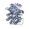
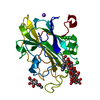
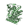
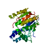
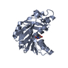
- PDB-3gwn: Crystal structure of the FAD binding domain from mimivirus sulfhy... -
+ Open data

- Basic information
| Entry | Database: PDB / ID: 3gwn | ||||||
|---|---|---|---|---|---|---|---|
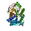
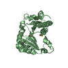
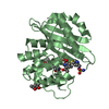
| Title | Crystal structure of the FAD binding domain from mimivirus sulfhydryl oxidase R596 | ||||||
 Components Components | Probable FAD-linked sulfhydryl oxidase R596 | ||||||
 Keywords Keywords | OXIDOREDUCTASE / FIVE HELIX BUNDLE / HOMODIMER / Disulfide bond / FAD / Flavoprotein / Virion | ||||||
| Function / homology |  Function and homology information Function and homology informationflavin-dependent sulfhydryl oxidase activity / thiol oxidase / virion component / flavin adenine dinucleotide binding Similarity search - Function | ||||||
| Biological species |   Acanthamoeba polyphaga mimivirus Acanthamoeba polyphaga mimivirus | ||||||
| Method |  X-RAY DIFFRACTION / MOLECULAR REPLACEMENT / Resolution: 1.78 Å X-RAY DIFFRACTION / MOLECULAR REPLACEMENT / Resolution: 1.78 Å | ||||||
 Authors Authors | Hakim, M. / Fass, D. | ||||||
 Citation Citation | Journal: J.Mol.Biol. / Year: 2009 Title: Dimer interface migration in a viral sulfhydryl oxidase Authors: Hakim, M. / Fass, D. | ||||||
| History |
|
- Structure visualization
| Structure viewer | Molecule: MolmilJmol/JSmol |
|---|
- Downloads & links
-Download
| PDBx/mmCIF format | 3gwn.cif.gz | 69 KB | Display | PDBx/mmCIF format |
|---|---|---|---|---|
| PDB format | pdb3gwn.ent.gz | 51.1 KB | Display | PDB format |
| PDBx/mmJSON format | 3gwn.json.gz | Tree view | PDBx/mmJSON format | |
| Others |  Other downloads Other downloads |
-Validation report
| Arichive directory | https://data.pdbj.org/pub/pdb/validation_reports/gw/3gwnftp://data.pdbj.org/pub/pdb/validation_reports/gw/3gwn | HTTPS FTP |
|---|
-Related structure data
| Related structure data |  3gwlC  1jr8S S: Starting model for refinement C: citing same article ( |
|---|---|
| Similar structure data |


-Links
 PDBj
PDBj- Assembly
| Deposited unit | 
| ||||||||
|---|---|---|---|---|---|---|---|---|---|
| 1 |
| ||||||||
| Unit cell |
|
-Components
| #1: Protein | Mass: 13297.046 Da / Num. of mol.: 2 / Fragment: mimivirus R596 FAD binding domain Source method: isolated from a genetically manipulated source Source: (gene. exp.) Acanthamoeba polyphaga mimivirus / Gene: MIMI_R596, R596 / Plasmid: pET15b / Production host:  #2: Chemical |   Mass: 785.550 Da / Num. of mol.: 2 / Source method: obtained synthetically / Formula: C27H33N9O15P2 / Comment: FAD*YM Mass: 785.550 Da / Num. of mol.: 2 / Source method: obtained synthetically / Formula: C27H33N9O15P2 / Comment: FAD*YM#3: Chemical |   Mass: 96.063 Da / Num. of mol.: 3 / Source method: obtained synthetically / Formula: SO4 Mass: 96.063 Da / Num. of mol.: 3 / Source method: obtained synthetically / Formula: SO4#4: Chemical | ChemComp-GOL / |   Mass: 92.094 Da / Num. of mol.: 1 / Source method: obtained synthetically / Formula: C3H8O3 Mass: 92.094 Da / Num. of mol.: 1 / Source method: obtained synthetically / Formula: C3H8O3#5: Water | ChemComp-HOH / |  Mass: 18.015 Da / Num. of mol.: 229 / Source method: isolated from a natural source / Formula: H2O Mass: 18.015 Da / Num. of mol.: 229 / Source method: isolated from a natural source / Formula: H2OHas protein modification | Y | |
|---|
-Experimental details
-Experiment
| Experiment | Method: X-RAY DIFFRACTION / Number of used crystals: 1 |
|---|
- Sample preparation
| Crystal | Density Matthews: 2.31 Å3/Da / Density % sol: 46.67 % |
|---|---|
| Crystal grow | Temperature: 293 K / Method: vapor diffusion, hanging drop / pH: 8 Details: 0.5M ammonium sulfate, 25% PEG 4K, pH 8, VAPOR DIFFUSION, HANGING DROP, temperature 293K |
-Data collection
| Diffraction | Mean temperature: 293 K |
|---|---|
| Diffraction source | Source: ROTATING ANODE / Type: RIGAKU RUH3R / Wavelength: 1.5418 Å |
| Detector | Type: RIGAKU RAXIS IV++ / Detector: IMAGE PLATE / Date: Sep 28, 2008 |
| Radiation | Protocol: SINGLE WAVELENGTH / Monochromatic (M) / Laue (L): M / Scattering type: x-ray |
| Radiation wavelength | Wavelength: 1.5418 Å / Relative weight: 1 |
| Reflection | Resolution: 1.78→50 Å / Num. all: 24453 / Num. obs: 24259 / % possible obs: 99.2 % / Observed criterion σ(I): -3 / Redundancy: 11.2 % / Biso Wilson estimate: 22.8 Å2 / Rsym value: 0.078 / Net I/σ(I): 12.5 |
| Reflection shell | Resolution: 1.78→1.81 Å / Redundancy: 4.5 % / Mean I/σ(I) obs: 2.3 / Rsym value: 0.407 / % possible all: 97.9 |
- Processing
| Software |
| |||||||||||||||||||||||||
|---|---|---|---|---|---|---|---|---|---|---|---|---|---|---|---|---|---|---|---|---|---|---|---|---|---|---|
| Refinement | Method to determine structure: MOLECULAR REPLACEMENT Starting model: PDB ENTRY 1JR8 Resolution: 1.78→50 Å / Cross valid method: THROUGHOUT / σ(F): 0
| |||||||||||||||||||||||||
| Refinement step | Cycle: LAST / Resolution: 1.78→50 Å
| |||||||||||||||||||||||||
| Refine LS restraints |
|
