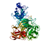
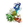
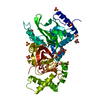
UV-A, blue light phototransduction / magnetoreception / detection of light stimulus involved in magnetoreception / gravitaxis / Phosphorylation of PER and TIM / Degradation of CRY / Degradation of TIM / response to magnetism / blue light signaling pathway / cellular response to light stimulus ...UV-A, blue light phototransduction / magnetoreception / detection of light stimulus involved in magnetoreception / gravitaxis / Phosphorylation of PER and TIM / Degradation of CRY / Degradation of TIM / response to magnetism / blue light signaling pathway / cellular response to light stimulus / blue light photoreceptor activity / response to blue light / regulation of circadian sleep/wake cycle, sleep / entrainment of circadian clock / circadian behavior / entrainment of circadian clock by photoperiod / locomotor rhythm / photoreceptor activity / phototransduction / response to light stimulus / FAD binding / circadian regulation of gene expression / circadian rhythm / regulation of circadian rhythm / flavin adenine dinucleotide binding / negative regulation of DNA-templated transcription / perinuclear region of cytoplasm / DNA binding / nucleoplasm / nucleus / cytosol / cytoplasm Similarity search - Function
Serine Threonine Protein Phosphatase 5, Tetratricopeptide repeat - #80 / DNA Cyclobutane Dipyrimidine Photolyase, subunit A; domain 3 / DNA Cyclobutane Dipyrimidine Photolyase, subunit A, domain 3 / Cryptochrome/DNA photolyase class 1 / Cryptochrome/DNA photolyase, FAD-binding domain / FAD binding domain of DNA photolyase / DNA photolyase, N-terminal / Cryptochrome/photolyase, N-terminal domain superfamily / DNA photolyase / Photolyase/cryptochrome alpha/beta domain profile. ...Serine Threonine Protein Phosphatase 5, Tetratricopeptide repeat - #80 / DNA Cyclobutane Dipyrimidine Photolyase, subunit A; domain 3 / DNA Cyclobutane Dipyrimidine Photolyase, subunit A, domain 3 / Cryptochrome/DNA photolyase class 1 / Cryptochrome/DNA photolyase, FAD-binding domain / FAD binding domain of DNA photolyase / DNA photolyase, N-terminal / Cryptochrome/photolyase, N-terminal domain superfamily / DNA photolyase / Photolyase/cryptochrome alpha/beta domain profile. / Cryptochrome/DNA photolyase, FAD-binding domain-like superfamily / HUPs / Rossmann-like alpha/beta/alpha sandwich fold / Serine Threonine Protein Phosphatase 5, Tetratricopeptide repeat / Alpha Horseshoe / Rossmann fold / Orthogonal Bundle / 3-Layer(aba) Sandwich / Mainly Alpha / Alpha Beta Similarity search - Domain/homology
Mass: 18.015 Da / Num. of mol.: 321 / Source method: isolated from a natural source / Formula: H2O
Has protein modification
Y
-
Experimental details
-
Experiment
Experiment
Method: X-RAY DIFFRACTION / Number of used crystals: 1
-
Sample preparation
Crystal
Density Matthews: 2.43 Å3/Da / Density % sol: 49.39 %
Crystal grow
Temperature: 298 K / pH: 8 Details: 50 MM HEPES PH 8.0; 150 MM NACL 5MG/ML PROTEIN MIXED 1:1 WITH 18% PDG 4K, 150 MM MGACETATE, 100 MM TRIS PH 8.5, VAPOR DIFFUSION, HANGING DROP, TEMPERATURE 298K
In the structure databanks used in Yorodumi, some data are registered as the other names, "COVID-19 virus" and "2019-nCoV". Here are the details of the virus and the list of structure data.
Jan 31, 2019. EMDB accession codes are about to change! (news from PDBe EMDB page)
EMDB accession codes are about to change! (news from PDBe EMDB page)
The allocation of 4 digits for EMDB accession codes will soon come to an end. Whilst these codes will remain in use, new EMDB accession codes will include an additional digit and will expand incrementally as the available range of codes is exhausted. The current 4-digit format prefixed with “EMD-” (i.e. EMD-XXXX) will advance to a 5-digit format (i.e. EMD-XXXXX), and so on. It is currently estimated that the 4-digit codes will be depleted around Spring 2019, at which point the 5-digit format will come into force.
The EM Navigator/Yorodumi systems omit the EMD- prefix.
Related info.:Q: What is EMD? / ID/Accession-code notation in Yorodumi/EM Navigator
Yorodumi is a browser for structure data from EMDB, PDB, SASBDB, etc.
This page is also the successor to EM Navigator detail page, and also detail information page/front-end page for Omokage search.
The word "yorodu" (or yorozu) is an old Japanese word meaning "ten thousand". "mi" (miru) is to see.
Related info.:EMDB / PDB / SASBDB / Comparison of 3 databanks / Yorodumi Search / Aug 31, 2016. New EM Navigator & Yorodumi / Yorodumi Papers / Jmol/JSmol / Function and homology information / Changes in new EM Navigator and Yorodumi
 Movie
Movie Controller


 Open data
 Basic information
 Components
 Keywords
 Function and homology information
 X-RAY DIFFRACTION /
  Authors
 Citation
 Structure visualization
 Downloads & links
 Other downloads


 PDBj
PDBj
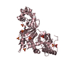
 Assembly
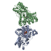
 Spodoptera frugiperda (fall armyworm) / Strain (production host): SF9 / References: UniProt: O77059
 Mass: 24.305 Da / Num. of mol.: 2 / Source method: obtained synthetically / Formula: Mg
 Mass: 785.550 Da / Num. of mol.: 2 / Source method: obtained synthetically / Formula: C27H33N9O15P2 / Comment: FAD*YM
 Mass: 18.015 Da / Num. of mol.: 321 / Source method: isolated from a natural source / Formula: H2O
 Sample preparation
 / Beamline: 24-ID-E / Wavelength: 0.9793
  Processing