 Movie
Movie Controller

[English] 日本語
 Yorodumi
- PDB-1tez: COMPLEX BETWEEN DNA AND THE DNA PHOTOLYASE FROM ANACYSTIS NIDULANS -
+ Open data

- Basic information
| Entry | Database: PDB / ID: 1tez | |||||||||
|---|---|---|---|---|---|---|---|---|---|---|
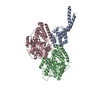
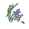
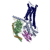
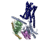
| Title | COMPLEX BETWEEN DNA AND THE DNA PHOTOLYASE FROM ANACYSTIS NIDULANS | |||||||||
 Components Components |
| |||||||||
 Keywords Keywords | LYASE/DNA / PHOTOLYASE / DNA REPAIR / LIGHT-DRIVEN ELECTRON TRANSFER / LYASE-DNA COMPLEX | |||||||||
| Function / homology |  Function and homology information Function and homology informationdeoxyribodipyrimidine photo-lyase / deoxyribodipyrimidine photo-lyase activity / nucleobase-containing compound metabolic process / response to stress / response to light stimulus / FAD binding / DNA repair / DNA binding Similarity search - Function | |||||||||
| Biological species |  Synechococcus elongatus PCC 6301 (bacteria) Synechococcus elongatus PCC 6301 (bacteria)synthetic construct (others) | |||||||||
| Method |  X-RAY DIFFRACTION / SYNCHROTRON / MOLECULAR REPLACEMENT / Resolution: 1.8 Å X-RAY DIFFRACTION / SYNCHROTRON / MOLECULAR REPLACEMENT / Resolution: 1.8 Å | |||||||||
 Authors Authors | Essen, L.-O. / Carell, T. / Mees, A. / Klar, T. | |||||||||
 Citation Citation | Journal: Science / Year: 2004 Title: Crystal structure of a photolyase bound to a CPD-like DNA lesion after in situ repair Authors: Mees, A. / Klar, T. / Gnau, P. / Hennecke, U. / Eker, A.P.M. / Carell, T. / Essen, L.-O. #1: Journal: Nat.Struct.Biol. / Year: 1997Title: Crystal Structure of DNA Photolyase from Anacystis Authors: Tamada, T. / Kitadokoro, K. / Higuchi, Y. / Inaka, K. / Yasui, A. / De Ruiter, P.E. / Eker, A.P. / Miki, K. | |||||||||
| History |
|
- Structure visualization
| Structure viewer | Molecule: MolmilJmol/JSmol |
|---|
- Downloads & links
-Download
| PDBx/mmCIF format | 1tez.cif.gz | 459.3 KB | Display | PDBx/mmCIF format |
|---|---|---|---|---|
| PDB format | pdb1tez.ent.gz | 368 KB | Display | PDB format |
| PDBx/mmJSON format | 1tez.json.gz | Tree view | PDBx/mmJSON format | |
| Others |  Other downloads Other downloads |
-Validation report
| Arichive directory | https://data.pdbj.org/pub/pdb/validation_reports/te/1tezftp://data.pdbj.org/pub/pdb/validation_reports/te/1tez | HTTPS FTP |
|---|
-Related structure data
| Related structure data | 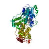 1qnfS S: Starting model for refinement |
|---|---|
| Similar structure data |


-Links
 PDBj
PDBj


- Assembly
| Deposited unit | 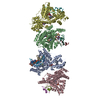
| ||||||||||||||||||||
|---|---|---|---|---|---|---|---|---|---|---|---|---|---|---|---|---|---|---|---|---|---|
| 1 | 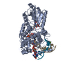
| ||||||||||||||||||||
| 2 | 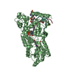
| ||||||||||||||||||||
| 3 | 
| ||||||||||||||||||||
| 4 | 
| ||||||||||||||||||||
| Unit cell |
| ||||||||||||||||||||
| Noncrystallographic symmetry (NCS) | NCS oper:
|
-Components
-DNA chain , 4 types, 8 molecules IKJLMONP
| #1: DNA chain | Mass: 3259.219 Da / Num. of mol.: 2 / Source method: obtained synthetically Details: Formacetal linkage replaces phosphate linkage between T7 and T8 of chains I and K Source: (synth.) synthetic construct (others) #2: DNA chain | Mass: 2749.825 Da / Num. of mol.: 2 / Source method: obtained synthetically / Source: (synth.) synthetic construct (others) #3: DNA chain | Mass: 1166.805 Da / Num. of mol.: 2 / Source method: obtained synthetically / Source: (synth.) synthetic construct (others) #4: DNA chain | Mass: 1505.025 Da / Num. of mol.: 2 / Source method: obtained synthetically / Source: (synth.) synthetic construct (others) |
|---|
-Protein , 1 types, 4 molecules ABCD
| #5: Protein | Mass: 53474.418 Da / Num. of mol.: 4 Source method: isolated from a genetically manipulated source Details: SYNTHETIC DNA OLIGONUCLEOTIDES ENGINEERED AS COUNTERSTRAND Source: (gene. exp.) Synechococcus elongatus PCC 6301 (bacteria)Species: Synechococcus elongatus / Strain: PCC 6301 / Cellular location: CYTOPLASM / Gene: Synpcc7942_0112 / Plasmid: PET3A / Cellular location (production host): CYTOPLASM / Production host: References: UniProt: Q31S25, UniProt: P05327*PLUS, deoxyribodipyrimidine photo-lyase |
|---|
-Non-polymers , 4 types, 1624 molecules 


| #6: Chemical |  Mass: 24.305 Da / Num. of mol.: 2 / Source method: obtained synthetically / Formula: Mg Mass: 24.305 Da / Num. of mol.: 2 / Source method: obtained synthetically / Formula: Mg#7: Chemical | ChemComp-FAD /  Mass: 785.550 Da / Num. of mol.: 4 / Source method: obtained synthetically / Formula: C27H33N9O15P2 / Comment: FAD*YM Mass: 785.550 Da / Num. of mol.: 4 / Source method: obtained synthetically / Formula: C27H33N9O15P2 / Comment: FAD*YM#8: Chemical | ChemComp-HDF /  Mass: 363.322 Da / Num. of mol.: 4 / Source method: obtained synthetically / Formula: C16H17N3O7 Mass: 363.322 Da / Num. of mol.: 4 / Source method: obtained synthetically / Formula: C16H17N3O7#9: Water | ChemComp-HOH / | Mass: 18.015 Da / Num. of mol.: 1614 / Source method: isolated from a natural source / Formula: H2O |
|---|
-Details
| Compound details | COMPOUND THE CYCLOBUTAN| Nonpolymer details | HETEROGEN DESOXYTHYM | |
|---|
-Experimental details
-Experiment
| Experiment | Method: X-RAY DIFFRACTION / Number of used crystals: 1 |
|---|
- Sample preparation
| Crystal | Density Matthews: 2.44 Å3/Da / Density % sol: 49.2 % | ||||||||||||||||||||||||||||
|---|---|---|---|---|---|---|---|---|---|---|---|---|---|---|---|---|---|---|---|---|---|---|---|---|---|---|---|---|---|
| Crystal grow | Temperature: 281 K / Method: vapor diffusion, hanging drop / pH: 8 Details: PEG 4000, magnesium chloride, Tris-HCl, pH 8.0, VAPOR DIFFUSION, HANGING DROP, temperature 281K | ||||||||||||||||||||||||||||
| Components of the solutions |
|
-Data collection
| Diffraction | Mean temperature: 100 K |
|---|---|
| Diffraction source | Source: SYNCHROTRON / Site: SLS  / Beamline: X06SA / Wavelength: 0.9796 / Wavelength: 0.9796 Å / Beamline: X06SA / Wavelength: 0.9796 / Wavelength: 0.9796 Å |
| Detector | Type: MARRESEARCH / Detector: CCD / Date: Jan 27, 2004 / Details: MIRRORS |
| Radiation | Monochromator: Si 111 CHANNEL / Protocol: SINGLE WAVELENGTH / Monochromatic (M) / Laue (L): M / Scattering type: x-ray |
| Radiation wavelength | Wavelength: 0.9796 Å / Relative weight: 1 |
| Reflection | Resolution: 1.8→30 Å / Num. all: 218105 / Num. obs: 218105 / % possible obs: 96.3 % / Observed criterion σ(F): 0 / Observed criterion σ(I): 0 / Redundancy: 3.3 % / Biso Wilson estimate: 22.3 Å2 / Rmerge(I) obs: 0.072 / Net I/σ(I): 13.9 |
| Reflection shell | Resolution: 1.8→1.86 Å / Redundancy: 2 % / Rmerge(I) obs: 0.399 / Mean I/σ(I) obs: 2.1 / % possible all: 81.7 |
- Processing
| Software |
| ||||||||||||||||||||||||||||||||||||||||||||||||||||||||||||
|---|---|---|---|---|---|---|---|---|---|---|---|---|---|---|---|---|---|---|---|---|---|---|---|---|---|---|---|---|---|---|---|---|---|---|---|---|---|---|---|---|---|---|---|---|---|---|---|---|---|---|---|---|---|---|---|---|---|---|---|---|---|
| Refinement | Method to determine structure: MOLECULAR REPLACEMENT Starting model: PDB ENTRY 1QNF Resolution: 1.8→29.92 Å / Rfactor Rfree error: 0.003 / Data cutoff high rms absF: 2423887.54 / Isotropic thermal model: RESTRAINED / Cross valid method: THROUGHOUT / σ(F): 0 / Stereochemistry target values: Engh & Huber
| ||||||||||||||||||||||||||||||||||||||||||||||||||||||||||||
| Solvent computation | Solvent model: FLAT MODEL / Bsol: 53.7551 Å2 / ksol: 0.37764 e/Å3 | ||||||||||||||||||||||||||||||||||||||||||||||||||||||||||||
| Displacement parameters | Biso mean: 30.7 Å2
| ||||||||||||||||||||||||||||||||||||||||||||||||||||||||||||
| Refine analyze |
| ||||||||||||||||||||||||||||||||||||||||||||||||||||||||||||
| Refinement step | Cycle: LAST / Resolution: 1.8→29.92 Å
| ||||||||||||||||||||||||||||||||||||||||||||||||||||||||||||
| Refine LS restraints |
| ||||||||||||||||||||||||||||||||||||||||||||||||||||||||||||
| LS refinement shell | Resolution: 1.8→1.91 Å / Rfactor Rfree error: 0.009 / Total num. of bins used: 6
| ||||||||||||||||||||||||||||||||||||||||||||||||||||||||||||
| Xplor file |
|
