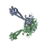
Movie
Movie Controller


+ Open data

- Basic information
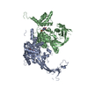
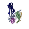
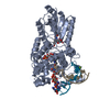
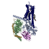
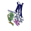
| Entry | Database: PDB / ID: 3j1u | ||||||
|---|---|---|---|---|---|---|---|
| Title | Low affinity dynein microtubule binding domain - tubulin complex | ||||||
 Components Components |
| ||||||
 Keywords Keywords | MOTOR PROTEIN/STRUCTURAL PROTEIN / MOTOR PROTEIN-STRUCTURAL PROTEIN complex | ||||||
| Function / homology |  Function and homology information Function and homology informationCOPI-independent Golgi-to-ER retrograde traffic / HSP90 chaperone cycle for steroid hormone receptors (SHR) in the presence of ligand / Amplification of signal from unattached kinetochores via a MAD2 inhibitory signal / COPI-mediated anterograde transport / cilium movement / Aggrephagy / Mitotic Prometaphase / EML4 and NUDC in mitotic spindle formation / Resolution of Sister Chromatid Cohesion / RHO GTPases Activate Formins ...COPI-independent Golgi-to-ER retrograde traffic / HSP90 chaperone cycle for steroid hormone receptors (SHR) in the presence of ligand / Amplification of signal from unattached kinetochores via a MAD2 inhibitory signal / COPI-mediated anterograde transport / cilium movement / Aggrephagy / Mitotic Prometaphase / EML4 and NUDC in mitotic spindle formation / Resolution of Sister Chromatid Cohesion / RHO GTPases Activate Formins / Loss of Nlp from mitotic centrosomes / Recruitment of mitotic centrosome proteins and complexes / Loss of proteins required for interphase microtubule organization from the centrosome / Anchoring of the basal body to the plasma membrane / Separation of Sister Chromatids / Recruitment of NuMA to mitotic centrosomes / AURKA Activation by TPX2 / dynein complex / manchette / Regulation of PLK1 Activity at G2/M Transition / positive regulation of intracellular transport / regulation of metaphase plate congression / positive regulation of spindle assembly / establishment of spindle localization / MHC class II antigen presentation / positive regulation of axon guidance / retrograde axonal transport / minus-end-directed microtubule motor activity / P-body assembly / dynein light intermediate chain binding / cytoplasmic dynein complex / nuclear migration / dynein intermediate chain binding / microtubule-based process / cytoplasmic microtubule / cytoplasmic microtubule organization / Neutrophil degranulation / axon cytoplasm / cellular response to interleukin-4 / stress granule assembly / mitotic spindle organization / regulation of mitotic spindle organization / filopodium / structural constituent of cytoskeleton / microtubule cytoskeleton organization / neuron migration / nuclear envelope / mitotic cell cycle / double-stranded RNA binding / positive regulation of cold-induced thermogenesis / microtubule cytoskeleton / cell cortex / microtubule / Hydrolases; Acting on acid anhydrides; Acting on GTP to facilitate cellular and subcellular movement / cilium / protein heterodimerization activity / axon / cell division / neuronal cell body / GTPase activity / ubiquitin protein ligase binding / centrosome / GTP binding / ATP binding / metal ion binding / identical protein binding / cytoplasm / cytosol Similarity search - Function | ||||||
| Biological species |  | ||||||
| Method | ELECTRON MICROSCOPY / helical reconstruction / cryo EM / Resolution: 9.7 Å | ||||||
 Authors Authors | Redwine, W.B. / Hernandez-Lopez, R. / Zou, S. / Huang, J. / Reck-Peterson, S.L. / Leschziner, A.E. | ||||||
 Citation Citation | Journal: Science / Year: 2012 Title: Structural basis for microtubule binding and release by dynein. Authors: W B Redwine / R Hernandez-Lopez / S Zou / J Huang / S L Reck-Peterson / A E Leschziner /  Abstract: Cytoplasmic dynein is a microtubule-based motor required for intracellular transport and cell division. Its movement involves coupling cycles of track binding and release with cycles of force- ...Cytoplasmic dynein is a microtubule-based motor required for intracellular transport and cell division. Its movement involves coupling cycles of track binding and release with cycles of force-generating nucleotide hydrolysis. How this is accomplished given the ~25 nanometers separating dynein's track- and nucleotide-binding sites is not understood. Here, we present a subnanometer-resolution structure of dynein's microtubule-binding domain bound to microtubules by cryo-electron microscopy that was used to generate a pseudo-atomic model of the complex with molecular dynamics. We identified large rearrangements triggered by track binding and specific interactions, confirmed by mutagenesis and single-molecule motility assays, which tune dynein's affinity for microtubules. Our results provide a molecular model for how dynein's binding to microtubules is communicated to the rest of the motor. | ||||||
| History |
|
- Structure visualization
| Movie |
Movie viewer |
|---|---|
| Structure viewer | Molecule: MolmilJmol/JSmol |
- Downloads & links
-Download
| PDBx/mmCIF format | 3j1u.cif.gz | 192.1 KB | Display | PDBx/mmCIF format |
|---|---|---|---|---|
| PDB format | pdb3j1u.ent.gz | 143 KB | Display | PDB format |
| PDBx/mmJSON format | 3j1u.json.gz | Tree view | PDBx/mmJSON format | |
| Others |  Other downloads Other downloads |
-Validation report
| Arichive directory | https://data.pdbj.org/pub/pdb/validation_reports/j1/3j1uftp://data.pdbj.org/pub/pdb/validation_reports/j1/3j1u | HTTPS FTP |
|---|
-Related structure data
| Related structure data | 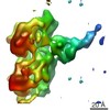 5439MC 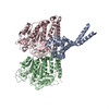 3j1tC C: citing same article ( M: map data used to model this data |
|---|---|
| Similar structure data |


-Links
 PDBj
PDBj


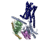
- Assembly
| Deposited unit | 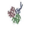
|
|---|---|
| 1 |
|
-Components
| #1: Protein | Mass: 18729.609 Da / Num. of mol.: 1 / Fragment: SEE REMARK 999 Source method: isolated from a genetically manipulated source Source: (gene. exp.)  |
|---|---|
| #2: Protein | Mass: 50107.238 Da / Num. of mol.: 1 / Source method: isolated from a natural source / Source: (natural) |
| #3: Protein | Mass: 47940.945 Da / Num. of mol.: 1 / Source method: isolated from a natural source / Source: (natural) |
| Sequence details | CHAINS B AND C ARE DERIVED FROM PDB ENTRY 1JFF. AS A RESULT, THE MODELED SEQUENCES ARE FROM SUS ...CHAINS B AND C ARE DERIVED FROM PDB ENTRY 1JFF. AS A RESULT, THE MODELED SEQUENCES ARE FROM SUS SCROFA (UNP P02550 AND P02554, RESPECTIVE |
-Experimental details
-Experiment
| Experiment | Method: ELECTRON MICROSCOPY |
|---|---|
| EM experiment | Aggregation state: FILAMENT / 3D reconstruction method: helical reconstruction |
- Sample preparation
| Component |
| ||||||||||||||||||||
|---|---|---|---|---|---|---|---|---|---|---|---|---|---|---|---|---|---|---|---|---|---|
| Buffer solution | Name: 50 mM Tris HCl, 1 mM MgCl2, 1mM EGTA, 1mM DTT / pH: 8 / Details: 50 mM Tris HCl, 1 mM MgCl2, 1mM EGTA, 1mM DTT | ||||||||||||||||||||
| Specimen | Conc.: 4 mg/ml / Embedding applied: NO / Shadowing applied: NO / Staining applied: NO / Vitrification applied: YES | ||||||||||||||||||||
| Specimen support | Details: C-flat 2/2-2C holey carbon grids (Protochips) were glow-discharged for 20 seconds at 30 mA in an Edwards carbon evaporator. | ||||||||||||||||||||
| Vitrification | Instrument: FEI VITROBOT MARK IV / Cryogen name: ETHANE / Humidity: 100 % / Chamber temperature: 295 K Details: The solution was blotted manually, and the process of addition and blotting of SRS-MTBD was repeated a total of three times. The final blotting was done inside the humidity chamber of a ...Details: The solution was blotted manually, and the process of addition and blotting of SRS-MTBD was repeated a total of three times. The final blotting was done inside the humidity chamber of a Vitrobot Mark IV (FEI) at 22 Celsius. |
- Electron microscopy imaging
| Experimental equipment |  Model: Tecnai F20 / Image courtesy: FEI Company |
|---|---|
| Microscopy | Model: FEI TECNAI F20 / Date: Apr 10, 2011 |
| Electron gun | Electron source:  FIELD EMISSION GUN / Accelerating voltage: 120 kV / Illumination mode: FLOOD BEAM FIELD EMISSION GUN / Accelerating voltage: 120 kV / Illumination mode: FLOOD BEAM |
| Electron lens | Mode: BRIGHT FIELD / Nominal magnification: 62000 X / Calibrated magnification: 63377 X / Nominal defocus max: 2500 nm / Nominal defocus min: 500 nm / Cs: 2.2 mm |
| Specimen holder | Temperature: 100 K |
| Image recording | Electron dose: 15 e/Å2 / Film or detector model: KODAK SO-163 FILM |
| Image scans | Num. digital images: 225 |
- Processing
| EM software |
| ||||||||||||||||||||||||
|---|---|---|---|---|---|---|---|---|---|---|---|---|---|---|---|---|---|---|---|---|---|---|---|---|---|
| CTF correction | Details: phase and amplitude correction using Frealign | ||||||||||||||||||||||||
| Helical symmerty | Angular rotation/subunit: -25.76 ° / Axial rise/subunit: 9.26 Å / Axial symmetry: C1 | ||||||||||||||||||||||||
| 3D reconstruction | Method: Projection matching and back-projection in Fourier space Resolution: 9.7 Å / Num. of particles: 10419 / Nominal pixel size: 1.988 Å / Actual pixel size: 1.988 Å / Magnification calibration: TMV images Details: Projection matching and Helical symmetry operator during reconstruction Symmetry type: HELICAL | ||||||||||||||||||||||||
| Atomic model building |
| ||||||||||||||||||||||||
| Atomic model building | Source name: PDB / Type: experimental model
| ||||||||||||||||||||||||
| Refinement step | Cycle: LAST
|