 Movie
Movie Controller

[English] 日本語
 Yorodumi
- PDB-1ha5: Structural features of a zinc-binding site in the superantigen st... -
+ Open data

- Basic information
| Entry | Database: PDB / ID: 1ha5 | ||||||
|---|---|---|---|---|---|---|---|
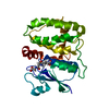
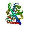
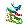
| Title | Structural features of a zinc-binding site in the superantigen streptococcal pyrogenic exotoxin A (SpeA1): implications for MHC class II recognition. | ||||||
 Components Components | STREPTOCOCCAL PYOGENIC EXOTOXIN A1 | ||||||
 Keywords Keywords | TOXIN / MOLECULAR RECOGNITION / SUPERANTIGEN / EXOTOXIN / ZINC BINDING | ||||||
| Function / homology |  Function and homology information Function and homology information | ||||||
| Biological species |  STREPTOCOCCUS PYOGENES (bacteria) STREPTOCOCCUS PYOGENES (bacteria) | ||||||
| Method |  X-RAY DIFFRACTION / SYNCHROTRON / MOLECULAR REPLACEMENT / Resolution: 2.82 Å X-RAY DIFFRACTION / SYNCHROTRON / MOLECULAR REPLACEMENT / Resolution: 2.82 Å | ||||||
 Authors Authors | Baker, M.D. / Gutman, D.M. / Papageorgiou, A.C. / Collins, C.M. / Acharya, K.R. | ||||||
 Citation Citation | Journal: Protein Sci. / Year: 2001 Title: Structural Features of a Zinc Binding Site in the Superantigen Strepococcal Pyrogenic Exotoxin a (Spea1): Implications for Mhc Class II Recognition. Authors: Baker, M.D. / Gutman, D.M. / Papageorgiou, A.C. / Collins, C.M. / Acharya, K.R. | ||||||
| History |
|
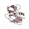
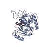
- Structure visualization
| Structure viewer | Molecule: MolmilJmol/JSmol |
|---|
- Downloads & links
-Download
| PDBx/mmCIF format | 1ha5.cif.gz | 175.5 KB | Display | PDBx/mmCIF format |
|---|---|---|---|---|
| PDB format | pdb1ha5.ent.gz | 139.4 KB | Display | PDB format |
| PDBx/mmJSON format | 1ha5.json.gz | Tree view | PDBx/mmJSON format | |
| Others |  Other downloads Other downloads |
-Validation report
| Arichive directory | https://data.pdbj.org/pub/pdb/validation_reports/ha/1ha5ftp://data.pdbj.org/pub/pdb/validation_reports/ha/1ha5 | HTTPS FTP |
|---|
-Related structure data
| Related structure data | 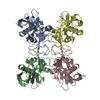 1b1zS S: Starting model for refinement |
|---|---|
| Similar structure data |


-Links
 PDBj
PDBj
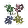
- Assembly
| Deposited unit | 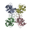
| ||||||||||||||||
|---|---|---|---|---|---|---|---|---|---|---|---|---|---|---|---|---|---|
| 1 | 
| ||||||||||||||||
| 2 | 
| ||||||||||||||||
| 3 | 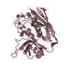
| ||||||||||||||||
| 4 | 
| ||||||||||||||||
| Unit cell |
| ||||||||||||||||
| Noncrystallographic symmetry (NCS) | NCS oper:
|
-Components
| #1: Protein | Mass: 25376.520 Da / Num. of mol.: 4 / Fragment: RESIDUES 33-250 Source method: isolated from a genetically manipulated source Source: (gene. exp.) STREPTOCOCCUS PYOGENES (bacteria) / Production host: #2: Chemical | ChemComp-ZN /   Mass: 65.409 Da / Num. of mol.: 4 / Source method: obtained synthetically / Formula: Zn Mass: 65.409 Da / Num. of mol.: 4 / Source method: obtained synthetically / Formula: Zn#3: Water | ChemComp-HOH / |  Mass: 18.015 Da / Num. of mol.: 142 / Source method: isolated from a natural source / Formula: H2O Mass: 18.015 Da / Num. of mol.: 142 / Source method: isolated from a natural source / Formula: H2OCompound details | SUPERANTIGEN STEPTOCOCCAL PYROGENIC EXOTOXIN A1 IS RECOGNISED BY MHC CLASSII MOLECULES AND T-CELL ...SUPERANTIG | Has protein modification | Y | |
|---|
-Experimental details
-Experiment
| Experiment | Method: X-RAY DIFFRACTION / Number of used crystals: 2 |
|---|
- Sample preparation
| Crystal | Density Matthews: 2.6 Å3/Da / Density % sol: 52.66 % |
|---|---|
| Crystal grow | pH: 6.5 / Details: pH 6.50 |
-Data collection
| Diffraction | Mean temperature: 100 K |
|---|---|
| Diffraction source | Source: SYNCHROTRON / Site: EMBL/DESY, HAMBURG  / Beamline: X11 / Wavelength: 0.9057 / Beamline: X11 / Wavelength: 0.9057 |
| Detector | Type: MARRESEARCH / Detector: CCD |
| Radiation | Protocol: SINGLE WAVELENGTH / Monochromatic (M) / Laue (L): M / Scattering type: x-ray |
| Radiation wavelength | Wavelength: 0.9057 Å / Relative weight: 1 |
| Reflection | Resolution: 2.8→40 Å / Num. obs: 26185 / % possible obs: 95.8 % / Redundancy: 5.7 % / Biso Wilson estimate: 5.7 Å2 / Rmerge(I) obs: 0.108 / Net I/σ(I): 6.4 |
| Reflection shell | Resolution: 2.8→2.9 Å / Rmerge(I) obs: 0.285 / Mean I/σ(I) obs: 5.3 / % possible all: 61.3 |
- Processing
| Software |
| ||||||||||||||||||||||||||||||||||||||||||||||||||||||||||||
|---|---|---|---|---|---|---|---|---|---|---|---|---|---|---|---|---|---|---|---|---|---|---|---|---|---|---|---|---|---|---|---|---|---|---|---|---|---|---|---|---|---|---|---|---|---|---|---|---|---|---|---|---|---|---|---|---|---|---|---|---|---|
| Refinement | Method to determine structure: MOLECULAR REPLACEMENT Starting model: 1B1Z Resolution: 2.82→39.6 Å / Rfactor Rfree error: 0.006 / Data cutoff high absF: 1727947.22 / Isotropic thermal model: RESTRAINED / Cross valid method: THROUGHOUT / σ(F): 0 Details: RESIDUES 5, 88, 112, 115, 179, 180 WERE MODELLED AS ALANINE IN ALL 4 MOLECULES DUE TO INSUFFICIENT DENSITY.
| ||||||||||||||||||||||||||||||||||||||||||||||||||||||||||||
| Solvent computation | Solvent model: FLAT MODEL / ksol: 0.317447 e/Å3 | ||||||||||||||||||||||||||||||||||||||||||||||||||||||||||||
| Displacement parameters | Biso mean: 31.1 Å2
| ||||||||||||||||||||||||||||||||||||||||||||||||||||||||||||
| Refine analyze |
| ||||||||||||||||||||||||||||||||||||||||||||||||||||||||||||
| Refinement step | Cycle: LAST / Resolution: 2.82→39.6 Å
| ||||||||||||||||||||||||||||||||||||||||||||||||||||||||||||
| Refine LS restraints |
| ||||||||||||||||||||||||||||||||||||||||||||||||||||||||||||
| LS refinement shell | Resolution: 2.82→2.98 Å / Rfactor Rfree error: 0.023 / Total num. of bins used: 6
| ||||||||||||||||||||||||||||||||||||||||||||||||||||||||||||
| Xplor file |
|
