 Movie
Movie Controller

[English] 日本語
 Yorodumi
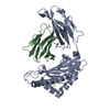
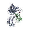
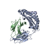
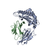
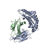
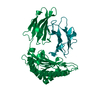
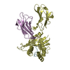
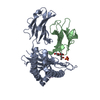
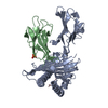
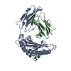
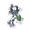
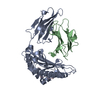
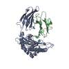
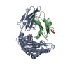
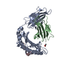
- PDB-1osz: MHC CLASS I H-2KB HEAVY CHAIN COMPLEXED WITH BETA-2 MICROGLOBULIN... -
+ Open data

- Basic information
| Entry | Database: PDB / ID: 1osz | ||||||
|---|---|---|---|---|---|---|---|
| Title | MHC CLASS I H-2KB HEAVY CHAIN COMPLEXED WITH BETA-2 MICROGLOBULIN AND AN (L4V) MUTANT OF THE VESICULAR STOMATITIS VIRUS NUCLEOPROTEIN | ||||||
 Components Components |
| ||||||
 Keywords Keywords | COMPLEX (MHC I/PEPTIDE) / MHC-PEPTIDE COMPLEX / TRANSMEMBRANE PROTEIN / THYMIC SELECTION / COMPLEX (MHC I-PEPTIDE) / COMPLEX (MHC I-PEPTIDE) complex | ||||||
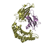
| Function / homology |  Function and homology information Function and homology informationMHC class Ib protein complex / natural killer cell lectin-like receptor binding / TAP2 binding / TAP1 binding / cis-Golgi network membrane / Endosomal/Vacuolar pathway / DAP12 interactions / Antigen Presentation: Folding, assembly and peptide loading of class I MHC / DAP12 signaling / ER-Phagosome pathway ...MHC class Ib protein complex / natural killer cell lectin-like receptor binding / TAP2 binding / TAP1 binding / cis-Golgi network membrane / Endosomal/Vacuolar pathway / DAP12 interactions / Antigen Presentation: Folding, assembly and peptide loading of class I MHC / DAP12 signaling / ER-Phagosome pathway / Immunoregulatory interactions between a Lymphoid and a non-Lymphoid cell / antigen processing and presentation of endogenous peptide antigen via MHC class I via ER pathway, TAP-dependent / TAP complex binding / antigen processing and presentation of exogenous peptide antigen via MHC class I / Golgi medial cisterna / regulation of membrane depolarization / inner ear development / CD8 receptor binding / beta-2-microglobulin binding / endoplasmic reticulum exit site / TAP binding / MHC class I protein binding / antigen processing and presentation of endogenous peptide antigen via MHC class Ib / antigen processing and presentation of endogenous peptide antigen via MHC class I via ER pathway, TAP-independent / cellular defense response / T cell receptor binding / Neutrophil degranulation / 14-3-3 protein binding / lumenal side of endoplasmic reticulum membrane / regulation of iron ion transport / cellular response to iron(III) ion / negative regulation of iron ion transport / negative regulation of forebrain neuron differentiation / antigen processing and presentation of exogenous protein antigen via MHC class Ib, TAP-dependent / iron ion transport / peptide antigen assembly with MHC class I protein complex / regulation of erythrocyte differentiation / response to molecule of bacterial origin / HFE-transferrin receptor complex / MHC class I peptide loading complex / transferrin transport / cellular response to iron ion / negative regulation of receptor-mediated endocytosis / positive regulation of T cell cytokine production / antigen processing and presentation of endogenous peptide antigen via MHC class I / MHC class I protein complex / peptide antigen assembly with MHC class II protein complex / negative regulation of neurogenesis / cellular response to nicotine / MHC class II protein complex / positive regulation of receptor-mediated endocytosis / multicellular organismal-level iron ion homeostasis / positive regulation of T cell mediated cytotoxicity / antigen processing and presentation of exogenous peptide antigen via MHC class II / positive regulation of immune response / peptide antigen binding / phagocytic vesicle membrane / positive regulation of T cell activation / negative regulation of epithelial cell proliferation / sensory perception of smell / positive regulation of cellular senescence / MHC class II protein complex binding / T cell differentiation in thymus / late endosome membrane / antimicrobial humoral immune response mediated by antimicrobial peptide / negative regulation of neuron projection development / antibacterial humoral response / protein refolding / protein-folding chaperone binding / cellular response to lipopolysaccharide / early endosome membrane / amyloid fibril formation / protein homotetramerization / defense response to Gram-negative bacterium / intracellular iron ion homeostasis / learning or memory / early endosome / defense response to bacterium / defense response to Gram-positive bacterium / immune response / receptor ligand activity / signaling receptor binding / Golgi membrane / external side of plasma membrane / innate immune response / lysosomal membrane / structural molecule activity / cell surface / endoplasmic reticulum / Golgi apparatus / protein homodimerization activity / : / identical protein binding / plasma membrane / cytosol Similarity search - Function | ||||||
| Biological species |  | ||||||
| Method |  X-RAY DIFFRACTION / SYNCHROTRON / MOLECULAR REPLACEMENT / Resolution: 2.1 Å X-RAY DIFFRACTION / SYNCHROTRON / MOLECULAR REPLACEMENT / Resolution: 2.1 Å | ||||||
 Authors Authors | Ghendler, Y. / Teng, M.-K. / Liu, J.-H. / Witte, T. / Liu, J. / Kim, K.S. / Kern, P. / Chang, H.-C. / Wang, J.-H. / Reinherz, E.L. | ||||||
 Citation Citation | Journal: Proc.Natl.Acad.Sci.USA / Year: 1998 Title: Differential thymic selection outcomes stimulated by focal structural alteration in peptide/major histocompatibility complex ligands. Authors: Ghendler, Y. / Teng, M.K. / Liu, J.H. / Witte, T. / Liu, J. / Kim, K.S. / Kern, P. / Chang, H.C. / Wang, J.H. / Reinherz, E.L. | ||||||
| History |
|
- Structure visualization
| Structure viewer | Molecule: MolmilJmol/JSmol |
|---|
- Downloads & links
-Download
| PDBx/mmCIF format | 1osz.cif.gz | 87.3 KB | Display | PDBx/mmCIF format |
|---|---|---|---|---|
| PDB format | pdb1osz.ent.gz | 67.3 KB | Display | PDB format |
| PDBx/mmJSON format | 1osz.json.gz | Tree view | PDBx/mmJSON format | |
| Others |  Other downloads Other downloads |
-Validation report
| Arichive directory | https://data.pdbj.org/pub/pdb/validation_reports/os/1oszftp://data.pdbj.org/pub/pdb/validation_reports/os/1osz | HTTPS FTP |
|---|
-Related structure data
| Related structure data |  2vaaS S: Starting model for refinement |
|---|---|
| Similar structure data |


-Links
 PDBj
PDBj


- Assembly
| Deposited unit | 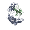
| ||||||||
|---|---|---|---|---|---|---|---|---|---|
| 1 |
| ||||||||
| Unit cell |
|
-Components
| #1: Protein | Mass: 31648.322 Da / Num. of mol.: 1 / Fragment: EXTRACELLULAR DOMAINS Source method: isolated from a genetically manipulated source Source: (gene. exp.)  |
|---|---|
| #2: Protein | Mass: 11704.359 Da / Num. of mol.: 1 Source method: isolated from a genetically manipulated source Source: (gene. exp.) |
| #3: Protein/peptide | Mass: 970.105 Da / Num. of mol.: 1 / Fragment: RESIDUES 52 - 59 / Mutation: L4V MUTATION AT THE P4 POSITION Source method: isolated from a genetically manipulated source |
| Has protein modification | Y |
-Experimental details
-Experiment
| Experiment | Method: X-RAY DIFFRACTION / Number of used crystals: 1 |
|---|
- Sample preparation
| Crystal | Density Matthews: 2.67 Å3/Da / Density % sol: 54 % | |||||||||||||||||||||||||
|---|---|---|---|---|---|---|---|---|---|---|---|---|---|---|---|---|---|---|---|---|---|---|---|---|---|---|
| Crystal grow | pH: 7 / Details: pH 7.0 | |||||||||||||||||||||||||
| Crystal grow | *PLUS Method: vapor diffusion, hanging dropDetails: drop contains equal volume of the reservoir solution | |||||||||||||||||||||||||
| Components of the solutions | *PLUS
|
-Data collection
| Diffraction | Mean temperature: 110 K |
|---|---|
| Diffraction source | Source: SYNCHROTRON / Site: NSLS  / Beamline: X8C / Wavelength: 1.009 / Beamline: X8C / Wavelength: 1.009 |
| Detector | Type: MARRESEARCH / Detector: IMAGE PLATE / Date: Dec 17, 1997 |
| Radiation | Monochromatic (M) / Laue (L): M / Scattering type: x-ray |
| Radiation wavelength | Wavelength: 1.009 Å / Relative weight: 1 |
| Reflection | Resolution: 2.1→15 Å / Num. obs: 32332 / % possible obs: 98.8 % / Redundancy: 7.1 % / Rsym value: 0.076 / Net I/σ(I): 15 |
| Reflection shell | Resolution: 2.1→2.2 Å / Redundancy: 4 % / Mean I/σ(I) obs: 7.8 / Rsym value: 0.29 / % possible all: 98.8 |
| Reflection | *PLUS Rmerge(I) obs: 0.076 |
| Reflection shell | *PLUS % possible obs: 98.8 % / Rmerge(I) obs: 0.29 |
- Processing
| Software |
| ||||||||||||||||||||||||||||||||||||||||||||||||||||||||||||
|---|---|---|---|---|---|---|---|---|---|---|---|---|---|---|---|---|---|---|---|---|---|---|---|---|---|---|---|---|---|---|---|---|---|---|---|---|---|---|---|---|---|---|---|---|---|---|---|---|---|---|---|---|---|---|---|---|---|---|---|---|---|
| Refinement | Method to determine structure: MOLECULAR REPLACEMENT Starting model: PDB ENTRY 2VAA Resolution: 2.1→15 Å / σ(F): 2
| ||||||||||||||||||||||||||||||||||||||||||||||||||||||||||||
| Displacement parameters | Biso mean: 24.1 Å2 | ||||||||||||||||||||||||||||||||||||||||||||||||||||||||||||
| Refine analyze | Luzzati coordinate error obs: 0.33 Å / Luzzati d res low obs: 15 Å | ||||||||||||||||||||||||||||||||||||||||||||||||||||||||||||
| Refinement step | Cycle: LAST / Resolution: 2.1→15 Å
| ||||||||||||||||||||||||||||||||||||||||||||||||||||||||||||
| Refine LS restraints |
| ||||||||||||||||||||||||||||||||||||||||||||||||||||||||||||
| LS refinement shell | Resolution: 2.1→2.2 Å / Total num. of bins used: 8
| ||||||||||||||||||||||||||||||||||||||||||||||||||||||||||||
| Xplor file |
| ||||||||||||||||||||||||||||||||||||||||||||||||||||||||||||
| Software | *PLUS Name: X-PLOR / Version: 3.8 / Classification: refinement | ||||||||||||||||||||||||||||||||||||||||||||||||||||||||||||
| Refinement | *PLUS | ||||||||||||||||||||||||||||||||||||||||||||||||||||||||||||
| Solvent computation | *PLUS | ||||||||||||||||||||||||||||||||||||||||||||||||||||||||||||
| Displacement parameters | *PLUS | ||||||||||||||||||||||||||||||||||||||||||||||||||||||||||||
| LS refinement shell | *PLUS Rfactor obs: 0.278 |
