 Movie
Movie Controller

[English] 日本語
 Yorodumi
- PDB-1vad: MHC CLASS I H-2KB HEAVY CHAIN COMPLEXED WITH BETA-2 MICROGLOBULIN... -
+ Open data

- Basic information
| Entry | Database: PDB / ID: 1vad | ||||||
|---|---|---|---|---|---|---|---|
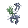
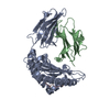
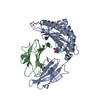
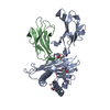
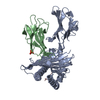
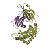
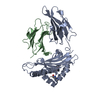
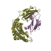
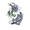
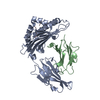
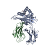
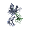
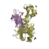
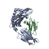
| Title | MHC CLASS I H-2KB HEAVY CHAIN COMPLEXED WITH BETA-2 MICROGLOBULIN AND YEAST ALPHA-GLUCOSIDASE | ||||||
 Components Components |
| ||||||
 Keywords Keywords | COMPLEX (MHC I/PEPTIDE) / HISTOCOMPATIBILITY ANTIGEN / CLASS I MAJOR HISTOCOMPATIBILITY COMPLEX / MHC I / COMPLEX (MHC I-PEPTIDE) COMPLEX | ||||||
| Function / homology |  Function and homology information Function and homology informationglucan 1,4-alpha-maltotriohydrolase activity / oligo-1,6-glucosidase activity / maltose catabolic process / sucrose catabolic process / alpha-glucosidase / sucrose alpha-glucosidase activity / alpha-1,4-glucosidase activity / MHC class Ib protein complex / natural killer cell lectin-like receptor binding / TAP2 binding ...glucan 1,4-alpha-maltotriohydrolase activity / oligo-1,6-glucosidase activity / maltose catabolic process / sucrose catabolic process / alpha-glucosidase / sucrose alpha-glucosidase activity / alpha-1,4-glucosidase activity / MHC class Ib protein complex / natural killer cell lectin-like receptor binding / TAP2 binding / TAP1 binding / cis-Golgi network membrane / Endosomal/Vacuolar pathway / DAP12 interactions / Antigen Presentation: Folding, assembly and peptide loading of class I MHC / alpha-amylase activity / ER-Phagosome pathway / DAP12 signaling / Immunoregulatory interactions between a Lymphoid and a non-Lymphoid cell / antigen processing and presentation of endogenous peptide antigen via MHC class I via ER pathway, TAP-dependent / TAP complex binding / antigen processing and presentation of exogenous peptide antigen via MHC class I / Golgi medial cisterna / regulation of membrane depolarization / inner ear development / CD8 receptor binding / endoplasmic reticulum exit site / TAP binding / beta-2-microglobulin binding / MHC class I protein binding / antigen processing and presentation of endogenous peptide antigen via MHC class Ib / antigen processing and presentation of endogenous peptide antigen via MHC class I via ER pathway, TAP-independent / cellular defense response / T cell receptor binding / Neutrophil degranulation / 14-3-3 protein binding / lumenal side of endoplasmic reticulum membrane / regulation of iron ion transport / cellular response to iron(III) ion / antigen processing and presentation of exogenous protein antigen via MHC class Ib, TAP-dependent / negative regulation of iron ion transport / negative regulation of forebrain neuron differentiation / regulation of erythrocyte differentiation / iron ion transport / peptide antigen assembly with MHC class I protein complex / response to molecule of bacterial origin / HFE-transferrin receptor complex / MHC class I peptide loading complex / transferrin transport / negative regulation of receptor-mediated endocytosis / cellular response to iron ion / positive regulation of T cell cytokine production / antigen processing and presentation of endogenous peptide antigen via MHC class I / MHC class I protein complex / peptide antigen assembly with MHC class II protein complex / negative regulation of neurogenesis / multicellular organismal-level iron ion homeostasis / cellular response to nicotine / MHC class II protein complex / positive regulation of receptor-mediated endocytosis / positive regulation of T cell mediated cytotoxicity / negative regulation of epithelial cell proliferation / antigen processing and presentation of exogenous peptide antigen via MHC class II / positive regulation of immune response / peptide antigen binding / phagocytic vesicle membrane / positive regulation of T cell activation / sensory perception of smell / positive regulation of cellular senescence / MHC class II protein complex binding / T cell differentiation in thymus / late endosome membrane / negative regulation of neuron projection development / antimicrobial humoral immune response mediated by antimicrobial peptide / antibacterial humoral response / cellular response to lipopolysaccharide / protein refolding / protein-folding chaperone binding / early endosome membrane / amyloid fibril formation / defense response to Gram-negative bacterium / protein homotetramerization / intracellular iron ion homeostasis / learning or memory / early endosome / defense response to bacterium / defense response to Gram-positive bacterium / immune response / receptor ligand activity / Golgi membrane / external side of plasma membrane / signaling receptor binding / innate immune response / lysosomal membrane / structural molecule activity / Golgi apparatus / cell surface / endoplasmic reticulum / protein homodimerization activity / : Similarity search - Function | ||||||
| Biological species |   | ||||||
| Method |  X-RAY DIFFRACTION / Resolution: 2.5 Å X-RAY DIFFRACTION / Resolution: 2.5 Å | ||||||
 Authors Authors | Fremont, D.H. / Wilson, I.A. | ||||||
 Citation Citation | Journal: Proc.Natl.Acad.Sci.USA / Year: 1995 Title: Crystal structure of an H-2Kb-ovalbumin peptide complex reveals the interplay of primary and secondary anchor positions in the major histocompatibility complex binding groove. Authors: Fremont, D.H. / Stura, E.A. / Matsumura, M. / Peterson, P.A. / Wilson, I.A. #1: Journal: Science / Year: 1992Title: Crystal Structures of Two Viral Peptides in Complex with Murine Mhc Class I H-2Kb Authors: Fremont, D.H. / Matsumura, M. / Stura, E.A. / Peterson, P.A. / Wilson, I.A. #2: Journal: Science / Year: 1992Title: Emerging Principles for the Recognition of Peptide Antigens by Mhc Class I Molecules Authors: Matsumura, M. / Fremont, D.H. / Peterson, P.A. / Wilson, I.A. | ||||||
| History |
|
- Structure visualization
| Structure viewer | Molecule: MolmilJmol/JSmol |
|---|
- Downloads & links
-Download
| PDBx/mmCIF format | 1vad.cif.gz | 91.4 KB | Display | PDBx/mmCIF format |
|---|---|---|---|---|
| PDB format | pdb1vad.ent.gz | 70.2 KB | Display | PDB format |
| PDBx/mmJSON format | 1vad.json.gz | Tree view | PDBx/mmJSON format | |
| Others |  Other downloads Other downloads |
-Validation report
| Arichive directory | https://data.pdbj.org/pub/pdb/validation_reports/va/1vadftp://data.pdbj.org/pub/pdb/validation_reports/va/1vad | HTTPS FTP |
|---|
-Related structure data


-Links
 PDBj
PDBj


- Assembly
| Deposited unit | 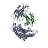
| ||||||||
|---|---|---|---|---|---|---|---|---|---|
| 1 |
| ||||||||
| Unit cell |
|
-Components
| #1: Protein | Mass: 31648.322 Da / Num. of mol.: 1 / Fragment: EXTRACELLULAR DOMAINS Source method: isolated from a genetically manipulated source Source: (gene. exp.)  |
|---|---|
| #2: Protein | Mass: 11704.359 Da / Num. of mol.: 1 Source method: isolated from a genetically manipulated source Source: (gene. exp.) |
| #3: Protein/peptide | Mass: 1089.208 Da / Num. of mol.: 1 / Fragment: RESIDUES 438 - 446 / Source method: isolated from a natural source / Source: (natural) |
| #4: Water | ChemComp-HOH /  Mass: 18.015 Da / Num. of mol.: 129 / Source method: isolated from a natural source / Formula: H2O Mass: 18.015 Da / Num. of mol.: 129 / Source method: isolated from a natural source / Formula: H2O |
| Has protein modification | Y |
| Sequence details | SOURCE MOLECULE_NAME: MALTASE, YEAST ALPHA-GLUCOSIDASE (RESIDUES 438 - 446). SOURCE OF PEPTIDE ...SOURCE MOLECULE_NAME: MALTASE, YEAST ALPHA-GLUCOSIDAS |
-Experimental details
-Experiment
| Experiment | Method: X-RAY DIFFRACTION |
|---|
- Sample preparation
| Crystal | Density Matthews: 3.19 Å3/Da / Density % sol: 61.4 % |
|---|
-Data collection
| Diffraction source | Wavelength: 1.5418 |
|---|---|
| Detector | Type: SIEMENS / Detector: AREA DETECTOR |
| Radiation | Monochromatic (M) / Laue (L): M / Scattering type: x-ray |
| Radiation wavelength | Wavelength: 1.5418 Å / Relative weight: 1 |
| Reflection | Num. obs: 18677 / % possible obs: 91.5 % / Redundancy: 1.7 % / Rmerge(I) obs: 0.068 |
- Processing
| Software |
| ||||||||||||||||||||||||||||||||||||||||||||||||||||||||||||
|---|---|---|---|---|---|---|---|---|---|---|---|---|---|---|---|---|---|---|---|---|---|---|---|---|---|---|---|---|---|---|---|---|---|---|---|---|---|---|---|---|---|---|---|---|---|---|---|---|---|---|---|---|---|---|---|---|---|---|---|---|---|
| Refinement | Resolution: 2.5→6 Å / σ(F): 2 /
| ||||||||||||||||||||||||||||||||||||||||||||||||||||||||||||
| Refinement step | Cycle: LAST / Resolution: 2.5→6 Å
| ||||||||||||||||||||||||||||||||||||||||||||||||||||||||||||
| Refine LS restraints |
|
