microtubule-dependent intracellular transport of viral material towards nucleus / T=4 icosahedral viral capsid / antigen processing and presentation of peptide antigen via MHC class I / symbiont-mediated perturbation of host defense response / early endosome lumen / Nef mediated downregulation of MHC class I complex cell surface expression / DAP12 interactions / Endosomal/Vacuolar pathway / T cell mediated cytotoxicity / Antigen Presentation: Folding, assembly and peptide loading of class I MHC ...microtubule-dependent intracellular transport of viral material towards nucleus / T=4 icosahedral viral capsid / antigen processing and presentation of peptide antigen via MHC class I / symbiont-mediated perturbation of host defense response / early endosome lumen / Nef mediated downregulation of MHC class I complex cell surface expression / DAP12 interactions / Endosomal/Vacuolar pathway / T cell mediated cytotoxicity / Antigen Presentation: Folding, assembly and peptide loading of class I MHC / lumenal side of endoplasmic reticulum membrane / regulation of iron ion transport / cellular response to iron(III) ion / negative regulation of iron ion transport / negative regulation of forebrain neuron differentiation / antigen processing and presentation of exogenous protein antigen via MHC class Ib, TAP-dependent / peptide antigen assembly with MHC class I protein complex / ER to Golgi transport vesicle membrane / regulation of erythrocyte differentiation / response to molecule of bacterial origin / HFE-transferrin receptor complex / MHC class I peptide loading complex / transferrin transport / cellular response to iron ion / negative regulation of receptor-mediated endocytosis / positive regulation of T cell cytokine production / antigen processing and presentation of endogenous peptide antigen via MHC class I / MHC class I protein complex / peptide antigen assembly with MHC class II protein complex / negative regulation of neurogenesis / MHC class II protein complex / cellular response to nicotine / positive regulation of receptor-mediated endocytosis / multicellular organismal-level iron ion homeostasis / positive regulation of T cell mediated cytotoxicity / viral penetration into host nucleus / specific granule lumen / antigen processing and presentation of exogenous peptide antigen via MHC class II / positive regulation of immune response / peptide antigen binding / phagocytic vesicle membrane / recycling endosome membrane / positive regulation of T cell activation / negative regulation of epithelial cell proliferation / Interferon gamma signaling / Immunoregulatory interactions between a Lymphoid and a non-Lymphoid cell / sensory perception of smell / Modulation by Mtb of host immune system / positive regulation of cellular senescence / tertiary granule lumen / MHC class II protein complex binding / T cell differentiation in thymus / DAP12 signaling / late endosome membrane / host cell / negative regulation of neuron projection development / protein refolding / ER-Phagosome pathway / early endosome membrane / amyloid fibril formation / protein homotetramerization / host cell cytoplasm / intracellular iron ion homeostasis / learning or memory / immune response / endoplasmic reticulum lumen / Amyloid fiber formation / Golgi membrane / external side of plasma membrane / lysosomal membrane / focal adhesion / Neutrophil degranulation / symbiont entry into host cell / host cell nucleus / SARS-CoV-2 activates/modulates innate and adaptive immune responses / structural molecule activity / cell surface / Golgi apparatus / endoplasmic reticulum / protein homodimerization activity / : / DNA binding / RNA binding / extracellular exosome / extracellular region / membrane / identical protein binding / plasma membrane / cytosol Similarity search - Function
Hepatitis B virus, capsid N-terminal / Hepatitis core protein, putative zinc finger / Hepatitis core antigen / Viral capsid core domain supefamily, Hepatitis B virus / Hepatitis core antigen / MHC class I, alpha chain, C-terminal / MHC_I C-terminus / MHC class I-like antigen recognition-like / Murine Class I Major Histocompatibility Complex, H2-DB; Chain A, domain 1 / MHC class I alpha chain, alpha1 alpha2 domains ...Hepatitis B virus, capsid N-terminal / Hepatitis core protein, putative zinc finger / Hepatitis core antigen / Viral capsid core domain supefamily, Hepatitis B virus / Hepatitis core antigen / MHC class I, alpha chain, C-terminal / MHC_I C-terminus / MHC class I-like antigen recognition-like / Murine Class I Major Histocompatibility Complex, H2-DB; Chain A, domain 1 / MHC class I alpha chain, alpha1 alpha2 domains / Class I Histocompatibility antigen, domains alpha 1 and 2 / Beta-2-Microglobulin / : / MHC class I-like antigen recognition-like / MHC class I-like antigen recognition-like superfamily / MHC classes I/II-like antigen recognition protein / : / Immunoglobulin/major histocompatibility complex, conserved site / Immunoglobulins and major histocompatibility complex proteins signature. / Immunoglobulin C-Type / Immunoglobulin C1-set / Immunoglobulin C1-set domain / Ig-like domain profile. / Immunoglobulin-like domain / Immunoglobulin-like domain superfamily / Immunoglobulin-like fold / Immunoglobulins / Immunoglobulin-like / Sandwich / 2-Layer Sandwich / Mainly Beta / Alpha Beta Similarity search - Domain/homology
External core antigen / Beta-2-microglobulin / HLA class I histocompatibility antigen A alpha chain / External core antigen Similarity search - Component
#200 - Aug 2016 Quasisymmetry in Icosahedral Viruses similarity (1)
-
Assembly
Deposited unit
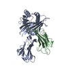
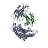
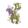
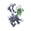
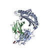
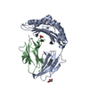
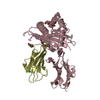
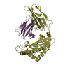
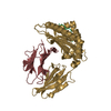
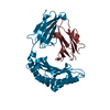
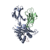
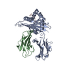
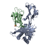
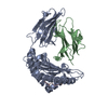
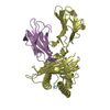
A: MHC class I antigen B: Beta-2-microglobulin C: 10mer peptide from Pre-core-protein D: MHC class I antigen E: Beta-2-microglobulin F: 10mer peptide from Pre-core-protein
Mass: 18.015 Da / Num. of mol.: 450 / Source method: isolated from a natural source / Formula: H2O
Has protein modification
Y
-
Experimental details
-
Experiment
Experiment
Method: X-RAY DIFFRACTION / Number of used crystals: 1
-
Sample preparation
Crystal
Density Matthews: 2.92 Å3/Da / Density % sol: 57.83 %
Crystal grow
Temperature: 298 K / Method: vapor diffusion, hanging drop / pH: 7.5 Details: 0.1M HEPSE pH7.5, 1.4M sodium citrate tribasic dihydrate, VAPOR DIFFUSION, HANGING DROP, temperature 298K
-
Data collection
Diffraction
Mean temperature: 298 K
Diffraction source
Source: ROTATING ANODE / Type: BRUKER AXS MICROSTAR / Wavelength: 1.5418 Å
Detector
Type: Bruker Platinum 135 / Detector: CCD / Date: Feb 5, 2010
Radiation
Protocol: SINGLE WAVELENGTH / Monochromatic (M) / Laue (L): M / Scattering type: x-ray
Radiation wavelength
Wavelength: 1.5418 Å / Relative weight: 1
Reflection
Resolution: 2.1→64.12 Å / Num. obs: 68748 / % possible obs: 95.76 % / Observed criterion σ(I): 2 / Redundancy: 4 % / Biso Wilson estimate: 11.062 Å2 / Rmerge(I) obs: 0.066 / Net I/σ(I): 4.68
Reflection shell
Resolution (Å)
Redundancy (%)
Rmerge(I) obs
Mean I/σ(I) obs
Num. unique all
Diffraction-ID
% possible all
2.16-2.22
1.71
0.3449
2.1
3517
1
80.7
2.22-2.28
2.22
0.34
2.43
3534
1
89.8
2.28-2.34
2.9
0.3119
2.97
3457
1
97.7
2.34-2.41
3.61
0.2826
3.7
3681
1
99.9
2.41-2.48
3.93
0.2701
4.2
3292
1
100
2.48-2.56
4.39
0.2473
4.93
3333
1
100
2.56-2.65
4.71
0.2311
5.73
3284
1
100
2.65-2.76
4.74
0.216
6.37
3434
1
100
2.76-2.89
4.72
0.1852
7.77
3466
1
100
2.89-3.05
4.71
0.1644
9.26
3387
1
100
3.05-3.24
4.7
0.1371
11.41
3244
1
100
-
Processing
Software
Name
Version
Classification
PROTEUM PLUS
PLUS
datacollection
AMoRE
phasing
REFMAC
5.5.0102
refinement
PROTEUM PLUS
PLUS
datareduction
PROTEUM PLUS
PLUS
datascaling
Refinement
Resolution: 2.16→64.12 Å / Cor.coef. Fo:Fc: 0.941 / Cor.coef. Fo:Fc free: 0.901 / SU B: 4.614 / SU ML: 0.121 / Cross valid method: THROUGHOUT / ESU R: 0.218 / ESU R Free: 0.19 / Stereochemistry target values: MAXIMUM LIKELIHOOD / Details: HYDROGENS HAVE BEEN ADDED IN THE RIDING POSITIONS
Rfactor
Num. reflection
% reflection
Selection details
Rfree
0.23409
2733
5.1 %
RANDOM
Rwork
0.18221
-
-
-
obs
0.1848
50693
95.88 %
-
Solvent computation
Ion probe radii: 0.8 Å / Shrinkage radii: 0.8 Å / VDW probe radii: 1.4 Å
Displacement parameters
Biso mean: 18.144 Å2
Baniso -1
Baniso -2
Baniso -3
1-
0.06 Å2
0.06 Å2
0.03 Å2
2-
-
0 Å2
-0.02 Å2
3-
-
-
-0.06 Å2
Refinement step
Cycle: LAST / Resolution: 2.16→64.12 Å
Protein
Nucleic acid
Ligand
Solvent
Total
Num. atoms
6354
0
0
450
6804
Refine LS restraints
Refine-ID
Type
Dev ideal
Dev ideal target
Number
X-RAY DIFFRACTION
r_bond_refined_d
0.012
0.021
6566
X-RAY DIFFRACTION
r_bond_other_d
X-RAY DIFFRACTION
r_angle_refined_deg
1.313
1.92
8916
X-RAY DIFFRACTION
r_angle_other_deg
X-RAY DIFFRACTION
r_dihedral_angle_1_deg
7.824
5
768
X-RAY DIFFRACTION
r_dihedral_angle_2_deg
35.764
23.073
358
X-RAY DIFFRACTION
r_dihedral_angle_3_deg
19.434
15
1060
X-RAY DIFFRACTION
r_dihedral_angle_4_deg
22.504
15
58
X-RAY DIFFRACTION
r_chiral_restr
0.125
0.2
896
X-RAY DIFFRACTION
r_gen_planes_refined
0.017
0.021
5202
X-RAY DIFFRACTION
r_gen_planes_other
X-RAY DIFFRACTION
r_nbd_refined
X-RAY DIFFRACTION
r_nbd_other
X-RAY DIFFRACTION
r_nbtor_refined
X-RAY DIFFRACTION
r_nbtor_other
X-RAY DIFFRACTION
r_xyhbond_nbd_refined
X-RAY DIFFRACTION
r_xyhbond_nbd_other
X-RAY DIFFRACTION
r_metal_ion_refined
X-RAY DIFFRACTION
r_metal_ion_other
X-RAY DIFFRACTION
r_symmetry_vdw_refined
X-RAY DIFFRACTION
r_symmetry_vdw_other
X-RAY DIFFRACTION
r_symmetry_hbond_refined
X-RAY DIFFRACTION
r_symmetry_hbond_other
X-RAY DIFFRACTION
r_symmetry_metal_ion_refined
X-RAY DIFFRACTION
r_symmetry_metal_ion_other
X-RAY DIFFRACTION
r_mcbond_it
1.62
1.5
3846
X-RAY DIFFRACTION
r_mcbond_other
X-RAY DIFFRACTION
r_mcangle_it
2.762
2
6208
X-RAY DIFFRACTION
r_scbond_it
4.462
3
2720
X-RAY DIFFRACTION
r_scangle_it
6.669
4.5
2706
X-RAY DIFFRACTION
r_rigid_bond_restr
X-RAY DIFFRACTION
r_sphericity_free
X-RAY DIFFRACTION
r_sphericity_bonded
LS refinement shell
Resolution: 2.16→2.216 Å / Total num. of bins used: 20
Rfactor
Num. reflection
% reflection
Rfree
0.308
179
-
Rwork
0.23
3171
-
obs
-
-
82.15 %
+
About Yorodumi
-
News
-
Feb 9, 2022. New format data for meta-information of EMDB entries
New format data for meta-information of EMDB entries
Version 3 of the EMDB header file is now the official format.
The previous official version 1.9 will be removed from the archive.
In the structure databanks used in Yorodumi, some data are registered as the other names, "COVID-19 virus" and "2019-nCoV". Here are the details of the virus and the list of structure data.
Jan 31, 2019. EMDB accession codes are about to change! (news from PDBe EMDB page)
EMDB accession codes are about to change! (news from PDBe EMDB page)
The allocation of 4 digits for EMDB accession codes will soon come to an end. Whilst these codes will remain in use, new EMDB accession codes will include an additional digit and will expand incrementally as the available range of codes is exhausted. The current 4-digit format prefixed with “EMD-” (i.e. EMD-XXXX) will advance to a 5-digit format (i.e. EMD-XXXXX), and so on. It is currently estimated that the 4-digit codes will be depleted around Spring 2019, at which point the 5-digit format will come into force.
The EM Navigator/Yorodumi systems omit the EMD- prefix.
Related info.:Q: What is EMD? / ID/Accession-code notation in Yorodumi/EM Navigator
Yorodumi is a browser for structure data from EMDB, PDB, SASBDB, etc.
This page is also the successor to EM Navigator detail page, and also detail information page/front-end page for Omokage search.
The word "yorodu" (or yorozu) is an old Japanese word meaning "ten thousand". "mi" (miru) is to see.
Related info.:EMDB / PDB / SASBDB / Comparison of 3 databanks / Yorodumi Search / Aug 31, 2016. New EM Navigator & Yorodumi / Yorodumi Papers / Jmol/JSmol / Function and homology information / Changes in new EM Navigator and Yorodumi
 Movie
Movie Controller


 Open data
 Basic information
 Components
 Keywords
 Function and homology information
 Homo sapiens (human)
 Hepatitis B virus
 X-RAY DIFFRACTION / Resolution: 2.16 Å
  Authors
 Citation
 Structure visualization
 Downloads & links
 Other downloads


 PDBj
PDBj


 Assembly

 Mass: 18.015 Da / Num. of mol.: 450 / Source method: isolated from a natural source / Formula: H2O
 Sample preparation
 Processing