 Movie
Movie Controller


+ Open data

- Basic information
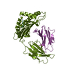
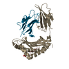
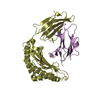
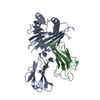
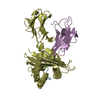
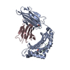
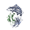
| Entry | Database: PDB / ID: 1qlf | ||||||
|---|---|---|---|---|---|---|---|
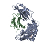
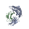
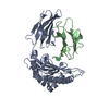
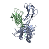
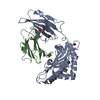
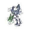
| Title | MHC CLASS I H-2DB COMPLEXED WITH GLYCOPEPTIDE K3G | ||||||
 Components Components |
| ||||||
 Keywords Keywords | IMMUNE SYSTEM/PEPTIDE / MURINE CLASS I MHC-PEPTIDE COMPLEX / MHC / GLYCOPEPTIDE / ANTIGEN / HISTOCOMPATIBILITY / IMMUNOLOGY / IMMUNE SYSTEM-PEPTIDE complex | ||||||
| Function / homology |  Function and homology information Function and homology information: / : / : / : / : / negative regulation of receptor binding / retina homeostasis / positive regulation of protein binding / helical viral capsid / regulation of membrane depolarization ...: / : / : / : / : / negative regulation of receptor binding / retina homeostasis / positive regulation of protein binding / helical viral capsid / regulation of membrane depolarization / antigen processing and presentation of peptide antigen via MHC class I / early endosome lumen / Nef mediated downregulation of MHC class I complex cell surface expression / DAP12 interactions / Endosomal/Vacuolar pathway / T cell mediated cytotoxicity / Antigen Presentation: Folding, assembly and peptide loading of class I MHC / lumenal side of endoplasmic reticulum membrane / regulation of iron ion transport / cellular response to iron(III) ion / negative regulation of iron ion transport / negative regulation of forebrain neuron differentiation / antigen processing and presentation of exogenous protein antigen via MHC class Ib, TAP-dependent / iron ion transport / peptide antigen assembly with MHC class I protein complex / ER to Golgi transport vesicle membrane / regulation of erythrocyte differentiation / response to molecule of bacterial origin / HFE-transferrin receptor complex / MHC class I peptide loading complex / transferrin transport / cellular response to iron ion / negative regulation of receptor-mediated endocytosis / positive regulation of T cell cytokine production / antigen processing and presentation of endogenous peptide antigen via MHC class I / MHC class I protein complex / peptide antigen assembly with MHC class II protein complex / negative regulation of neurogenesis / MHC class II protein complex / cellular response to nicotine / positive regulation of receptor-mediated endocytosis / multicellular organismal-level iron ion homeostasis / positive regulation of T cell mediated cytotoxicity / specific granule lumen / antigen processing and presentation of exogenous peptide antigen via MHC class II / positive regulation of immune response / peptide antigen binding / phagocytic vesicle membrane / recycling endosome membrane / positive regulation of T cell activation / negative regulation of epithelial cell proliferation / Interferon gamma signaling / Immunoregulatory interactions between a Lymphoid and a non-Lymphoid cell / sensory perception of smell / Modulation by Mtb of host immune system / positive regulation of cellular senescence / tertiary granule lumen / MHC class II protein complex binding / T cell differentiation in thymus / DAP12 signaling / late endosome membrane / antimicrobial humoral immune response mediated by antimicrobial peptide / negative regulation of neuron projection development / antibacterial humoral response / protein refolding / viral nucleocapsid / cellular response to lipopolysaccharide / ER-Phagosome pathway / early endosome membrane / amyloid fibril formation / protein homotetramerization / defense response to Gram-negative bacterium / host cell cytoplasm / intracellular iron ion homeostasis / learning or memory / defense response to Gram-positive bacterium / immune response / endoplasmic reticulum lumen / ribonucleoprotein complex / Amyloid fiber formation / Golgi membrane / external side of plasma membrane / innate immune response / lysosomal membrane / focal adhesion / Neutrophil degranulation / SARS-CoV-2 activates/modulates innate and adaptive immune responses / structural molecule activity / Golgi apparatus / endoplasmic reticulum / protein homodimerization activity / : / RNA binding / extracellular exosome / extracellular region / membrane / identical protein binding / plasma membrane / cytosol Similarity search - Function | ||||||
| Biological species |   HOMO SAPIENS (human) HOMO SAPIENS (human) SENDAI VIRUS SENDAI VIRUS | ||||||
| Method |  X-RAY DIFFRACTION / SYNCHROTRON / MOLECULAR REPLACEMENT / Resolution: 2.65 Å X-RAY DIFFRACTION / SYNCHROTRON / MOLECULAR REPLACEMENT / Resolution: 2.65 Å | ||||||
 Authors Authors | Tormo, J. / Jones, E.Y. | ||||||
 Citation Citation | Journal: Immunity / Year: 1999 Title: Crystal Structures of Two H-2Db/Glycopeptide Complexes Suggest a Molecular Basis for Ctl Cross-Reactivity Authors: Glithero, A. / Tormo, J. / Haurum, J.S. / Arsequell, G. / Valencia, G. / Edwards, J. / Springer, S. / Townsend, A. / Pao, Y.-L. / Wormald, M. / Dwek, R.A. / Jones, E.Y. / Elliot, T. | ||||||
| History |
|
- Structure visualization
| Structure viewer | Molecule: MolmilJmol/JSmol |
|---|
- Downloads & links
-Download
| PDBx/mmCIF format | 1qlf.cif.gz | 96.4 KB | Display | PDBx/mmCIF format |
|---|---|---|---|---|
| PDB format | pdb1qlf.ent.gz | 72.6 KB | Display | PDB format |
| PDBx/mmJSON format | 1qlf.json.gz | Tree view | PDBx/mmJSON format | |
| Others |  Other downloads Other downloads |
-Validation report
| Arichive directory | https://data.pdbj.org/pub/pdb/validation_reports/ql/1qlfftp://data.pdbj.org/pub/pdb/validation_reports/ql/1qlf | HTTPS FTP |
|---|
-Related structure data
| Related structure data | 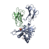 1ce6SC S: Starting model for refinement C: citing same article ( |
|---|---|
| Similar structure data |


-Links
 PDBj
PDBj


- Assembly
| Deposited unit | 
| ||||||||
|---|---|---|---|---|---|---|---|---|---|
| 1 | 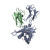
| ||||||||
| Unit cell |
|
-Components
-Protein , 2 types, 2 molecules AB
| #1: Protein | Mass: 32087.703 Da / Num. of mol.: 1 / Fragment: EXTRACELLULAR DOMAINS Source method: isolated from a genetically manipulated source Details: FORMS HETEROTRIMER WITH CHAIN B (BETA-2-MICROGLOBULIN) AND CHAIN C (PEPTIDE ANTIGEN) Source: (gene. exp.)  CRICETULUS GRISEUS (Chinese hamster) / References: UniProt: P01899 CRICETULUS GRISEUS (Chinese hamster) / References: UniProt: P01899 |
|---|---|
| #2: Protein | Mass: 11748.160 Da / Num. of mol.: 1 / Fragment: MHC ASSOCIATED LIGHT CHAIN Source method: isolated from a genetically manipulated source Details: FORMS HETEROTRIMER WITH CHAIN A AND CHAIN C / Source: (gene. exp.) HOMO SAPIENS (human) / Cell: MOST NUCLEATED CELLS / Cellular location: CELL SURFACE / Cell (production host): CHO K1 CELLS / Production host: CRICETULUS GRISEUS (Chinese hamster) / References: UniProt: P01884, UniProt: P61769*PLUS |
-Protein/peptide / Sugars , 2 types, 2 molecules C
| #3: Protein/peptide | Mass: 979.086 Da / Num. of mol.: 1 Fragment: H-2DB-BOUND GLYCOPEPTIDE FROM NUCLEOCAPSID PROTEIN Source method: obtained synthetically Details: PEPTIDE DERIVED FROM SENDAI VIRUS NUCLEOPROTEIN RESIDUES 324-332, GLY327 HAS BEEN REPLACED BY AN O-GLCNAC SUBSTITUTED SERINE Source: (synth.) SENDAI VIRUS / References: UniProt: P04857 |
|---|---|
| #6: Sugar | ChemComp-NAG /  Type: D-saccharide, beta linking / Mass: 221.208 Da / Num. of mol.: 1 Type: D-saccharide, beta linking / Mass: 221.208 Da / Num. of mol.: 1Source method: isolated from a genetically manipulated source Formula: C8H15NO6 |
-Non-polymers , 3 types, 163 molecules 


| #4: Chemical |  Mass: 92.094 Da / Num. of mol.: 2 / Source method: obtained synthetically / Formula: C3H8O3 Mass: 92.094 Da / Num. of mol.: 2 / Source method: obtained synthetically / Formula: C3H8O3#5: Chemical | ChemComp-SO4 / |  Mass: 96.063 Da / Num. of mol.: 1 / Source method: obtained synthetically / Formula: SO4 Mass: 96.063 Da / Num. of mol.: 1 / Source method: obtained synthetically / Formula: SO4#7: Water | ChemComp-HOH / | Mass: 18.015 Da / Num. of mol.: 160 / Source method: isolated from a natural source / Formula: H2O |
|---|
-Details
| Has protein modification | Y |
|---|
-Experimental details
-Experiment
| Experiment | Method: X-RAY DIFFRACTION / Number of used crystals: 1 |
|---|
- Sample preparation
| Crystal | Density Matthews: 2.8 Å3/Da / Density % sol: 55 % | ||||||||||||||||||||||||||||||
|---|---|---|---|---|---|---|---|---|---|---|---|---|---|---|---|---|---|---|---|---|---|---|---|---|---|---|---|---|---|---|---|
| Crystal grow | pH: 5 / Details: pH 5.00 | ||||||||||||||||||||||||||||||
| Crystal grow | *PLUS Temperature: 4 ℃ / pH: 7.5 / Method: vapor diffusion, sitting drop | ||||||||||||||||||||||||||||||
| Components of the solutions | *PLUS
|
-Data collection
| Diffraction | Mean temperature: 100 K |
|---|---|
| Diffraction source | Source: SYNCHROTRON / Site: ESRF  / Beamline: BM14 / Wavelength: 1 / Beamline: BM14 / Wavelength: 1 |
| Detector | Type: MARRESEARCH / Detector: IMAGE PLATE |
| Radiation | Protocol: SINGLE WAVELENGTH / Monochromatic (M) / Laue (L): M / Scattering type: x-ray |
| Radiation wavelength | Wavelength: 1 Å / Relative weight: 1 |
| Reflection | Resolution: 2.65→10 Å / Num. obs: 14363 / % possible obs: 97 % / Biso Wilson estimate: 39.3 Å2 / Rsym value: 0.06 |
| Reflection shell | Resolution: 2.65→2.73 Å / Rsym value: 0.17 / % possible all: 95.1 |
| Reflection | *PLUS % possible obs: 97 % / Rmerge(I) obs: 0.06 |
| Reflection shell | *PLUS % possible obs: 95.1 % / Rmerge(I) obs: 0.176 |
- Processing
| Software |
| ||||||||||||||||||||||||||||||||||||||||||||||||||||||||||||||||||||||||||||||||
|---|---|---|---|---|---|---|---|---|---|---|---|---|---|---|---|---|---|---|---|---|---|---|---|---|---|---|---|---|---|---|---|---|---|---|---|---|---|---|---|---|---|---|---|---|---|---|---|---|---|---|---|---|---|---|---|---|---|---|---|---|---|---|---|---|---|---|---|---|---|---|---|---|---|---|---|---|---|---|---|---|---|
| Refinement | Method to determine structure: MOLECULAR REPLACEMENT Starting model: PDB ENTRY 1CE6 Resolution: 2.65→20 Å / Rfactor Rfree error: 0.007 / Isotropic thermal model: RESTRAINED / Cross valid method: THROUGHOUT / σ(F): 0 Details: THE CARBOHYDRATE ATTACHED TO THE GLYCOPEPTIDE IS DISORDERED. ONLY ONE OF THE TWO MAJOR CONFORMATIONS HAS BEEN MODELLED WITH HALF OCCUPANCY.
| ||||||||||||||||||||||||||||||||||||||||||||||||||||||||||||||||||||||||||||||||
| Solvent computation | Solvent model: FLAT MODEL / Bsol: 23.7963 Å2 / ksol: 0.32 e/Å3 | ||||||||||||||||||||||||||||||||||||||||||||||||||||||||||||||||||||||||||||||||
| Displacement parameters | Biso mean: 33.7 Å2
| ||||||||||||||||||||||||||||||||||||||||||||||||||||||||||||||||||||||||||||||||
| Refine analyze |
| ||||||||||||||||||||||||||||||||||||||||||||||||||||||||||||||||||||||||||||||||
| Refinement step | Cycle: LAST / Resolution: 2.65→20 Å
| ||||||||||||||||||||||||||||||||||||||||||||||||||||||||||||||||||||||||||||||||
| Refine LS restraints |
| ||||||||||||||||||||||||||||||||||||||||||||||||||||||||||||||||||||||||||||||||
| LS refinement shell | Resolution: 2.65→2.74 Å / Rfactor Rfree error: 0.028 / Total num. of bins used: 10
| ||||||||||||||||||||||||||||||||||||||||||||||||||||||||||||||||||||||||||||||||
| Xplor file |
| ||||||||||||||||||||||||||||||||||||||||||||||||||||||||||||||||||||||||||||||||
| Software | *PLUS Name: CNS / Version: 0.5 / Classification: refinement | ||||||||||||||||||||||||||||||||||||||||||||||||||||||||||||||||||||||||||||||||
| Refine LS restraints | *PLUS
|
