 Movie
Movie Controller


+ Open data

- Basic information
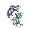
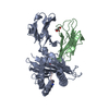
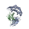
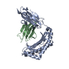
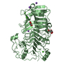
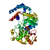
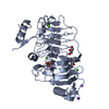
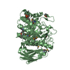
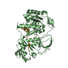
| Entry | Database: PDB / ID: 1zs8 | ||||||
|---|---|---|---|---|---|---|---|
| Title | Crystal Structure of the Murine MHC Class Ib Molecule M10.5 | ||||||
 Components Components |
| ||||||
 Keywords Keywords | IMMUNE SYSTEM / major histocompatibility complex / MHC / vomeronasal organ / VNO / V2R receptors / pheromone receptors / beta-2-microglobulin / peptides | ||||||
| Function / homology |  Function and homology information Function and homology informationantigen processing and presentation of peptide antigen via MHC class I / early endosome lumen / Nef mediated downregulation of MHC class I complex cell surface expression / DAP12 interactions / T cell mediated cytotoxicity / Endosomal/Vacuolar pathway / Antigen Presentation: Folding, assembly and peptide loading of class I MHC / lumenal side of endoplasmic reticulum membrane / antigen processing and presentation of exogenous protein antigen via MHC class Ib, TAP-dependent / regulation of iron ion transport ...antigen processing and presentation of peptide antigen via MHC class I / early endosome lumen / Nef mediated downregulation of MHC class I complex cell surface expression / DAP12 interactions / T cell mediated cytotoxicity / Endosomal/Vacuolar pathway / Antigen Presentation: Folding, assembly and peptide loading of class I MHC / lumenal side of endoplasmic reticulum membrane / antigen processing and presentation of exogenous protein antigen via MHC class Ib, TAP-dependent / regulation of iron ion transport / cellular response to iron(III) ion / negative regulation of iron ion transport / negative regulation of forebrain neuron differentiation / response to molecule of bacterial origin / regulation of erythrocyte differentiation / peptide antigen assembly with MHC class I protein complex / ER to Golgi transport vesicle membrane / HFE-transferrin receptor complex / MHC class I peptide loading complex / transferrin transport / negative regulation of receptor-mediated endocytosis / cellular response to iron ion / positive regulation of T cell cytokine production / multicellular organismal-level iron ion homeostasis / antigen processing and presentation of endogenous peptide antigen via MHC class I / MHC class I protein complex / peptide antigen assembly with MHC class II protein complex / positive regulation of T cell mediated cytotoxicity / negative regulation of epithelial cell proliferation / cellular response to nicotine / MHC class II protein complex / positive regulation of receptor-mediated endocytosis / negative regulation of neurogenesis / specific granule lumen / antigen processing and presentation of exogenous peptide antigen via MHC class II / positive regulation of immune response / peptide antigen binding / recycling endosome membrane / phagocytic vesicle membrane / positive regulation of T cell activation / Interferon gamma signaling / Immunoregulatory interactions between a Lymphoid and a non-Lymphoid cell / T cell differentiation in thymus / sensory perception of smell / Modulation by Mtb of host immune system / tertiary granule lumen / positive regulation of cellular senescence / MHC class II protein complex binding / DAP12 signaling / negative regulation of neuron projection development / late endosome membrane / protein refolding / ER-Phagosome pathway / early endosome membrane / amyloid fibril formation / protein homotetramerization / intracellular iron ion homeostasis / learning or memory / endoplasmic reticulum lumen / Amyloid fiber formation / external side of plasma membrane / Golgi membrane / lysosomal membrane / focal adhesion / Neutrophil degranulation / SARS-CoV-2 activates/modulates innate and adaptive immune responses / Golgi apparatus / structural molecule activity / endoplasmic reticulum / protein homodimerization activity / : / extracellular exosome / extracellular region / membrane / identical protein binding / plasma membrane / cytosol Similarity search - Function | ||||||
| Biological species |   Homo sapiens (human) Homo sapiens (human) | ||||||
| Method |  X-RAY DIFFRACTION / SYNCHROTRON / MOLECULAR REPLACEMENT / Resolution: 3 Å X-RAY DIFFRACTION / SYNCHROTRON / MOLECULAR REPLACEMENT / Resolution: 3 Å | ||||||
 Authors Authors | Olson, R. / Huey-Tubman, K.E. / Dulac, C. / Bjorkman, P.J. | ||||||
 Citation Citation | Journal: Plos Biol. / Year: 2005 Title: Structure of a pheromone receptor-associated MHC molecule with an open and empty groove. Authors: Olson, R. / Huey-Tubman, K.E. / Dulac, C. / Bjorkman, P.J. | ||||||
| History |
|
- Structure visualization
| Structure viewer | Molecule: MolmilJmol/JSmol |
|---|
- Downloads & links
-Download
| PDBx/mmCIF format | 1zs8.cif.gz | 352.3 KB | Display | PDBx/mmCIF format |
|---|---|---|---|---|
| PDB format | pdb1zs8.ent.gz | 282.6 KB | Display | PDB format |
| PDBx/mmJSON format | 1zs8.json.gz | Tree view | PDBx/mmJSON format | |
| Others |  Other downloads Other downloads |
-Validation report
| Arichive directory | https://data.pdbj.org/pub/pdb/validation_reports/zs/1zs8ftp://data.pdbj.org/pub/pdb/validation_reports/zs/1zs8 | HTTPS FTP |
|---|
-Related structure data


-Links
 PDBj
PDBj

- Assembly
| Deposited unit | 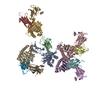
| ||||||||
|---|---|---|---|---|---|---|---|---|---|
| 1 | 
| ||||||||
| 2 | 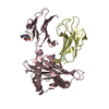
| ||||||||
| 3 | 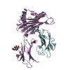
| ||||||||
| 4 | 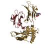
| ||||||||
| 5 | 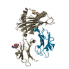
| ||||||||
| 6 | 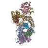
| ||||||||
| Unit cell |
| ||||||||
| Details | The biological assembly consists of chains A and B. To generate the asymmetric unit, the following transformations are required: MTRIX1 1 1.000000 0.000000 0.000000 0.00000 MTRIX2 1 0.000000 1.000000 0.000000 0.00000 MTRIX3 1 0.000000 0.000000 1.000000 0.00000 MTRIX1 2 0.983930 -0.015240 -0.177890 21.04371 MTRIX2 2 0.157760 -0.392300 0.906210 15.20531 MTRIX3 2 -0.083600 -0.919710 -0.383590 123.20857 MTRIX1 3 -0.995850 0.007030 0.090700 21.34146 MTRIX2 3 0.050020 -0.790450 0.610480 41.21530 MTRIX3 3 0.075980 0.612480 0.786820 -23.46491 MTRIX1 4 -0.990940 -0.026570 0.131680 6.34136 MTRIX2 4 -0.118920 -0.282390 -0.951900 47.17054 MTRIX3 4 0.062480 -0.958930 0.276670 112.45370 MTRIX1 5 0.986590 0.035450 -0.159320 33.74422 MTRIX2 5 -0.062000 -0.821580 -0.566710 38.04500 MTRIX3 5 -0.150980 0.568990 -0.808360 91.82664 |
-Components
| #1: Protein | Mass: 31421.334 Da / Num. of mol.: 5 Source method: isolated from a genetically manipulated source Source: (gene. exp.)  Trichoplusia ni (cabbage looper) / Strain (production host): Tn5 / References: GenBank: 29244030, UniProt: Q860W6*PLUS Trichoplusia ni (cabbage looper) / Strain (production host): Tn5 / References: GenBank: 29244030, UniProt: Q860W6*PLUS#2: Protein | Mass: 11748.160 Da / Num. of mol.: 5 Source method: isolated from a genetically manipulated source Source: (gene. exp.) Homo sapiens (human) / Gene: B2M / Plasmid: pACUW31 / Production host: Trichoplusia ni (cabbage looper) / Strain (production host): Tn5 / References: UniProt: P61769#3: Sugar | ChemComp-NAG /   Type: D-saccharide, beta linking / Mass: 221.208 Da / Num. of mol.: 5 Type: D-saccharide, beta linking / Mass: 221.208 Da / Num. of mol.: 5Source method: isolated from a genetically manipulated source Formula: C8H15NO6 Has protein modification | Y | |
|---|
-Experimental details
-Experiment
| Experiment | Method: X-RAY DIFFRACTION / Number of used crystals: 2 |
|---|
- Sample preparation
| Crystal | Density Matthews: 2.5 Å3/Da / Density % sol: 50.4 % |
|---|---|
| Crystal grow | Temperature: 298 K / Method: vapor diffusion, hanging drop / pH: 8 Details: 0.1 M imidazole, 20% PEG 1000, 0.2 M calcium acetate, pH 8.0, VAPOR DIFFUSION, HANGING DROP, temperature 298K |
-Data collection
| Diffraction |
| |||||||||||||||
|---|---|---|---|---|---|---|---|---|---|---|---|---|---|---|---|---|
| Diffraction source |
| |||||||||||||||
| Detector |
| |||||||||||||||
| Radiation | Monochromator: double crystal / Protocol: SINGLE WAVELENGTH / Monochromatic (M) / Laue (L): M / Scattering type: x-ray | |||||||||||||||
| Radiation wavelength |
| |||||||||||||||
| Reflection | Resolution: 3→99 Å / Num. all: 46977 / Num. obs: 46977 / % possible obs: 91.2 % / Observed criterion σ(F): 0 / Observed criterion σ(I): 0 / Redundancy: 5.7 % / Biso Wilson estimate: 76.6 Å2 / Rmerge(I) obs: 0.156 / Rsym value: 0.156 / Χ2: 1.106 / Net I/σ(I): 9 | |||||||||||||||
| Reflection shell | Resolution: 3→3.11 Å / % possible obs: 64.8 % / Redundancy: 2.7 % / Rmerge(I) obs: 0.535 / Mean I/σ(I) obs: 2.7 / Num. measured obs: 3287 / Num. unique all: 3287 / Rsym value: 0.535 / Χ2: 0.814 / % possible all: 64.8 |

- Processing
| Software |
| |||||||||||||||||||||||||
|---|---|---|---|---|---|---|---|---|---|---|---|---|---|---|---|---|---|---|---|---|---|---|---|---|---|---|
| Refinement | Method to determine structure: MOLECULAR REPLACEMENT Starting model: PDB ENTRY 1K8D (confirmed with 3FRU) Resolution: 3→56.36 Å / Rfactor Rfree error: 0.006 / Data cutoff high absF: 2322336.75 / Data cutoff low absF: 0 / Isotropic thermal model: GROUP / Cross valid method: THROUGHOUT / σ(F): 0 / σ(I): 0 / Stereochemistry target values: Engh & Huber / Details: BULK SOLVENT MODEL USED
| |||||||||||||||||||||||||
| Solvent computation | Solvent model: FLAT MODEL / Bsol: 98.338 Å2 / ksol: 0.424 e/Å3 | |||||||||||||||||||||||||
| Displacement parameters | Biso mean: 59.1 Å2
| |||||||||||||||||||||||||
| Refine analyze |
| |||||||||||||||||||||||||
| Refinement step | Cycle: LAST / Resolution: 3→56.36 Å
| |||||||||||||||||||||||||
| Refine LS restraints |
| |||||||||||||||||||||||||
| Refine LS restraints NCS | NCS model details: RESTRAINTS | |||||||||||||||||||||||||
| LS refinement shell | Resolution: 3→3.19 Å / Rfactor Rfree error: 0.026 / Total num. of bins used: 6
| |||||||||||||||||||||||||
| Xplor file |
|
