 Movie
Movie Controller

[English] 日本語
 Yorodumi
- PDB-4f7t: Crystal Structure of HLA-A*2402 Complexed with a Newly Identified... -
+ Open data

- Basic information
| Entry | Database: PDB / ID: 4f7t | ||||||
|---|---|---|---|---|---|---|---|
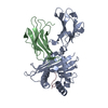
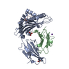
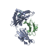
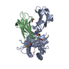
| Title | Crystal Structure of HLA-A*2402 Complexed with a Newly Identified Peptide from 2009 H1N1 PB1 (498-505) | ||||||
 Components Components |
| ||||||
 Keywords Keywords | IMMUNE SYSTEM / HLA-A*2402 / 2009H1N1 / HLA-A3 supertype / cross-allele recognition | ||||||
| Function / homology |  Function and homology information Function and homology informationregulation of natural killer cell mediated immunity / positive regulation of memory T cell activation / T cell mediated cytotoxicity directed against tumor cell target / antigen processing and presentation of exogenous peptide antigen via MHC class Ib / positive regulation of CD8-positive, alpha-beta T cell activation / CD8-positive, alpha-beta T cell activation / positive regulation of CD8-positive, alpha-beta T cell proliferation / antigen processing and presentation of endogenous peptide antigen via MHC class I via ER pathway, TAP-dependent / TAP complex binding / antigen processing and presentation of exogenous peptide antigen via MHC class I ...regulation of natural killer cell mediated immunity / positive regulation of memory T cell activation / T cell mediated cytotoxicity directed against tumor cell target / antigen processing and presentation of exogenous peptide antigen via MHC class Ib / positive regulation of CD8-positive, alpha-beta T cell activation / CD8-positive, alpha-beta T cell activation / positive regulation of CD8-positive, alpha-beta T cell proliferation / antigen processing and presentation of endogenous peptide antigen via MHC class I via ER pathway, TAP-dependent / TAP complex binding / antigen processing and presentation of exogenous peptide antigen via MHC class I / Golgi medial cisterna / CD8 receptor binding / viral transcription / symbiont-mediated suppression of host mRNA transcription via inhibition of RNA polymerase II activity / protection from natural killer cell mediated cytotoxicity / endoplasmic reticulum exit site / TAP binding / detection of bacterium / antigen processing and presentation of endogenous peptide antigen via MHC class Ib / antigen processing and presentation of endogenous peptide antigen via MHC class I via ER pathway, TAP-independent / beta-2-microglobulin binding / T cell receptor binding / early endosome lumen / Nef mediated downregulation of MHC class I complex cell surface expression / DAP12 interactions / T cell mediated cytotoxicity / Endosomal/Vacuolar pathway / Antigen Presentation: Folding, assembly and peptide loading of class I MHC / lumenal side of endoplasmic reticulum membrane / antigen processing and presentation of exogenous protein antigen via MHC class Ib, TAP-dependent / regulation of iron ion transport / cellular response to iron(III) ion / negative regulation of iron ion transport / negative regulation of forebrain neuron differentiation / response to molecule of bacterial origin / regulation of erythrocyte differentiation / peptide antigen assembly with MHC class I protein complex / ER to Golgi transport vesicle membrane / HFE-transferrin receptor complex / MHC class I peptide loading complex / transferrin transport / negative regulation of receptor-mediated endocytosis / cellular response to iron ion / positive regulation of T cell cytokine production / multicellular organismal-level iron ion homeostasis / antigen processing and presentation of endogenous peptide antigen via MHC class I / MHC class I protein complex / peptide antigen assembly with MHC class II protein complex / positive regulation of T cell mediated cytotoxicity / negative regulation of epithelial cell proliferation / cellular response to nicotine / MHC class II protein complex / positive regulation of receptor-mediated endocytosis / negative regulation of neurogenesis / specific granule lumen / antigen processing and presentation of exogenous peptide antigen via MHC class II / positive regulation of immune response / positive regulation of type II interferon production / peptide antigen binding / recycling endosome membrane / phagocytic vesicle membrane / positive regulation of T cell activation / Interferon gamma signaling / Immunoregulatory interactions between a Lymphoid and a non-Lymphoid cell / T cell differentiation in thymus / sensory perception of smell / Interferon alpha/beta signaling / Modulation by Mtb of host immune system / tertiary granule lumen / positive regulation of cellular senescence / MHC class II protein complex binding / DAP12 signaling / T cell receptor signaling pathway / negative regulation of neuron projection development / late endosome membrane / E3 ubiquitin ligases ubiquitinate target proteins / protein refolding / antibacterial humoral response / ER-Phagosome pathway / early endosome membrane / amyloid fibril formation / protein homotetramerization / host cell cytoplasm / intracellular iron ion homeostasis / learning or memory / defense response to Gram-positive bacterium / immune response / endoplasmic reticulum lumen / Amyloid fiber formation / symbiont-mediated suppression of host gene expression / external side of plasma membrane / signaling receptor binding / Golgi membrane / innate immune response / RNA-directed RNA polymerase / lysosomal membrane / focal adhesion / viral RNA genome replication / nucleotide binding / RNA-directed RNA polymerase activity Similarity search - Function | ||||||
| Biological species |  Homo sapiens (human) Homo sapiens (human) Influenza A virus H3N2 Influenza A virus H3N2 | ||||||
| Method |  X-RAY DIFFRACTION / MOLECULAR REPLACEMENT / Resolution: 1.7 Å X-RAY DIFFRACTION / MOLECULAR REPLACEMENT / Resolution: 1.7 Å | ||||||
 Authors Authors | Liu, J. / Zhang, S. / Tan, S. / Yi, Y. / Wu, B. / Zhu, F. / Wang, H. / Qi, J. / Gao, G.F. | ||||||
 Citation Citation | Journal: J.Virol. / Year: 2012 Title: Cross-Allele Cytotoxic T Lymphocyte Responses against 2009 Pandemic H1N1 Influenza A Virus among HLA-A24 and HLA-A3 Supertype-Positive Individuals. Authors: Liu, J. / Zhang, S. / Tan, S. / Yi, Y. / Wu, B. / Cao, B. / Zhu, F. / Wang, C. / Wang, H. / Qi, J. / Gao, G.F. | ||||||
| History |
|
- Structure visualization
| Structure viewer | Molecule: MolmilJmol/JSmol |
|---|
- Downloads & links
-Download
| PDBx/mmCIF format | 4f7t.cif.gz | 350.1 KB | Display | PDBx/mmCIF format |
|---|---|---|---|---|
| PDB format | pdb4f7t.ent.gz | 286 KB | Display | PDB format |
| PDBx/mmJSON format | 4f7t.json.gz | Tree view | PDBx/mmJSON format | |
| Others |  Other downloads Other downloads |
-Validation report
| Arichive directory | https://data.pdbj.org/pub/pdb/validation_reports/f7/4f7tftp://data.pdbj.org/pub/pdb/validation_reports/f7/4f7t | HTTPS FTP |
|---|
-Related structure data
| Related structure data |  4f7mC  4f7pC  3i6lS S: Starting model for refinement C: citing same article ( |
|---|---|
| Similar structure data |

-Links
 PDBj
PDBj

- Assembly
| Deposited unit | 
| ||||||||
|---|---|---|---|---|---|---|---|---|---|
| 1 | 
| ||||||||
| 2 | 
| ||||||||
| Unit cell |
|
-Components
| #1: Protein | Mass: 31683.086 Da / Num. of mol.: 2 / Fragment: unp residues 25-298 Source method: isolated from a genetically manipulated source Source: (gene. exp.) Homo sapiens (human) / Gene: HLA-A, HLAA / Production host:  #2: Protein | Mass: 11879.356 Da / Num. of mol.: 2 / Fragment: unp residues 21-119 Source method: isolated from a genetically manipulated source Source: (gene. exp.) Homo sapiens (human) / Gene: B2M, CDABP0092, HDCMA22P / Production host: #3: Protein/peptide | Mass: 974.094 Da / Num. of mol.: 2 / Fragment: unp residues 498-505 / Source method: obtained synthetically Details: in vitro synthesized peptide from 2009H1N1 PB1(498-505) Source: (synth.) Influenza A virus H3N2 / References: UniProt: Q9YXL6, RNA-directed RNA polymerase#4: Water | ChemComp-HOH / |  Mass: 18.015 Da / Num. of mol.: 881 / Source method: isolated from a natural source / Formula: H2O Mass: 18.015 Da / Num. of mol.: 881 / Source method: isolated from a natural source / Formula: H2OHas protein modification | Y | |
|---|
-Experimental details
-Experiment
| Experiment | Method: X-RAY DIFFRACTION / Number of used crystals: 1 |
|---|
- Sample preparation
| Crystal | Density Matthews: 2.47 Å3/Da / Density % sol: 50.14 % |
|---|---|
| Crystal grow | Temperature: 293 K / pH: 5.5 Details: 0.1 M Bis-Tris and 10% (w/v) polyethylene glycol 3,350, pH 5.5, VAPOR DIFFUSION, HANGING DROP, temperature 293K |
-Data collection
| Diffraction | Mean temperature: 100 K |
|---|---|
| Diffraction source | Source: ROTATING ANODE / Type: RIGAKU MICROMAX-007 / Wavelength: 1.5418 |
| Detector | Type: RIGAKU RAXIS VII / Detector: IMAGE PLATE / Date: Nov 13, 2010 |
| Radiation | Monochromator: SI 111 CHANNEL / Protocol: SINGLE WAVELENGTH / Monochromatic (M) / Laue (L): M / Scattering type: x-ray |
| Radiation wavelength | Wavelength: 1.5418 Å / Relative weight: 1 |
| Reflection | Resolution: 1.7→50 Å / Num. obs: 90267 / % possible obs: 96.1 % / Observed criterion σ(I): -3 / Redundancy: 3.3 % / Rmerge(I) obs: 0.045 / Rsym value: 0.045 / Net I/σ(I): 25.58 |
| Reflection shell | Resolution: 1.7→1.76 Å / Redundancy: 3.1 % / Rmerge(I) obs: 0.289 / Mean I/σ(I) obs: 4.264 / Rsym value: 0.289 / % possible all: 93.8 |
- Processing
| Software |
| |||||||||||||||||||||||||||||||||||||||||||||||||||||||||||||||||||||||||||||||||||||||||||||||||||||||||||||||||||||||||||||||||||||||||||||||||||||||||||||||||||||||||||||||||||||||||||||||||||||||||||||||||||||||||
|---|---|---|---|---|---|---|---|---|---|---|---|---|---|---|---|---|---|---|---|---|---|---|---|---|---|---|---|---|---|---|---|---|---|---|---|---|---|---|---|---|---|---|---|---|---|---|---|---|---|---|---|---|---|---|---|---|---|---|---|---|---|---|---|---|---|---|---|---|---|---|---|---|---|---|---|---|---|---|---|---|---|---|---|---|---|---|---|---|---|---|---|---|---|---|---|---|---|---|---|---|---|---|---|---|---|---|---|---|---|---|---|---|---|---|---|---|---|---|---|---|---|---|---|---|---|---|---|---|---|---|---|---|---|---|---|---|---|---|---|---|---|---|---|---|---|---|---|---|---|---|---|---|---|---|---|---|---|---|---|---|---|---|---|---|---|---|---|---|---|---|---|---|---|---|---|---|---|---|---|---|---|---|---|---|---|---|---|---|---|---|---|---|---|---|---|---|---|---|---|---|---|---|---|---|---|---|---|---|---|---|---|---|---|---|---|---|---|---|
| Refinement | Method to determine structure: MOLECULAR REPLACEMENT Starting model: 3I6L Resolution: 1.7→24.42 Å / SU ML: 0.23 / σ(F): 0.09 / Phase error: 23.95 / Stereochemistry target values: ML
| |||||||||||||||||||||||||||||||||||||||||||||||||||||||||||||||||||||||||||||||||||||||||||||||||||||||||||||||||||||||||||||||||||||||||||||||||||||||||||||||||||||||||||||||||||||||||||||||||||||||||||||||||||||||||
| Solvent computation | Shrinkage radii: 0.9 Å / VDW probe radii: 1.11 Å / Solvent model: FLAT BULK SOLVENT MODEL / Bsol: 48.93 Å2 / ksol: 0.41 e/Å3 | |||||||||||||||||||||||||||||||||||||||||||||||||||||||||||||||||||||||||||||||||||||||||||||||||||||||||||||||||||||||||||||||||||||||||||||||||||||||||||||||||||||||||||||||||||||||||||||||||||||||||||||||||||||||||
| Displacement parameters |
| |||||||||||||||||||||||||||||||||||||||||||||||||||||||||||||||||||||||||||||||||||||||||||||||||||||||||||||||||||||||||||||||||||||||||||||||||||||||||||||||||||||||||||||||||||||||||||||||||||||||||||||||||||||||||
| Refinement step | Cycle: LAST / Resolution: 1.7→24.42 Å
| |||||||||||||||||||||||||||||||||||||||||||||||||||||||||||||||||||||||||||||||||||||||||||||||||||||||||||||||||||||||||||||||||||||||||||||||||||||||||||||||||||||||||||||||||||||||||||||||||||||||||||||||||||||||||
| Refine LS restraints |
| |||||||||||||||||||||||||||||||||||||||||||||||||||||||||||||||||||||||||||||||||||||||||||||||||||||||||||||||||||||||||||||||||||||||||||||||||||||||||||||||||||||||||||||||||||||||||||||||||||||||||||||||||||||||||
| LS refinement shell |
| |||||||||||||||||||||||||||||||||||||||||||||||||||||||||||||||||||||||||||||||||||||||||||||||||||||||||||||||||||||||||||||||||||||||||||||||||||||||||||||||||||||||||||||||||||||||||||||||||||||||||||||||||||||||||
| Refinement TLS params. | Method: refined / Origin x: -16.6265 Å / Origin y: -14.8161 Å / Origin z: -24.1268 Å
| |||||||||||||||||||||||||||||||||||||||||||||||||||||||||||||||||||||||||||||||||||||||||||||||||||||||||||||||||||||||||||||||||||||||||||||||||||||||||||||||||||||||||||||||||||||||||||||||||||||||||||||||||||||||||
| Refinement TLS group | Selection details: all |
