 Movie
Movie Controller

[English] 日本語
 Yorodumi
- PDB-6kwk: Crystal structure of pSLA-1*0401 complex with FMDV-derived epitop... -
+ Open data

- Basic information
| Entry | Database: PDB / ID: 6kwk | ||||||
|---|---|---|---|---|---|---|---|
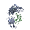
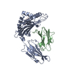
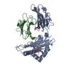
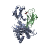
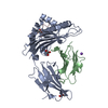
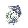
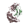
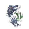
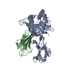
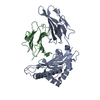
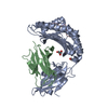
| Title | Crystal structure of pSLA-1*0401 complex with FMDV-derived epitope MTAHITVPY | ||||||
 Components Components |
| ||||||
 Keywords Keywords | STRUCTURAL PROTEIN / MHC class I structure / A single-amino acid mutation / Peptide motifs / Random peptide library | ||||||
| Function / homology |  Function and homology information Function and homology informationER-Phagosome pathway / Endosomal/Vacuolar pathway / Immunoregulatory interactions between a Lymphoid and a non-Lymphoid cell / DAP12 signaling / Antigen Presentation: Folding, assembly and peptide loading of class I MHC / L-peptidase / symbiont-mediated perturbation of host chromatin organization / Neutrophil degranulation / response to metal ion / antigen processing and presentation of peptide antigen via MHC class I ...ER-Phagosome pathway / Endosomal/Vacuolar pathway / Immunoregulatory interactions between a Lymphoid and a non-Lymphoid cell / DAP12 signaling / Antigen Presentation: Folding, assembly and peptide loading of class I MHC / L-peptidase / symbiont-mediated perturbation of host chromatin organization / Neutrophil degranulation / response to metal ion / antigen processing and presentation of peptide antigen via MHC class I / lumenal side of endoplasmic reticulum membrane / picornain 3C / T=pseudo3 icosahedral viral capsid / MHC class I protein complex / peptide antigen assembly with MHC class II protein complex / MHC class II protein complex / host cell cytoplasmic vesicle membrane / antigen processing and presentation of exogenous peptide antigen via MHC class II / positive regulation of immune response / peptide antigen binding / phagocytic vesicle membrane / positive regulation of T cell activation / MHC class II protein complex binding / ribonucleoside triphosphate phosphatase activity / late endosome membrane / nucleoside-triphosphate phosphatase / channel activity / monoatomic ion transmembrane transport / clathrin-dependent endocytosis of virus by host cell / RNA helicase activity / immune response / viral protein processing / host cell endoplasmic reticulum membrane / symbiont-mediated activation of host autophagy / RNA-directed RNA polymerase / lysosomal membrane / cysteine-type endopeptidase activity / viral RNA genome replication / RNA-directed RNA polymerase activity / virion attachment to host cell / host cell nucleus / structural molecule activity / DNA-templated transcription / proteolysis / RNA binding / extracellular region / ATP binding Similarity search - Function | ||||||
| Biological species |    Foot-and-mouth disease virus Foot-and-mouth disease virus | ||||||
| Method |  X-RAY DIFFRACTION / SYNCHROTRON / MOLECULAR REPLACEMENT / Resolution: 2.5 Å X-RAY DIFFRACTION / SYNCHROTRON / MOLECULAR REPLACEMENT / Resolution: 2.5 Å | ||||||
 Authors Authors | Wei, X.H. / Wang, S. / Zhang, N.Z. / Xia, C. | ||||||
| Funding support |  China, 1items China, 1items
| ||||||
 Citation Citation | Journal: Front Immunol / Year: 2021 Title: Peptidomes and Structures Illustrate Two Distinguishing Mechanisms of Alternating the Peptide Plasticity Caused by Swine MHC Class I Micropolymorphism. Authors: Wei, X. / Wang, S. / Li, Z. / Li, Z. / Qu, Z. / Wang, S. / Zou, B. / Liang, R. / Xia, C. / Zhang, N. | ||||||
| History |
|
- Structure visualization
| Structure viewer | Molecule: MolmilJmol/JSmol |
|---|
- Downloads & links
-Download
| PDBx/mmCIF format | 6kwk.cif.gz | 96.7 KB | Display | PDBx/mmCIF format |
|---|---|---|---|---|
| PDB format | pdb6kwk.ent.gz | 72.1 KB | Display | PDB format |
| PDBx/mmJSON format | 6kwk.json.gz | Tree view | PDBx/mmJSON format | |
| Others |  Other downloads Other downloads |
-Validation report
| Arichive directory | https://data.pdbj.org/pub/pdb/validation_reports/kw/6kwkftp://data.pdbj.org/pub/pdb/validation_reports/kw/6kwk | HTTPS FTP |
|---|
-Related structure data
| Related structure data | 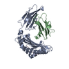 6kwlC  6kwnC  6kwoC  3qq3S S: Starting model for refinement C: citing same article ( |
|---|---|
| Similar structure data |


-Links
 PDBj
PDBj


- Assembly
| Deposited unit | 
| ||||||||
|---|---|---|---|---|---|---|---|---|---|
| 1 |
| ||||||||
| Unit cell |
|
-Components
| #1: Protein | Mass: 31681.988 Da / Num. of mol.: 1 Source method: isolated from a genetically manipulated source Source: (gene. exp.)  |
|---|---|
| #2: Protein | Mass: 11579.093 Da / Num. of mol.: 1 Source method: isolated from a genetically manipulated source Source: (gene. exp.) |
| #3: Protein/peptide | Mass: 1033.220 Da / Num. of mol.: 1 / Source method: obtained synthetically / Source: (synth.) Foot-and-mouth disease virus / References: UniProt: P03311*PLUS |
| #4: Water | ChemComp-HOH /  Mass: 18.015 Da / Num. of mol.: 207 / Source method: isolated from a natural source / Formula: H2O Mass: 18.015 Da / Num. of mol.: 207 / Source method: isolated from a natural source / Formula: H2O |
| Has protein modification | Y |
-Experimental details
-Experiment
| Experiment | Method: X-RAY DIFFRACTION / Number of used crystals: 1 |
|---|
- Sample preparation
| Crystal | Density Matthews: 2.1 Å3/Da / Density % sol: 41.31 % |
|---|---|
| Crystal grow | Temperature: 277 K / Method: vapor diffusion, sitting drop / pH: 6.5 Details: 0.05 M Calcium Chloride dihydrate, 0.1 M Bis-Tris pH 6.5, 30% v/v Polyethylene Glycol Monomethyl ether 550 |
-Data collection
| Diffraction | Mean temperature: 100 K / Serial crystal experiment: N |
|---|---|
| Diffraction source | Source: SYNCHROTRON / Site: SSRF / Beamline: BL17U / Wavelength: 0.97931 Å |
| Detector | Type: ADSC QUANTUM 315r / Detector: CCD / Date: Aug 17, 2016 |
| Radiation | Protocol: SINGLE WAVELENGTH / Monochromatic (M) / Laue (L): M / Scattering type: x-ray |
| Radiation wavelength | Wavelength: 0.97931 Å / Relative weight: 1 |
| Reflection | Resolution: 2→28.59 Å / Num. obs: 23996 / % possible obs: 96.8 % / Redundancy: 6 % / Rmerge(I) obs: 0.057 / Rsym value: 0.057 / Net I/σ(I): 26.535 |
| Reflection shell | Resolution: 2→2.07 Å / Redundancy: 6.2 % / Rmerge(I) obs: 0.157 / Mean I/σ(I) obs: 12.486 / Num. unique obs: 23996 / Rsym value: 0.157 / % possible all: 96.8 |
- Processing
| Software |
| |||||||||||||||||||||||||||||||||||
|---|---|---|---|---|---|---|---|---|---|---|---|---|---|---|---|---|---|---|---|---|---|---|---|---|---|---|---|---|---|---|---|---|---|---|---|---|
| Refinement | Method to determine structure: MOLECULAR REPLACEMENT Starting model: 3QQ3 Resolution: 2.5→28.577 Å / SU ML: 0.33 / Cross valid method: NONE / σ(F): 1.39 / Phase error: 29.82 / Stereochemistry target values: ML
| |||||||||||||||||||||||||||||||||||
| Solvent computation | Shrinkage radii: 0.9 Å / VDW probe radii: 1.11 Å / Solvent model: FLAT BULK SOLVENT MODEL | |||||||||||||||||||||||||||||||||||
| Refinement step | Cycle: LAST / Resolution: 2.5→28.577 Å
| |||||||||||||||||||||||||||||||||||
| Refine LS restraints |
| |||||||||||||||||||||||||||||||||||
| LS refinement shell |
|
